

Hyperfibrinolysis as an Underrecognized Cause of Bleeding
Bleeding disorders are commonly investigated through platelet function, coagulation factors and routine hemostatic tests, while abnormalities of fibrinolysis may receive less attention.
Fibrinolysis is the physiological process responsible for breaking down fibrin clots once they have fulfilled their hemostatic function. However, when this process becomes excessively active, clots may dissolve prematurely, resulting in persistent or recurrent bleeding.
Hyperfibrinolysis can arise from inherited abnormalities, including deficiencies of α2-antiplasmin or plasminogen activator inhibitor-1 (PAI-1), as well as from acquired conditions.
Clinically, affected individuals may experience delayed bleeding after surgery, trauma, or dental procedures, alongside mucosal bleeding and heavy menstrual bleeding.
Diagnosis can be challenging because routine coagulation tests may remain normal, while specialized fibrinolysis assays are not universally available.
Consequently, hyperfibrinolysis remains a potentially underrecognized cause of bleeding, highlighting the need to consider impaired clot stability when standard investigations fail to explain the clinical presentation.
From Plasmin Generation to Fibrin Degradation
Fibrinolysis is a proteolytic process centered on the conversion of plasminogen into plasmin, the principal enzyme responsible for fibrin degradation.
Its activity is spatially regulated by interactions among fibrin, plasminogen, and tissue-type plasminogen activator (tPA), which allow proteolysis to occur preferentially on the surface of a formed clot.
Following thrombin-mediated cleavage of fibrinogen, fibrin monomers polymerize into a network that is subsequently stabilized by activated coagulation factor XIII (FXIIIa).
Endothelial-derived tPA binds to fibrin and catalyzes the conversion of fibrin-associated plasminogen into plasmin through cleavage of a specific peptide bond.
Plasmin then degrades fibrin into soluble fibrin degradation products, including D-dimer, progressively disrupting the structural integrity of the clot.
This process is amplified by the exposure of new C-terminal lysine residues during fibrin degradation, which recruit additional plasminogen and tPA to the fibrin surface and facilitate further plasmin generation.
The extent of fibrinolysis is determined by a balance between proteolytic activation and several regulatory mechanisms.
Plasminogen activator inhibitor-1 (PAI-1) inhibits tPA and urokinase-type plasminogen activator (uPA), limiting further plasmin generation.
In contrast, α2-antiplasmin directly neutralizes free plasmin by forming inactive enzyme–inhibitor complexes and is incorporated into the fibrin network through FXIIIa-mediated cross-linking.
This incorporation is particularly important because it provides localized protection against premature clot dissolution.
A complementary mechanism is mediated by thrombin-activatable fibrinolysis inhibitor (TAFI), which is activated efficiently by the thrombin–thrombomodulin complex.
Activated TAFI removes C-terminal lysine residues from partially degraded fibrin, reducing the binding of plasminogen and tPA and attenuating the positive-feedback amplification of fibrinolysis.
Together, these mechanisms ensure that fibrin degradation remains appropriately regulated rather than progressing uncontrollably.
The clinical significance of this regulatory network lies in its ability to preserve clot integrity after initial hemostasis has been achieved.
Reduced α2-antiplasmin activity, impaired PAI-1 function, excessive plasminogen activation, or inadequate antifibrinolytic protection can shift the system toward accelerated fibrin degradation.
Importantly, fibrinolytic activity depends not only on the concentrations of individual proteins but also on their functional interactions, localization, and the structural properties of the fibrin network.
Consequently, hyperfibrinolysis may arise from distinct molecular abnormalities that converge on a common endpoint: premature loss of fibrin-dependent clot stability.
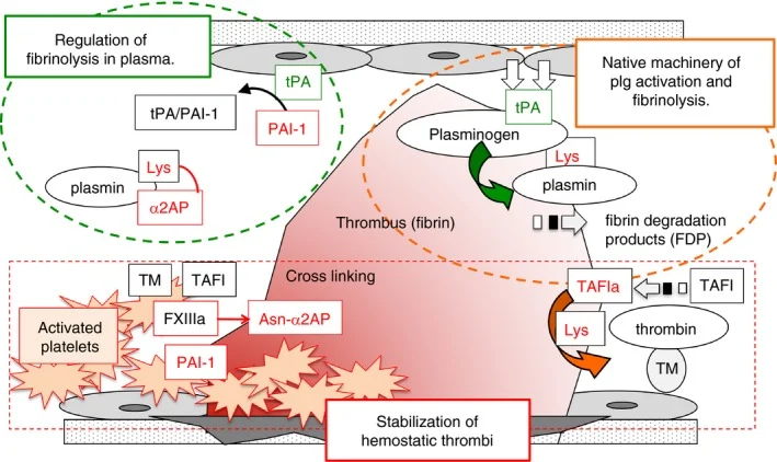
Figure 1: Urano T, Castellino FJ, Suzuki Y. Regulation of plasminogen activation on cell surfaces and fibrin. J Thromb Haemost. 2018 May 20;16(8):1487–97. doi: 10.1111/jth.14157. Epub ahead of print. PMID: 29779246; PMCID: PMC6099326.
Pathological Hyperfibrinolysis: When Clot Breakdown Becomes Excessive
Pathological hyperfibrinolysis develops when plasmin generation and fibrin degradation exceed the capacity of endogenous mechanisms to preserve clot integrity.
This imbalance can result from excessive plasminogen activation, inadequate inhibition of plasmin or its activators, or increased susceptibility of the fibrin network to proteolytic degradation.
At the molecular level, these abnormalities converge on accelerated loss of fibrin, compromising the mechanical stability of hemostatic plugs and promoting recurrent bleeding.
Importantly, hyperfibrinolysis is a functional phenotype rather than a single molecular defect, and its underlying mechanisms differ between inherited disorders and acquired coagulopathies.
1. One major mechanism is:
Excessive tissue-type plasminogen activator (tPA) activity, which increases the rate of plasmin generation on fibrin and accelerates clot dissolution.
This mechanism is particularly relevant in trauma-associated coagulopathy, where endothelial activation and injury, together with tissue hypoperfusion, can trigger substantial tPA release into the circulation.
Reduced functional availability of plasminogen activator inhibitor-1 (PAI-1) may further amplify this response by weakening inhibition of circulating and fibrin-associated tPA.
However, traumatic hyperfibrinolysis cannot be attributed to a single pathway: endothelial dysfunction, shock, platelet abnormalities, and depletion or dysfunction of other fibrinolytic regulators can contribute simultaneously.
2. A second mechanism involves:
Insufficient inhibition of plasmin after it has been generated.
α2-Antiplasmin normally neutralizes free plasmin and limits its proteolytic activity, while its incorporation into fibrin by activated factor XIII provides localized protection against clot degradation.
Reduced α2-antiplasmin activity or impaired incorporation into the fibrin network therefore increases susceptibility to premature fibrinolysis.
Similarly, reduced PAI-1 activity permits continued activation of plasminogen by tPA and urokinase-type plasminogen activator (uPA).
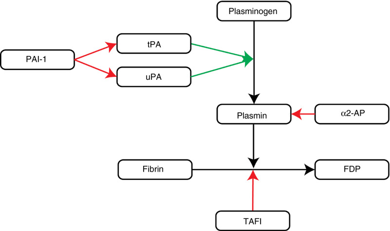
Figure 2: Saes JL, Schols SEM, van Heerde WL, Nijziel MR. Hemorrhagic disorders of fibrinolysis: a clinical review. J Thromb Haemost. 2018 May 30. doi: 10.1111/jth.14160. Epub ahead of print. PMID: 29847021.
These defects are mechanistically distinct:
PAI-1 deficiency primarily compromises the control of plasmin generation, whereas α2-antiplasmin deficiency impairs the neutralization of plasmin that has already formed.
Clot architecture and the regulation of fibrin-associated plasminogen activation also influence the severity of hyperfibrinolysis.
Reduced factor XIII activity, impaired fibrin cross-linking, or inadequate incorporation of α2-antiplasmin can produce a fibrin network that is less resistant to enzymatic degradation.
In addition, excessive activity of the fibrinolytic system may be compounded by depletion or dysfunction of thrombin-activatable fibrinolysis inhibitor (TAFI), which normally limits the recruitment of plasminogen and tPA to partially degraded fibrin.
These mechanisms demonstrate that pathological clot dissolution may reflect both increased proteolytic activity and inadequate structural or biochemical resistance of the clot.
Acquired hyperfibrinolysis is particularly complex because the balance between fibrinolytic activation and inhibition can change rapidly during systemic illness.
In severe trauma, early tPA-mediated fibrinolytic activation may evolve into fibrinolysis shutdown, characterized by markedly reduced clot lysis and often associated with increased plasminogen activator inhibitor activity.
Consequently, hyperfibrinolysis and fibrinolysis shutdown represent distinct, potentially time-dependent phenotypes rather than interchangeable features of trauma-induced coagulopathy.
Recognizing this distinction is essential because the same clinical context can involve different mechanisms of hemostatic failure, requiring careful assessment of the patient’s current fibrinolytic state.
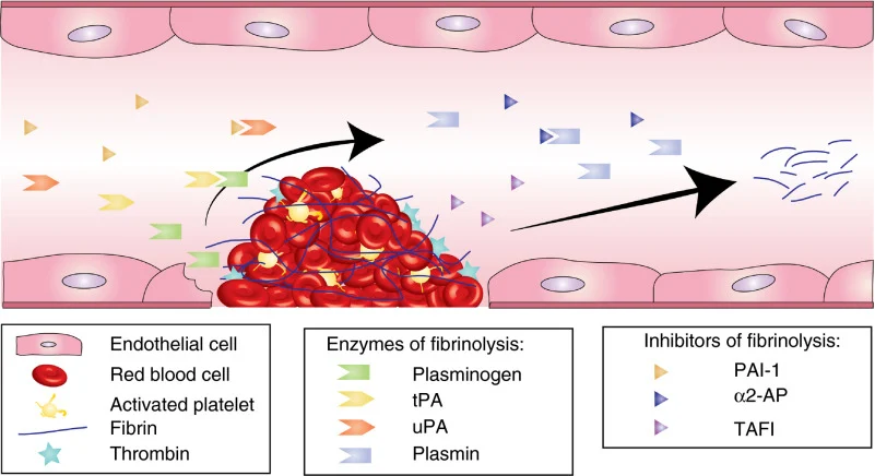
Figure 3: Saes JL, Schols SEM, van Heerde WL, Nijziel MR. Hemorrhagic disorders of fibrinolysis: a clinical review. J Thromb Haemost. 2018 May 30. doi: 10.1111/jth.14160. Epub ahead of print. PMID: 29847021.
Inherited Disorders of Fibrinolysis
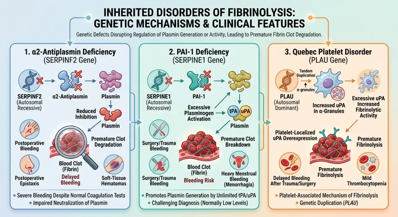
Inherited hyperfibrinolytic disorders arise from genetic abnormalities that disrupt the regulation of plasmin generation or activity, resulting in premature degradation of hemostatic clots.
The three principal disorders are:
- α2-antiplasmin deficiency
- plasminogen activator inhibitor-1 (PAI-1) deficiency
- Quebec platelet disorder (QPD)
Although their molecular mechanisms differ, all three demonstrate how abnormalities in fibrinolytic regulation can produce clinically significant bleeding despite normal or near-normal results on routine coagulation testing.
1. α2-Antiplasmin deficiency is caused by pathogenic variants in SERPINF2, the gene encoding the principal physiological inhibitor of plasmin.
The disorder is usually inherited in an autosomal recessive manner, and severe deficiency is associated with impaired neutralization of plasmin and increased susceptibility of fibrin clots to premature degradation.
Because α2-antiplasmin is also incorporated into the fibrin network through factor XIIIa-mediated cross-linking, its deficiency compromises both the systemic inhibition of plasmin and the localized protection of the clot.
Clinical manifestations include delayed postoperative bleeding, recurrent epistaxis, soft-tissue hematomas, and bleeding following dental extraction or trauma.
The severity varies with residual inhibitor activity, and severe cases may develop deep tissue or intramuscular hemorrhage.
Diagnosis relies on specific α2-antiplasmin activity testing, supplemented by antigen measurement and genetic analysis when indicated.
2. PAI-1 deficiency results from pathogenic variants in SERPINE1, which encodes the principal physiological inhibitor of tissue-type plasminogen activator (tPA) and urokinase-type plasminogen activator (uPA).
Unlike α2-antiplasmin deficiency, which impairs plasmin neutralization, PAI-1 deficiency promotes excessive plasmin generation by allowing continued plasminogen activation.
Complete congenital deficiency is generally inherited in an autosomal recessive manner and may produce delayed bleeding following surgery, trauma, or dental procedures, as well as heavy menstrual bleeding and obstetric complications.
The diagnostic evaluation is challenging because circulating PAI-1 concentrations are normally low and some functional assays have limited sensitivity near their lower detection limits.
Consequently, a single undetectable or low PAI-1 result is insufficient to establish the diagnosis without appropriate clinical and laboratory correlation.
Repeat testing under suitable conditions and molecular confirmation can help distinguish true inherited deficiency from assay-related uncertainty.
3. Quebec platelet disorder is an autosomal dominant bleeding disorder associated with a tandem duplication involving PLAU, the gene encoding uPA.
This genetic alteration causes increased expression and storage of uPA in platelet α-granules, producing excessive local fibrinolytic activity following platelet activation.
Released uPA promotes plasmin generation, which contributes to fibrin degradation and the proteolysis of platelet α-granule proteins.
The resulting phenotype combines abnormal platelet protein content with accelerated clot breakdown.
Patients typically experience delayed bleeding after surgery, trauma, or dental extraction, and mild thrombocytopenia may also be present.
Unlike α2-antiplasmin or PAI-1 deficiency, QPD is characterized by a platelet-associated source of excessive fibrinolytic activity, emphasizing the importance of the cellular localization of fibrinolytic regulators.
Diagnosis requires specialized platelet investigations and, where available, molecular confirmation of the characteristic PLAU duplication.
These three disorders illustrate distinct mechanisms through which inherited abnormalities can destabilize hemostatic clots:
- Impaired plasmin inhibition in α2-antiplasmin deficiency
- Excessive plasminogen activation in PAI-1 deficiency
- Platelet-localized uPA overexpression in QPD
Their clinical manifestations overlap with those of von Willebrand disease, platelet function disorders, and coagulation-factor deficiencies, making phenotype-based diagnosis alone unreliable.
Because routine prothrombin time and activated partial thromboplastin time do not directly measure fibrinolytic activity, recognition depends on clinical suspicion and targeted laboratory investigations.
Establishing the specific molecular defect is important not only for diagnosis but also for selecting appropriate management and identifying affected relatives.
Recognizing the Bleeding Phenotype
The clinical presentation of hyperfibrinolysis reflects the premature degradation of fibrin after initial clot formation.
Unlike disorders that primarily impair platelet adhesion or coagulation-factor activity, fibrinolytic abnormalities may permit apparently adequate initial hemostasis followed by recurrent or delayed bleeding.
This pattern is particularly characteristic of inherited α2-antiplasmin deficiency, plasminogen activator inhibitor-1 (PAI-1) deficiency, and Quebec platelet disorder, although the severity and distribution of bleeding vary among affected individuals.
The temporal relationship between an initial hemostatic response and subsequent bleeding is therefore an important clinical clue, particularly when conventional investigations fail to identify a cause.
Mucocutaneous bleeding is a frequent manifestation and may include recurrent epistaxis, gingival bleeding, heavy menstrual bleeding, and prolonged bleeding following dental extraction.
More severe phenotypes can involve postoperative hemorrhage, intramuscular hematomas, and deep soft-tissue bleeding.
In α2-antiplasmin deficiency, bleeding may be substantial because plasmin inhibition is impaired throughout the circulation and within the fibrin network.
PAI-1 deficiency commonly produces delayed bleeding following trauma or surgical procedures, while Quebec platelet disorder is particularly associated with delayed bleeding after hemostatic challenges because excessive urokinase-type plasminogen activator activity is concentrated in platelets.
Nevertheless, these patterns overlap considerably, and the clinical presentation alone rarely identifies the underlying molecular defect.
The age at onset, family history, and circumstances of previous bleeding episodes can help distinguish inherited fibrinolytic disorders from acquired abnormalities.
Symptoms beginning in childhood, recurrent bleeding after minor procedures, and similar manifestations in relatives support consideration of an inherited condition.
Conversely, new-onset bleeding in a patient with major trauma, severe systemic illness, or advanced liver disease may suggest an acquired disturbance of fibrinolytic regulation.
A history of bleeding that recurs after apparently successful local treatment is also relevant, particularly when platelet counts and routine coagulation studies do not adequately explain its severity.
However, delayed bleeding is not specific to hyperfibrinolysis and can also occur in factor XIII deficiency, von Willebrand disease, and other inherited or acquired hemostatic disorders.
Recognition therefore depends on evaluating the bleeding phenotype rather than relying on an isolated symptom or laboratory result.
The timing of bleeding, its anatomical distribution, its relationship to hemostatic challenges, and the presence of associated abnormalities should guide further investigation.
A normal prothrombin time or activated partial thromboplastin time does not exclude a fibrinolytic disorder because these tests do not directly assess the rate of fibrin degradation.
When the clinical history remains suggestive, targeted evaluation of α2-antiplasmin activity, PAI-1, and other relevant components of the fibrinolytic system may be warranted.
Identifying this clinical pattern is an essential step toward investigating hyperfibrinolysis in patients whose bleeding remains unexplained by conventional hemostatic testing.
Acquired Hyperfibrinolysis: Trauma, Surgery and Systemic Disease
Unlike inherited fibrinolytic disorders, acquired hyperfibrinolysis develops in response to acute injury, systemic disease, or disturbances in hemostatic regulation.
Its pathogenesis varies according to the underlying condition and may involve excessive tissue plasminogen activator (tPA) release, reduced activity of endogenous fibrinolysis inhibitors, or impaired regulation of plasmin generation.
The resulting increase in fibrin degradation can compromise clot stability and contribute to persistent bleeding, even when initial clot formation appears adequate.
Trauma-induced hyperfibrinolysis is one of the most extensively studied clinical forms.
Severe tissue injury and hemorrhagic shock can promote endothelial release of tPA, accelerating the conversion of plasminogen to plasmin and increasing fibrin breakdown.
This process may develop early after injury and contribute to uncontrolled hemorrhage, particularly in patients with severe shock.
However, trauma-associated fibrinolysis is dynamic: excessive fibrinolysis may be followed by fibrinolytic shutdown, in which clot breakdown becomes suppressed.
These contrasting phenotypes underscore the importance of recognizing the temporal and biological variability of trauma-induced coagulopathy rather than assuming that all severely injured patients have the same fibrinolytic abnormality.
Liver disease presents a more complex disturbance of fibrinolytic balance.
The liver synthesizes several proteins involved in fibrinolysis, including plasminogen, α2-antiplasmin, and thrombin-activatable fibrinolysis inhibitor (TAFI).
Impaired hepatic synthesis can therefore alter both fibrin degradation and its inhibition.
At the same time, changes in tissue plasminogen activator, plasminogen activator inhibitor-1 (PAI-1), and other regulatory proteins may shift the balance in either direction.
Consequently, patients with advanced liver disease cannot be assumed to have uniformly increased fibrinolysis; their hemostatic state may range from enhanced fibrin breakdown to reduced fibrinolytic activity, depending on disease severity and clinical context.
Other acquired settings, including major surgery and selected systemic disorders, may also disturb fibrinolytic regulation.
Surgical tissue injury can trigger substantial tPA release, while the magnitude and duration of the response depend on the procedure and the patient’s underlying condition.
In disseminated intravascular coagulation (DIC), fibrinolytic activity varies with the initiating disease and the balance between plasmin generation and inhibition.
Some patients develop a bleeding-predominant phenotype associated with enhanced fibrin degradation, whereas others exhibit suppressed fibrinolysis alongside widespread coagulation activation.
The presence of bleeding or elevated fibrin degradation products alone is therefore insufficient to establish hyperfibrinolysis as the dominant mechanism.
Distinguishing acquired from inherited hyperfibrinolysis has practical diagnostic implications.
A history of recurrent or delayed bleeding beginning early in life, particularly with a compatible family history, raises suspicion for an inherited defect such as α2-antiplasmin deficiency or PAI-1 deficiency.
By contrast, abrupt bleeding associated with trauma, surgery, hepatic dysfunction, or critical illness favors an acquired disturbance, although these categories are not mutually exclusive.
Identifying the underlying context helps guide further testing and treatment, particularly because fibrinolytic abnormalities may evolve over time and routine coagulation tests do not directly quantify clot breakdown.
Diagnostic Challenges and Differential Diagnosis
The laboratory diagnosis of hyperfibrinolysis remains challenging because routine coagulation assays assess clot formation rather than the subsequent kinetics of fibrin degradation.
Prothrombin time (PT), activated partial thromboplastin time (aPTT), and platelet count may therefore remain normal in patients with inherited fibrinolytic disorders, including α2-antiplasmin deficiency and plasminogen activator inhibitor-1 (PAI-1) deficiency.
Although elevated D-dimer concentrations may indicate increased cross-linked fibrin degradation, they are nonspecific and cannot independently establish pathological hyperfibrinolysis.
Conversely, a normal D-dimer concentration does not reliably exclude an inherited fibrinolytic disorder, particularly in the absence of active clot formation and degradation.
Consequently, diagnostic assessment requires a combination of clinical phenotyping, targeted measurements of fibrinolytic proteins, and functional assays that evaluate the susceptibility of fibrin clots to lysis.
Specific laboratory investigations can help identify defects at different levels of the fibrinolytic pathway.
Measurement of α2-antiplasmin activity is particularly important when impaired plasmin inhibition is suspected, while antigen assays may help distinguish quantitative deficiency from certain functional abnormalities.
Investigation of PAI-1 deficiency is more complex because circulating PAI-1 concentrations are low under physiological conditions, and activity assays may have limited sensitivity near their lower detection limits.
Interpretation must account for sample handling, collection conditions, and assay-specific limitations; an isolated low result should not be considered diagnostic without appropriate clinical correlation and, when indicated, repeat testing or genetic confirmation.
Additional measurements, including tissue-type plasminogen activator (tPA), plasminogen, and thrombin-activatable fibrinolysis inhibitor (TAFI), may be appropriate in selected cases, but their clinical utility depends on the suspected mechanism and the analytical performance of the available assay.
Global fibrinolysis assays provide complementary information by assessing clot dissolution under defined experimental conditions.
The euglobulin clot lysis time (ECLT) evaluates fibrinolytic activity in a plasma fraction from which many endogenous inhibitors have been removed or substantially reduced.
Although this approach can reveal accelerated fibrinolysis, its sensitivity depends on the preparation method and the relative recovery of individual fibrinolytic proteins.
In particular, conventional ECLT methods may fail to detect some deficiencies of α2-antiplasmin or TAFI. Plasma-based turbidity assays measure clot formation and subsequent lysis, commonly after the addition of an exogenous fibrinolytic activator such as tPA.
These assays allow investigators to characterize overall clot susceptibility to fibrinolysis, but results depend on reagent concentrations, fibrinogen levels, assay conditions, and the structural properties of the clot.
Consequently, results from different laboratories may not be directly comparable, and universally accepted diagnostic thresholds for inherited hyperfibrinolytic disorders remain lacking.
Viscoelastic tests, including thromboelastography (TEG) and rotational thromboelastometry (ROTEM), assess changes in clot firmness over time and can detect substantial fibrinolysis in selected clinical settings, particularly trauma and major surgery.
However, these methods may be insufficiently sensitive to mild or moderate fibrinolytic abnormalities, and a normal tracing does not exclude an inherited fibrinolytic disorder.
Differential diagnosis must therefore extend beyond conventional coagulation screening. Factor XIII deficiency can produce delayed bleeding despite normal PT and aPTT because fibrin cross-linking and clot stabilization are impaired.
Von Willebrand disease and platelet function disorders primarily affect platelet adhesion or aggregation, while coagulation-factor deficiencies compromise thrombin generation or fibrin formation.
Disseminated intravascular coagulation (DIC), by contrast, involves systemic coagulation activation and consumption, with fibrinolytic activity varying according to the underlying condition.
Distinguishing these disorders requires interpretation of the bleeding phenotype alongside targeted testing, rather than reliance on a single laboratory parameter.
A practical diagnostic strategy begins with documenting the timing, severity, and recurrence of bleeding and reviewing previous hemostatic investigations.
Initial testing generally includes a complete blood count, peripheral blood smear when indicated, PT, aPTT, fibrinogen, and selected tests for von Willebrand disease and platelet dysfunction.
If these investigations fail to explain a convincing bleeding phenotype, evaluation for rare coagulation-factor deficiencies, including factor XIII deficiency, and disorders of fibrinolysis should be considered.
Measurement of α2-antiplasmin activity and PAI-1 antigen or activity can be prioritized according to the clinical presentation, followed by global fibrinolysis assays and genetic testing where appropriate.
Because no single test reliably excludes all forms of hyperfibrinolysis, diagnosis depends on integrating clinical findings, individual factor measurements, and functional assessment of clot lysis.
This approach may improve recognition of fibrinolytic disorders in patients previously classified as having bleeding of unknown cause.
Therapeutic Strategies: Antifibrinolytic Treatment and Individualized Management
1. Therapeutic Rationale: Stabilizing the Fibrin Clot
The treatment of hyperfibrinolysis aims to preserve the hemostatic clot long enough for vascular repair and tissue healing to occur.
Unlike therapies that increase coagulation factor activity or promote platelet function, antifibrinolytic agents primarily interfere with the degradation of fibrin that has already formed.
Their clinical value therefore depends on whether excessive fibrinolysis contributes meaningfully to the bleeding phenotype.
Tranexamic acid (TXA) and ε-aminocaproic acid are the principal antifibrinolytic agents used in clinical practice, although their indications and supporting evidence differ across inherited bleeding disorders, trauma, surgery, and obstetric hemorrhage.
TXA is a synthetic lysine analogue that reversibly binds to lysine-binding sites on plasminogen, reducing its interaction with fibrin and limiting the assembly of the fibrinolytic machinery on the clot surface.
At higher concentrations, it can also inhibit plasmin activity.
By interfering with plasminogen binding to fibrin, TXA reduces the efficiency of fibrin degradation without directly correcting the underlying abnormality in plasminogen activator regulation or plasmin inhibition.
ε-Aminocaproic acid acts through a related mechanism, competing for lysine-binding sites and limiting plasminogen association with fibrin.
Both agents therefore provide functional protection against clot breakdown rather than replacing deficient endogenous inhibitors such as α2-antiplasmin or plasminogen activator inhibitor-1 (PAI-1).
This distinction has important implications for treatment selection.
In α2-antiplasmin deficiency, the principal defect is impaired neutralization of plasmin, whereas PAI-1 deficiency results in inadequate inhibition of tissue-type and urokinase-type plasminogen activators.
Quebec platelet disorder (QPD), by contrast, involves increased platelet urokinase-type plasminogen activator activity, promoting localized fibrinolysis and degradation of platelet α-granule proteins.
Although these disorders converge on the final pathway of excessive fibrin degradation, their molecular defects differ.
Antifibrinolytic therapy can counteract the resulting clot instability, but the degree of response depends on the severity of the defect, the anatomical site of bleeding, and the hemostatic challenge.
2. Inherited Hyperfibrinolytic Disorders: Treatment of Bleeding and Prevention of Recurrence
For patients with inherited fibrinolytic disorders, treatment is generally directed toward controlling active bleeding and preventing hemorrhage during predictable hemostatic challenges.
Because α2-antiplasmin deficiency, PAI-1 deficiency, and QPD are exceptionally rare, management recommendations rely predominantly on case reports, small case series, expert experience, and clinical reviews rather than large randomized controlled trials.
Consequently, the mechanistic rationale for antifibrinolytic treatment is well established, but the comparative efficacy of different regimens and their long-term outcomes remain incompletely characterized.
In α2-antiplasmin deficiency, TXA or ε-aminocaproic acid may be used for mucosal bleeding and as prophylaxis around dental procedures, surgery, and other interventions associated with substantial fibrinolytic activity.
The clinical phenotype can include delayed postoperative hemorrhage, recurrent epistaxis, menorrhagia, and soft-tissue or intramuscular bleeding. Because initial clot formation may be adequate, bleeding can recur after an apparently successful period of hemostasis.
Antifibrinolytic therapy is particularly relevant when premature clot dissolution is the dominant mechanism; however, severe hemorrhage may require additional hemostatic interventions according to the patient’s clinical condition and the nature of the underlying deficiency.
PAI-1 deficiency presents a related therapeutic challenge.
Reduced inhibition of tPA and uPA permits excessive plasmin generation, predisposing affected individuals to delayed bleeding after trauma, surgery, or dental extraction.
TXA is commonly used to manage bleeding episodes and provide prophylaxis for selected procedures.
Patients with heavy menstrual bleeding or bleeding associated with pregnancy and childbirth may require individualized gynecological and obstetric management, including assessment of the severity and recurrence of bleeding.
Because the disorder is rare and available laboratory assays have limitations, clinical history and confirmation of the underlying diagnosis are important when developing a long-term treatment plan.
Evidence remains insufficient to establish a single optimal prophylactic regimen for all affected patients [1,3].
QPD requires particular attention to its platelet-centered pathophysiology.
Increased uPA stored in platelet α-granules promotes localized plasmin generation and degradation of platelet proteins, producing a distinctive pattern of delayed bleeding that may be accompanied by mild thrombocytopenia.
Antifibrinolytic agents are central to the reported management of bleeding in QPD and may be used around surgical or dental procedures.
Nevertheless, treatment decisions should account for the severity of the platelet abnormality, the nature of the planned intervention, and the patient’s previous bleeding history.
The limited number of affected individuals means that evidence is derived mainly from reported clinical experience rather than controlled comparisons of treatment strategies.
3. Acquired Hyperfibrinolysis: Treating the Underlying Clinical Process
The management of acquired hyperfibrinolysis differs from that of inherited disorders because the fibrinolytic abnormality develops within a broader pathological process.
In trauma, major hemorrhage, obstetric bleeding, and selected surgical settings, antifibrinolytic treatment may be beneficial when administered within an evidence-supported clinical pathway.
However, the presence of bleeding alone does not establish that excessive fibrinolysis is the dominant mechanism. The underlying condition, timing of presentation, severity of hemorrhage, and possibility of concurrent thrombosis must all inform treatment decisions.
In major trauma, early TXA administration has been evaluated in large randomized controlled trials.
The CRASH-2 trial enrolled 20,211 adult trauma patients with significant hemorrhage or a risk of significant bleeding.
TXA reduced all-cause mortality and death due to bleeding, with the benefit strongly dependent on treatment timing.
Subsequent analyses demonstrated that early administration was associated with better outcomes, whereas treatment given more than three hours after injury was associated with increased death due to bleeding.
These findings support prompt TXA administration when indicated under established trauma protocols rather than waiting for laboratory confirmation of hyperfibrinolysis.
However, CRASH-2 was not restricted to patients with laboratory-confirmed excessive fibrinolysis; its findings therefore support treatment in the studied trauma population rather than proving that every treated patient had demonstrable hyperfibrinolysis.
The CRASH-3 trial investigated TXA in patients with traumatic brain injury.
Among patients treated within three hours of injury, the primary analysis of head-injury-related death did not demonstrate a statistically conclusive reduction across the entire study population.
However, prespecified analyses suggested benefit in patients with mild-to-moderate injury, while no apparent benefit was observed in patients with severe head injury.
These findings indicate that the effectiveness of antifibrinolytic treatment depends on injury characteristics and clinical context.
Evidence supporting TXA in extracranial traumatic hemorrhage should not automatically be extrapolated to every form or severity of intracranial bleeding.
In postpartum hemorrhage, the WOMAN trial evaluated early TXA administration alongside usual care in 20,060 women.
TXA reduced death due to bleeding, particularly when administered within three hours of childbirth, although it did not significantly reduce hysterectomy.
The findings support early administration for diagnosed postpartum hemorrhage within recommended treatment protocols.
As in trauma, the trial did not require proof of a specific fibrinolytic abnormality for enrollment.
Its relevance to hyperfibrinolysis lies in demonstrating the clinical benefit of inhibiting fibrinolysis in a defined hemorrhagic setting, not in establishing a universal treatment effect across all causes of excessive fibrin degradation.
By contrast, the HALT-IT trial evaluated high-dose TXA in 12,009 patients with acute gastrointestinal bleeding and found no reduction in death due to bleeding.
TXA was associated with an increased risk of venous thromboembolic events, and there was also a signal for increased seizures.
These findings demonstrate why antifibrinolytics should not be used indiscriminately whenever significant bleeding occurs.
A plausible biological mechanism is insufficient to establish clinical benefit: treatment must be supported by evidence from the relevant patient population, and the potential for harm must be considered alongside the anticipated hemostatic effect.
4. What Randomized Trials Establish-and What They Do Not
The major antifibrinolytic trials provide strong evidence that TXA can improve outcomes in certain clinical settings, but they do not establish the efficacy of a uniform treatment strategy for every patient with hyperfibrinolysis.
CRASH-2, CRASH-3, WOMAN, and HALT-IT investigated distinct populations with different bleeding mechanisms, clinical endpoints, treatment windows, and risk profiles.
Their results cannot be interpreted as interchangeable evidence because the biological contribution of fibrinolysis and the balance between bleeding and thrombosis differ among these conditions.
Taken together, the CRASH-2 and CRASH-3 trials demonstrate that the effects of TXA in trauma depend on the type and severity of injury and the timing of administration.
The WOMAN trial supports early TXA in postpartum hemorrhage, whereas HALT-IT shows that routine high-dose administration does not improve outcomes in acute gastrointestinal bleeding and may increase the risk of adverse events.
These results argue against treating antifibrinolytic therapy as a universal intervention for hemorrhage.
Instead, its use should be based on evidence specific to the clinical setting, with particular attention to treatment timing, the underlying mechanism of bleeding, and potential complications.
The distinction is especially important for rare inherited fibrinolytic disorders.
Although the mechanistic rationale for TXA and ε-aminocaproic acid is strong, the large trauma and obstetric trials do not directly establish their comparative efficacy, optimal dose, duration, or long-term safety in α2-antiplasmin deficiency, PAI-1 deficiency, or QPD.
Management in these disorders is therefore based on a combination of biological plausibility, clinical experience, reported outcomes, and extrapolation from better-studied indications.
This evidence gap should be acknowledged explicitly rather than presenting the results of unrelated clinical trials as direct proof of efficacy in inherited hyperfibrinolysis.
5. Safety, Contraindications, and Individualized Treatment
The decision to use an antifibrinolytic agent requires consideration of thrombotic risk, renal function, bleeding location, concomitant treatment, and the clinical setting.
Although TXA inhibits fibrinolysis rather than directly activating coagulation, excessive or inappropriate suppression of fibrin breakdown may be undesirable in patients with particular thrombotic risks or conditions in which clot clearance is important.
The negative findings of HALT-IT further demonstrate that the safety and efficacy of TXA depend on the indication and treatment regimen, not simply on the presence of hemorrhage.
Renal function is relevant because TXA is predominantly eliminated by the kidneys; impaired clearance may require dose adjustment according to the applicable prescribing information.
Seizures have been reported particularly in association with high exposures in certain clinical settings, and the risk should be considered when selecting a regimen.
Route of administration is also critical: TXA intended for intravenous use must never be administered intrathecally or epidurally, as inadvertent neuraxial administration can cause catastrophic neurological injury.
These considerations are especially important when treatment is given rapidly in perioperative or emergency settings.
Antifibrinolytic therapy should also be integrated with treatment of the underlying disorder.
In trauma, this includes hemorrhage control and appropriate resuscitation; in postpartum hemorrhage, it must accompany standard obstetric measures; and in inherited disorders, the plan should account for the specific defect and the anticipated hemostatic challenge.
Severe or persistent bleeding may require additional interventions, including targeted replacement therapy or other hemostatic support, depending on the diagnosis.
Antifibrinolytics should not replace definitive management of the cause of bleeding or be used as a substitute for appropriate assessment of thrombosis risk.
Unmet Needs and Future Directions: Toward Precision Diagnosis and Treatment
Despite advances in understanding fibrinolytic regulation, the diagnosis and treatment of hyperfibrinolysis remain limited by the rarity of inherited disorders, the lack of standardized diagnostic assays, and insufficient prospective clinical evidence.
For α2-antiplasmin deficiency, PAI-1 deficiency, and Quebec platelet disorder, treatment recommendations rely predominantly on case reports, small case series, and clinical experience rather than randomized controlled trials.
Consequently, the optimal antifibrinolytic regimens, prophylactic strategies, and long-term safety profiles remain incompletely defined.
Future research should prioritize standardized fibrinolysis assays, clinically meaningful diagnostic thresholds, and improved integration of functional testing with molecular analysis.
Better genotype–phenotype correlations could clarify how specific variants in genes such as SERPINF2 and SERPINE1 influence bleeding severity and treatment response.
In parallel, the development of biomarkers capable of distinguishing pathological hyperfibrinolysis from physiological fibrin turnover and fibrinolytic shutdown could improve patient stratification, particularly in trauma and other acute clinical settings.
International patient registries and multicenter prospective studies will be essential to address these evidence gaps.
By integrating clinical phenotype, laboratory findings, genetic data, and treatment outcomes, future studies may support more reliable diagnosis and individualized management.
Ultimately, progress will depend on identifying patients whose bleeding is genuinely driven by excessive fibrinolysis and translating that mechanistic understanding into evidence-based therapeutic decisions.
Frequently Asked Questions (FAQ)
1. What is hyperfibrinolysis?
Hyperfibrinolysis is a pathological increase in fibrin degradation caused by excessive plasmin activity or inadequate regulation of the fibrinolytic system. It can destabilize hemostatic clots and contribute to persistent or recurrent bleeding.
2. How does hyperfibrinolysis cause bleeding?
Plasmin degrades fibrin, the structural framework that stabilizes a blood clot. When fibrinolytic activity exceeds the body’s regulatory capacity, the clot may dissolve prematurely, resulting in delayed or recurrent hemorrhage.
3. Which inherited disorders are associated with hyperfibrinolysis?
The principal inherited disorders include α2-antiplasmin deficiency, plasminogen activator inhibitor-1 (PAI-1) deficiency, and Quebec platelet disorder. Although they involve different molecular abnormalities, each can increase pathological fibrin degradation.
4. What are the typical clinical manifestations of hyperfibrinolysis?
Clinical manifestations include delayed bleeding after surgery, trauma, or dental procedures, recurrent epistaxis, heavy menstrual bleeding, and soft-tissue hematomas. The presentation varies according to the underlying disorder and the severity of the fibrinolytic abnormality.
5. Can routine coagulation tests detect hyperfibrinolysis?
Not reliably. Prothrombin time and activated partial thromboplastin time primarily assess aspects of clot formation rather than clot breakdown. Specialized fibrinolysis assays, including plasmin inhibitor measurements and clot-lysis assays, may be necessary when hyperfibrinolysis is suspected.
6. How is hyperfibrinolysis diagnosed?
Diagnosis combines clinical history, bleeding phenotype, routine hemostatic investigations, and targeted testing of the fibrinolytic system. Depending on the suspected disorder, investigations may include α2-antiplasmin activity, PAI-1 testing, specialized clot-lysis assays, platelet studies, and genetic analysis.
7. What is the difference between inherited and acquired hyperfibrinolysis?
Inherited hyperfibrinolysis results from genetic abnormalities affecting fibrinolytic regulation, whereas acquired hyperfibrinolysis develops in association with clinical conditions such as severe trauma or selected forms of systemic illness. The distinction is important because the underlying mechanisms and treatment strategies differ.
8. How is hyperfibrinolysis treated?
Treatment depends on the underlying cause, bleeding severity, and clinical setting. Tranexamic acid and ε-aminocaproic acid inhibit fibrinolysis and are used in selected bleeding disorders and clinical situations. Severe bleeding may require additional interventions directed at the underlying hemostatic defect.
9. Are there clinical trials investigating antifibrinolytic treatment?
Yes. Large randomized trials, including CRASH-2 in traumatic hemorrhage, CRASH-3 in traumatic brain injury, WOMAN in postpartum hemorrhage, and HALT-IT in gastrointestinal bleeding, have investigated tranexamic acid. Their findings differ by clinical setting. Large randomized trials specifically evaluating treatment of rare inherited hyperfibrinolytic disorders remain lacking.
10. Why is hyperfibrinolysis considered an underrecognized cause of bleeding?
Hyperfibrinolysis can remain undetected because routine coagulation tests may be normal, specialized assays are not universally available, and its clinical manifestations overlap with other bleeding disorders. Recognizing delayed or recurrent bleeding and investigating clot stability when initial testing is unrevealing may improve diagnostic accuracy.
Written by Robert Tadevosyan
Stay updated with Hemostasis Today.
-
Oct 19, 2026, 15:19Why Hematologists Should Care About Diminished Ovarian Reserve in SCD – ASH Education
-
Oct 19, 2026, 13:36Shobhna Ladva: Aspen Pharma EME Raises Awareness of Blood Clot Risks During Hormonal Transitions
-
Oct 19, 2026, 13:11Giulia Furnari: Connecting With Global Hematology Experts at IACH 2026
-
Oct 19, 2026, 12:46Andrew Choong: Recognizing Thrombosis Before It Becomes Life-Threatening
-
Oct 19, 2026, 11:37Beth Faiman: APSHO Fellows Celebrated for Advancing Hematology and Oncology Practice
-
Oct 19, 2026, 11:26Emanuele Pacini: Myelodysplastic Syndromes Research Featured at JSH Annual Meeting in Kyoto
-
Oct 19, 2026, 10:57Jesmond Dalli: How Erythroblasts Shape Neutrophil Development and Function
-
Oct 19, 2026, 10:41Hisham Fahmi: SCD – Should Gene Editing Begin Before Organ Damage?
-
Oct 19, 2026, 10:33Pallavi Madhiraju: YARTEMLEA Faces Another European Rejection Despite FDA Approval