

Platelet Secretion Defects and Their Role in the Diagnosis of Bleeding Disorders
Platelet function disorders are an important cause of inherited and acquired bleeding. In some patients, recurrent epistaxis, gingival bleeding, easy bruising, heavy menstrual bleeding or excessive bleeding after surgery remains unexplained despite a normal platelet count and routine coagulation tests.
In these cases, platelet secretion defects may provide an important diagnostic clue.
Platelet aggregation is a central component of primary hemostasis, but it does not capture every aspect of platelet function. Activated platelets must also release biologically active substances stored in intracellular granules to recruit additional platelets, amplify activation and support thrombus formation.
Impairment of this process can compromise hemostasis even when aggregation responses appear relatively preserved.
For clinicians, recognizing this distinction is important when investigating patients with a convincing bleeding history and inconclusive initial laboratory results.
How Platelet Secretion Supports Hemostasis
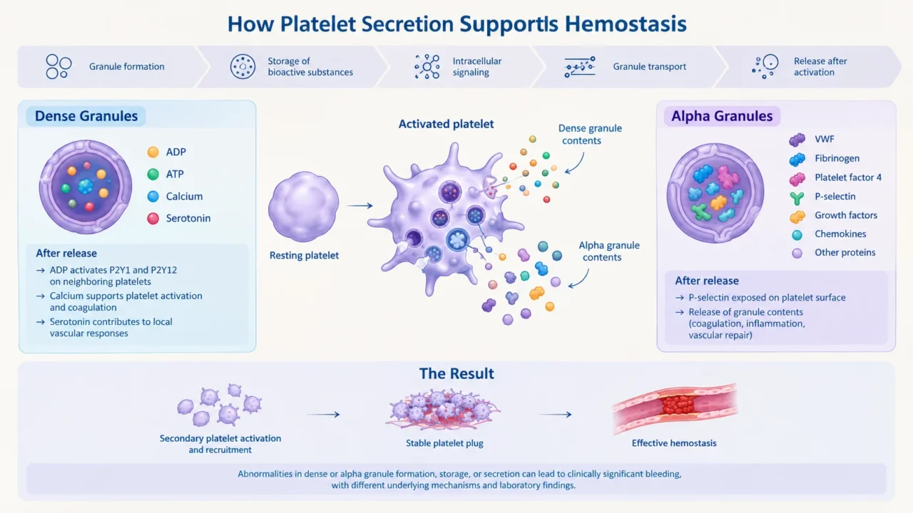
Platelet secretion is a coordinated process involving granule formation, storage of biologically active substances, intracellular signaling, granule transport and release of granule contents following platelet activation.
Two major granule populations contribute to this process: dense granules and alpha granules. Their contents serve different but complementary functions in hemostasis.
Dense granules contain adenosine diphosphate (ADP), adenosine triphosphate (ATP), calcium, and serotonin. Following platelet activation, released ADP stimulates P2Y1 and P2Y12 receptors on neighboring platelets, promoting recruitment and amplification of the platelet response. Calcium supports platelet activation and coagulation-related reactions, while serotonin contributes to local vascular responses.
Deficient dense-granule formation, storage, or secretion can therefore impair secondary platelet activation and reduce the stability of the developing platelet plug.
Alpha granules contain von Willebrand factor (VWF), fibrinogen, platelet factor 4, P-selectin, growth factors, chemokines, and other proteins involved in coagulation, inflammation, and vascular repair. Following activation, alpha-granule membranes fuse with the platelet membrane, exposing P-selectin on the platelet surface and releasing granule contents.
Abnormalities affecting either granule population can produce clinically significant bleeding, although the underlying mechanisms and associated laboratory findings differ.
Inherited and Acquired Secretion Defects
Inherited platelet secretion disorders may arise from abnormalities in granule biogenesis, intracellular trafficking, signaling, or membrane fusion. Some occur as isolated platelet disorders, while others form part of multisystem syndromes.
Delta-storage-pool deficiency is characterized by reduced dense-granule numbers or abnormal granule contents. Patients may present with mucocutaneous bleeding, excessive bleeding after dental procedures, or postoperative hemorrhage. Platelet aggregation may show reduced secondary responses, but the findings are not uniformly diagnostic.
Hermansky–Pudlak syndrome is an inherited disorder associated with dense-granule deficiency and oculocutaneous albinism. Depending on the genetic subtype, patients may also develop pulmonary fibrosis, granulomatous colitis, or immunodeficiency.
Chediak–Higashi syndrome is another syndromic disorder associated with abnormal granule formation and additional systemic manifestations.
Gray platelet syndrome, in contrast, is associated with alpha-granule deficiency. Affected patients typically have large, pale platelets on peripheral blood smear and bleeding of variable severity.
Acquired platelet secretion abnormalities also require consideration. Aspirin, nonsteroidal anti-inflammatory drugs, P2Y12 inhibitors, and selective serotonin reuptake inhibitors can affect platelet function.
Uremia, liver disease, myelodysplastic syndromes, myeloproliferative neoplasms, hematologic malignancies, and systemic inflammatory or autoimmune conditions may also contribute.
Medication exposure and underlying disease should therefore be assessed before an abnormal secretion result is attributed to an inherited disorder.
Why Normal Aggregation Does Not Exclude a Secretion Defect
Light transmission aggregometry (LTA) remains a core laboratory method for evaluating platelet function. It measures changes in light transmission as platelets aggregate after stimulation with agonists such as ADP, collagen, epinephrine, arachidonic acid and ristocetin.
However, aggregation and secretion are related but distinct processes. LTA does not directly quantify the amount, composition, or timing of granule release.
Depending on the underlying defect, patients may demonstrate reduced secondary aggregation, reversible aggregation responses, abnormalities limited to selected agonists, or apparently normal aggregation despite significant dense-granule deficiency.
Consequently, normal LTA results should not automatically end the investigation when the bleeding phenotype remains convincing. Platelet secretion testing may provide additional information that conventional aggregation studies cannot supply.
The diagnostic question is not simply whether platelets aggregate, but whether they release the mediators required to sustain an effective hemostatic response.
Laboratory Methods for Assessing Platelet Secretion
No single assay identifies every type of platelet secretion defect. The choice of investigation depends on the suspected abnormality, available laboratory methods, and the clinical presentation.
Lumi-aggregometry combines light transmission aggregometry with a luciferin–luciferase reaction to measure ATP release from dense granules. It allows aggregation and secretion to be assessed simultaneously. Reduced ATP release suggests impaired dense-granule output but does not, by itself, distinguish between reduced granule numbers, depleted nucleotide content, abnormal signaling, and defective exocytosis.
Whole-mount transmission electron microscopy allows direct visualization and quantification of platelet dense granules. A reduced number of dense bodies supports a quantitative dense-granule deficiency. However, a normal granule count does not exclude abnormal granule contents or a functional release defect.
Granule-content analysis can help distinguish deficient storage from impaired release. Measurements of platelet and released ATP, ADP, or serotonin may provide additional mechanistic information, although these techniques are technically demanding and generally restricted to specialized laboratories.
Flow cytometry provides another approach to platelet secretion assessment. Surface P-selectin expression following agonist stimulation is used as a marker of alpha-granule membrane externalization. Its interpretation requires attention to agonist selection, incubation conditions, sample preparation, and laboratory-specific reference intervals.
These methods provide complementary information. Functional testing can identify impaired secretion, structural analysis can demonstrate granule deficiency, and biochemical studies may help characterize the underlying defect. Results should be interpreted together rather than as independent proof of a specific diagnosis.
A Practical Diagnostic Approach
The investigation should begin with a detailed bleeding history. Recurrent epistaxis, gingival bleeding, easy bruising, heavy menstrual bleeding, postpartum hemorrhage, and excessive bleeding after dental or surgical procedures should be documented alongside family history. A validated bleeding-assessment tool can help standardize clinical evaluation.
Initial laboratory assessment generally includes a complete blood count, platelet indices, peripheral blood smear, prothrombin time, activated partial thromboplastin time, and VWF testing when indicated. Renal and liver function, fibrinogen, medication exposure, and relevant systemic conditions should also be considered.
If the clinical history continues to suggest a platelet disorder, LTA should be interpreted according to the agonists used, response patterns, reversibility, and laboratory reference intervals. Dense-granule secretion testing should be considered when the bleeding phenotype remains persuasive, including when aggregation results are normal or only mildly abnormal.
Abnormal secretion results require further characterization. Lumi-aggregometry can assess dense-granule release, electron microscopy can investigate granule deficiency, and biochemical analysis may help distinguish storage abnormalities from impaired release. Flow cytometry can provide additional information about alpha-granule secretion or platelet glycoproteins.
Repeat testing under controlled conditions is important because results may be influenced by sample age, temperature, platelet count, medication exposure, agonist concentration, and preanalytical variation.
A platelet secretion disorder should not be diagnosed from a single abnormal assay. The most reliable interpretation integrates the bleeding phenotype, laboratory findings, platelet morphology, medication history, and, where appropriate, genetic testing.
When Platelets Fail to Release: What Does the Laboratory Reveal?
A patient presents with lifelong bleeding, yet routine coagulation studies do not adequately explain the clinical history.
Should the investigation stop when initial tests fail to identify an abnormality, or should clinicians look more closely at platelet function?
A published case of Hermansky–Pudlak syndrome illustrates why the distinction matters. Whole-blood lumi-aggregometry demonstrated absent or markedly reduced ATP secretion, while whole-mount platelet transmission electron microscopy showed virtually absent dense granules.
Together with the patient’s hypopigmentation, these findings supported the diagnosis, which was further investigated by molecular analysis that identified a likely pathogenic genetic variant.
What did each test contribute to the diagnosis?
Lumi-aggregometry demonstrated impaired dense-granule output, but could not independently establish its cause. Electron microscopy provided structural evidence of dense-granule deficiency, helping distinguish a storage-pool abnormality from an isolated defect in the final release mechanism. The clinical phenotype and molecular findings added further diagnostic context.
This case raises a broader question:
How many patients with unexplained bleeding might remain undiagnosed if platelet aggregation is treated as the final step in the investigation?
Recognizing Hermansky–Pudlak syndrome also has implications beyond bleeding. Depending on the genetic subtype, patients may develop pulmonary fibrosis, granulomatous colitis, or immunodeficiency. Identifying a platelet secretion defect may therefore prompt systemic evaluation and genetic counseling, rather than simply providing an explanation for the bleeding history.
From Diagnosis to Clinical Decisions
Identifying a platelet secretion defect is not merely a laboratory exercise. It can influence planning for surgery, dental procedures, childbirth, and other situations associated with increased bleeding risk.
But does an abnormal secretion assay automatically mean that preventive treatment is necessary?
Not necessarily. Management should be guided by the patient’s bleeding history, the severity of the disorder, previous procedural outcomes, and the anticipated bleeding risk. Depending on the clinical situation, management may include local hemostatic measures, antifibrinolytic therapy, desmopressin in selected patients following specialist assessment, or platelet transfusion for severe bleeding and selected high-risk procedures. Avoiding unnecessary platelet-inhibiting medications and addressing iron deficiency or menstrual blood loss may also be important.
The key distinction is between identifying a laboratory abnormality and determining its clinical significance. Which patients are most likely to benefit from preventive treatment, and which laboratory findings should actually change management? These questions remain important when translating platelet function testing into individualized care.
What Remains Unanswered?
Despite advances in platelet function testing, important diagnostic challenges persist. Assays differ in availability, technical requirements, and sensitivity to preanalytical conditions. Differences in protocols and reference intervals can also complicate interpretation across laboratories.
Future studies need to establish how specific secretion abnormalities correlate with bleeding severity, procedural outcomes, and treatment response. Better methods for distinguishing granule storage deficiencies from functional release defects could also improve diagnostic classification.
Ultimately, the objective is not simply to detect impaired secretion. It is to determine which abnormalities explain the patient’s bleeding, predict clinically important outcomes, and justify a change in management.
Looking Beyond Aggregation
If platelet aggregation is normal, can a clinically significant platelet disorder truly be excluded?
The answer is no. Platelet secretion is an essential component of primary hemostasis, and defects in granule formation, storage, intracellular signaling, trafficking, or release may cause bleeding despite normal platelet counts and inconclusive routine investigations.
A phenotype-driven approach combining aggregation studies, secretion assays, platelet morphology, electron microscopy, and selected biochemical or molecular investigations provides a more complete assessment of platelet function.
The Hermansky–Pudlak syndrome case illustrates the value of this approach: functional testing identified impaired secretion, structural analysis demonstrated the underlying granule deficiency, and the wider clinical phenotype helped establish the diagnosis.
The clinically relevant question is not only whether platelets can aggregate, but whether they can release the mediators needed to sustain effective hemostasis.
FAQ
1. What is a platelet secretion defect?
A platelet secretion defect is an abnormality in the formation, storage, or release of substances contained in platelet granules. It can impair platelet recruitment and stabilization of the primary hemostatic plug.
2. Can platelet secretion defects occur with a normal platelet count?
Yes. Platelet secretion disorders can occur despite a normal platelet count. Routine coagulation tests may also be normal because these tests do not directly assess platelet granule release.
3. What is the difference between platelet aggregation and secretion?
Aggregation describes the formation of platelet aggregates following activation. Secretion involves the release of biologically active substances from platelet granules, which amplify activation and support hemostasis.
4. What are dense-granule storage-pool disorders?
These disorders involve reduced numbers of dense granules or abnormalities in their contents. Reduced availability or release of ADP and other mediators can impair secondary platelet activation.
5. Which tests are used to diagnose platelet secretion defects?
Investigations may include lumi-aggregometry, whole-mount transmission electron microscopy, granule-content analysis, and flow cytometry. The appropriate combination depends on the suspected defect.
6. Does normal light transmission aggregometry exclude a platelet secretion disorder?
No. Some secretion abnormalities may be present despite normal or minimally abnormal aggregation responses. Additional testing may be appropriate when the clinical bleeding history remains convincing.
7. What is the role of electron microscopy?
Whole-mount transmission electron microscopy allows direct assessment of dense-granule numbers. It can help distinguish quantitative granule deficiency from abnormalities that primarily affect secretion or granule contents.
8. Which medications can affect platelet secretion?
Aspirin, nonsteroidal anti-inflammatory drugs, P2Y12 inhibitors, and selective serotonin reuptake inhibitors can affect platelet function. Medication exposure should be considered when interpreting abnormal results.
9. How are platelet secretion disorders managed?
Management depends on the specific disorder, bleeding phenotype, and procedural risk. Options may include local hemostatic measures, antifibrinolytic therapy, selected use of desmopressin, or platelet transfusion when clinically indicated.
10. Why is identifying the specific secretion defect important?
Characterizing the defect can clarify the cause of bleeding, identify associated systemic conditions, guide procedural planning, and support appropriate family assessment or genetic counseling.
Written by Elen Avetisyan, MD.
Stay updated with Hemostasis Today.
-
Oct 23, 2026, 16:00Hemophilia Alliance team in New Orleans at the ATHN Data Summit
-
Oct 23, 2026, 15:49Brian Sapp: Why Exact Calf Vein Naming Is Critical for DVT Diagnosis?
-
Oct 23, 2026, 15:33Smitirupa Mishra: A Systematic Approach to Bone Marrow Examination
-
Oct 23, 2026, 15:29José Antonio García Erce: Preoperative Anemia Emerges as a Modifiable Surgical Risk in New POWER Study
-
Oct 23, 2026, 15:13Reza Shojaei: Manufactured Red Blood Cells vs Blood Donation
-
Oct 23, 2026, 14:29Jean Jacques Kiladjian: International Scientific Exchange at IHCAMS in Tianjin
-
Oct 23, 2026, 14:11Aravind Palraj: The Prolonged aPTT Puzzle in SLE
-
Oct 23, 2026, 13:56Chokri Ben Lamine: The Evolving Treatment Landscape of Persistent and Chronic ITP
-
Oct 23, 2026, 13:51Julia Barr: Back to Donating – One Pint, One Good Deed