

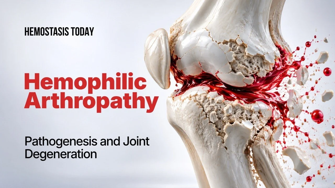
Blood-Induced Joint Damage in Hemophilia: Beyond Recurrent Hemarthrosis
Blood-Induced Joint Damage in Hemophilia has traditionally been viewed as the consequence of repeated hemarthroses: blood enters a joint, bleeding recurs, and progressive cartilage and bone destruction eventually produces chronic arthropathy.
This model remains fundamentally correct-but it is no longer sufficient.
Increasing evidence indicates that blood itself is biologically active within the joint. Iron deposition, oxidative stress, synovial proliferation, inflammatory cytokines, angiogenesis, cartilage injury and alterations in subchondral bone can create a self-perpetuating process in which joint damage may continue even when clinically recognized bleeding episodes become less frequent.
This shift in understanding has important implications for hemophilia care. The goal of treatment can no longer be limited to preventing overt hemarthrosis. It must increasingly encompass the prevention, detection and interruption of the biological processes that transform an episode of intra-articular bleeding into chronic joint disease.
From Hemarthrosis to a Self-Perpetuating Joint Disorder
The knees, ankles and elbows are particularly susceptible to hemarthrosis in hemophilia. In severe disease, joint bleeding is a dominant manifestation of the bleeding phenotype, and repeated episodes can lead to synovial hypertrophy, cartilage degradation, fibrosis, loss of range of motion and ultimately severe structural destruction.
The classical sequence is familiar: hemarthrosis is followed by synovitis; synovitis promotes further bleeding; recurrent bleeding produces progressive cartilage and bone damage; and the resulting arthropathy causes pain, stiffness, muscle wasting and disability.
But the relationship is more complex than simply ‘more bleeds equal more damage.’
Some patients develop structural abnormalities despite relatively few clinically documented hemarthroses. Conversely, the reduction of annualized bleeding rates with modern prophylaxis does not necessarily mean that the biological consequences of previous bleeding have disappeared. Subclinical or unrecognized joint bleeding may contribute to this discrepancy.
This is one reason why the concept of blood-induced joint damage is increasingly important.
Blood Is More Than a Passive Mechanical Insult
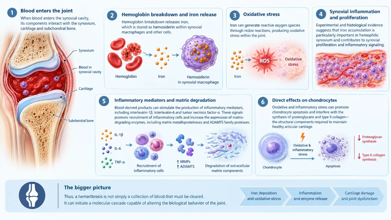
When blood enters the synovial cavity, its components interact with the synovium, cartilage and subchondral bone. Hemoglobin breakdown releases iron, which is subsequently stored as hemosiderin within synovial macrophages and other cells.
Iron can generate reactive oxygen species through redox reactions, producing oxidative stress within the joint. Experimental and histological evidence suggests that iron accumulation is particularly important in hemophilic synovium and contributes to synovial proliferation and inflammatory signaling.
The consequences extend beyond iron deposition.
Blood-derived products can stimulate the production of inflammatory mediators, including interleukin-1β, interleukin-6 and tumor necrosis factor-α. These signals promote recruitment of inflammatory cells and increase the expression of matrix-degrading enzymes, including matrix metalloproteinases and ADAMTS family proteases. The result is degradation of extracellular matrix components and impaired maintenance of cartilage.
Chondrocytes are also affected directly. Oxidative and inflammatory stress can promote chondrocyte apoptosis and interfere with the synthesis of proteoglycans and type II collagen—the structural components required to maintain healthy articular cartilage.
Thus, a hemarthrosis is not simply a collection of blood that must be cleared. It can initiate a molecular cascade capable of altering the biological behavior of the joint.
The ‘Two-Hit’ Hypothesis
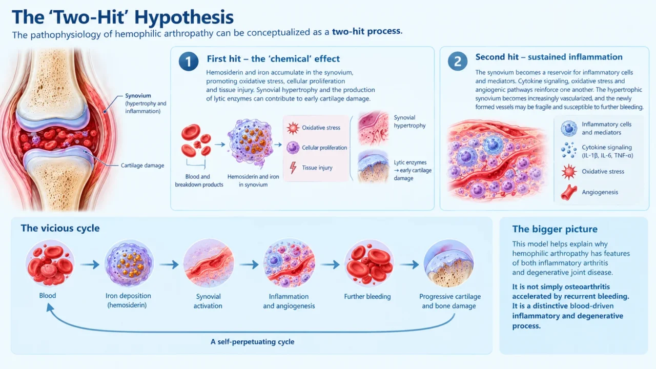
The pathophysiology described in the literature can be conceptualized as a two-hit process.
The first hit is the direct or ‘chemical’ effect of blood and its breakdown products. Hemosiderin and iron accumulate in the synovium, promoting oxidative stress, cellular proliferation and tissue injury. Synovial hypertrophy and the production of lytic enzymes can then contribute to early cartilage damage.
The second hit is the development of a sustained inflammatory response.
The synovium becomes a reservoir for inflammatory cells and mediators. Cytokine signaling, oxidative stress and angiogenic pathways reinforce one another. The hypertrophic synovium becomes increasingly vascularized, and the newly formed vessels may be fragile and susceptible to further bleeding.
The result is a vicious cycle:
blood-iron deposition-synovial activation-inflammation and angiogenesis-further bleeding-progressive cartilage and bone damage.
This model helps explain why hemophilic arthropathy has features of both inflammatory arthritis and degenerative joint disease. It is not simply osteoarthritis accelerated by recurrent bleeding. It is a distinctive blood-driven inflammatory and degenerative process.
Synovitis: The Critical Early Target
Synovitis may represent one of the most important-and potentially modifiable-stages in the evolution of hemophilic arthropathy.
Following blood exposure, the synovium becomes thickened and hypervascular. Inflammatory signaling stimulates further proliferation and angiogenesis. This enlarged synovium can become mechanically vulnerable, increasing the likelihood of additional bleeding and perpetuating the cycle.
Importantly, synovitis may precede obvious radiographic evidence of advanced arthropathy.
This makes early identification of synovial abnormalities particularly relevant. Clinical examination remains essential, but it may not detect the earliest structural changes. Ultrasound and magnetic resonance imaging (MRI) can provide additional information, with ultrasound offering a practical approach for monitoring synovial hypertrophy and early osteochondral abnormalities, while MRI provides a more comprehensive assessment of cartilage, bone and other intra-articular structures.
The implication is straightforward: waiting for severe pain, restricted movement or established radiographic destruction means waiting too long.
The Emerging Role of Oxidative Stress and Ferroptosis
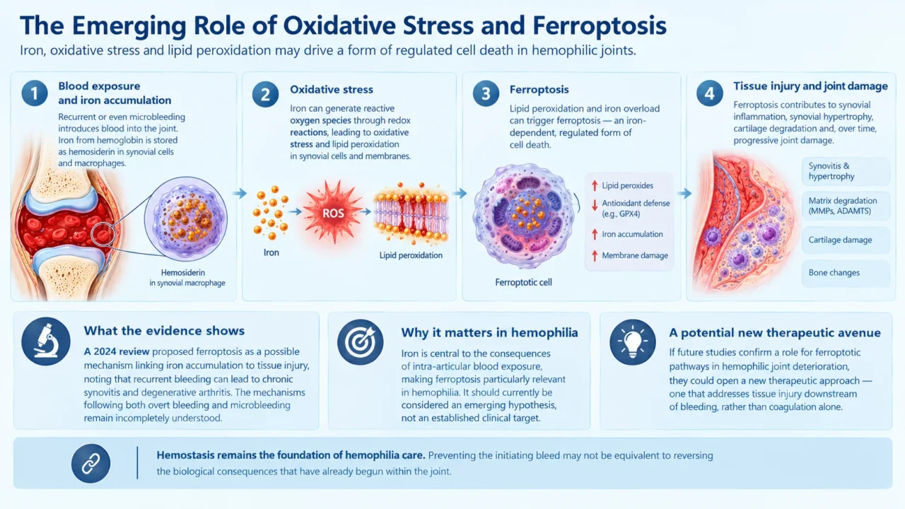
A particularly interesting development is the investigation of ferroptosis, an iron-dependent form of regulated cell death characterized by lipid peroxidation.
A 2024 review examined how blood exposure affects synovial joint tissues and proposed ferroptosis as a possible mechanism linking iron accumulation to tissue injury. The authors emphasized that recurrent bleeding can produce chronic synovitis and degenerative arthritis and that the biological mechanisms responsible for damage following both overt bleeding and microbleeding remain incompletely understood.
The ferroptosis hypothesis is especially relevant to hemophilia because iron is central to the consequences of intra-articular blood exposure. However, it should currently be regarded as an emerging mechanistic hypothesis rather than an established clinical target.
If future studies confirm that ferroptotic pathways contribute materially to hemophilic joint deterioration, they could open an entirely new therapeutic avenue-one that addresses tissue injury downstream of bleeding rather than coagulation alone.
That distinction is important. Hemostasis remains the foundation of hemophilia care, but preventing the initiating bleed may not be equivalent to reversing the biological consequences that have already begun within the joint.
Why ‘Zero Bleeds’ May Not Automatically Mean ‘Zero Arthropathy’
The evolution of prophylaxis and the emergence of extended-half-life and non-factor therapies have substantially changed hemophilia management. Early and sustained prophylaxis remains central to protecting joint health.
Yet arthropathy continues to occur.
The 2026 real-world study by Ardila and colleagues provides an important contemporary perspective. In a mixed-methods analysis of patients with severe hemophilia A in Colombia, the investigators identified prophylaxis strategy, bleeding episodes, physical activity and target joints among factors associated with hemophilic arthropathy. Primary prophylaxis was associated with protection against joint damage, while recent bleeding episodes were associated with a substantially higher likelihood of arthropathy. The study also found that patients with joint damage had experienced delays in starting prophylaxis compared with those without joint disease.
These findings reinforce an important principle: timing matters.
Once structural and inflammatory changes are established, simply reducing subsequent bleeding may not restore normal joint biology. Prevention therefore needs to begin before irreversible damage develops.
The same study also identified physical activity as a potentially protective factor. This is consistent with the modern view that joint protection does not mean avoiding movement. Rather, appropriately selected physical activity and rehabilitation can support muscle strength, joint stability and function while prophylaxis is individualized to bleeding risk and lifestyle.
Detecting The Joint Before the Patient Feels the Damage
The concept of subclinical joint disease is increasingly important.
Traditional clinical surveillance relies heavily on reported bleeding episodes, pain and physical findings. These remain valuable, but they do not capture every biological event occurring inside a joint.
A patient may have synovial hypertrophy, hemosiderin deposition or early osteochondral abnormalities without reporting a classic hemarthrosis. The 2014 review by Lobet and colleagues noted that significant joint damage can occasionally be identified despite relatively few clinically diagnosed hemarthroses, supporting the possibility that subclinical intra-articular bleeding contributes to disease progression.
This creates a diagnostic challenge-and an opportunity.
Joint surveillance increasingly needs to combine:
- bleeding history and bleeding patterns
- physical examination and range of motion
- validated clinical tools such as the Hemophilia Joint Health Score
- ultrasound for accessible, repeatable assessment of synovial and osteochondral abnormalities
- MRI when a more detailed structural assessment is required
- emerging biomarkers, although these are not yet validated for routine clinical use
The objective is not simply to document established arthropathy. It is to identify the transition from a healthy joint to a biologically activated joint.
Preventing Further Blood Exposure
Early, regular prophylaxis with factor VIII or factor IX concentrates, or appropriate non-factor therapies, is central to preventing hemarthrosis and preserving musculoskeletal health. The World Federation of Hemophilia (WFH) recommends prophylaxis as the standard of care for people with a severe hemophilia phenotype, particularly when initiated early in life. Episodic treatment alone cannot provide equivalent long-term protection against joint damage.
For patients who continue to experience joint bleeding despite prophylaxis, clinicians should reassess adherence, dosing, pharmacokinetic coverage, breakthrough bleeding patterns and inhibitor status when clinically indicated. Treatment should be individualized according to the patient’s bleeding phenotype, joint status, lifestyle and available therapies.
Prompt recognition and treatment of acute hemarthrosis are equally important. Delayed control of bleeding can prolong synovial exposure to blood and increase the risk of subsequent inflammation and tissue injury. Acute joint symptoms should therefore prompt clinical assessment and timely hemostatic treatment according to the individual’s treatment plan.
Identifying and Managing Persistent Synovitis
Persistent synovitis deserves particular attention because hypertrophic, inflamed synovium can contribute to recurrent bleeding and progressive joint damage. Clinical examination and appropriate imaging can help distinguish an acute bleed from persistent synovial disease or established arthropathy. Ultrasound and MRI are useful complementary tools when examination alone does not adequately characterize the joint.
When recurrent bleeding or chronic synovitis persists despite optimized hemostatic management, specialist assessment is warranted. Selected patients may benefit from synovial interventions, including radiosynovectomy or surgical synovectomy, depending on the degree of synovial disease, joint damage and local expertise. These procedures require coordinated planning with the hemophilia treatment team and appropriate hemostatic coverage.
Although targeting synovitis is biologically attractive, current clinical management should not assume that suppression of inflammation will reverse established cartilage destruction. More research is needed to determine which interventions can alter the long-term course of blood-induced joint disease.
Rehabilitation and Preservation of Joint Function
Physical therapy is an essential component of care after joint bleeding and in established hemophilic arthropathy. Rehabilitation may include range-of-motion exercises, progressive muscle strengthening, proprioceptive training, balance exercises and functional retraining.
The aim is to restore function after a bleed and maintain mobility, muscle strength and joint stability over time.
Rehabilitation should be individualized and supervised by a physiotherapist experienced in hemophilia. During recovery from an acute bleed, exercise intensity and loading should be adjusted to symptoms, clinical findings and hemostatic treatment. Gradual mobilization helps avoid the loss of muscle strength and joint function associated with prolonged immobilization.
Regular physical activity should be encouraged, with the type and intensity selected according to joint status and individual bleeding risk. Low-impact activities may be appropriate for many patients, while patients with target joints or advanced arthropathy may require specific adaptations, protective equipment or additional prophylactic planning.
Pain Management and Established Arthropathy
Pain in hemophilic arthropathy can arise from acute bleeding, synovial inflammation, cartilage loss, deformity and altered joint mechanics. Management should therefore address the underlying cause rather than treating pain in isolation.
Non-pharmacological measures include physiotherapy, activity modification, assistive devices and interventions aimed at maintaining function. Analgesic selection requires particular care because some medications, including non-selective nonsteroidal anti-inflammatory drugs, can increase bleeding risk. Selected cyclooxygenase-2 inhibitors may be considered in appropriate patients following an individualized assessment of gastrointestinal, cardiovascular, renal and other risks.
For patients with persistent pain, restricted movement or progressive structural damage despite conservative treatment, orthopedic consultation is appropriate.
Depending on the clinical situation, surgical options may include synovectomy, joint debridement, release of contractures, corrective osteotomy, ankle arthrodesis or joint replacement for end-stage disease. Such procedures require experienced multidisciplinary care, adequate hemostatic support and postoperative rehabilitation.
Monitoring Outcomes Beyond Bleeding Rates
A comprehensive assessment of joint health should not rely exclusively on annualized bleeding rates. Clinical examination, the Hemophilia Joint Health Score, patient-reported symptoms and functional assessments can help identify changes that may warrant further investigation. Ultrasound or MRI may provide additional information when subclinical synovitis or structural damage is suspected.
Longitudinal monitoring is especially important in patients with previous target joints, recurrent breakthrough bleeding, persistent synovitis or established arthropathy. The purpose is to identify deterioration early, review the effectiveness of prophylaxis and adapt rehabilitation or specialist care before additional functional loss occurs.
Ultimately, management should address two interconnected but distinct outcomes: preventing new bleeding and preserving the health and function of the joint. The first remains indispensable, but the second requires a broader, multidisciplinary approach.
Looking Ahead
The evolution of hemophilia treatment has made dramatic progress toward reducing spontaneous bleeding. The next frontier may be the prevention of the biological consequences that remain after blood reaches the joint.
Research into iron-mediated injury, inflammatory signaling, angiogenesis and ferroptosis could help identify new therapeutic targets. Improved imaging may allow clinicians to recognize synovitis and osteochondral abnormalities before patients develop clinically obvious arthropathy. Biomarker research could eventually provide minimally invasive indicators of joint health or disease activity.
At the same time, real-world evidence reinforces the importance of early prophylaxis, individualized treatment, adherence and appropriate physical activity.
For clinicians, the message is increasingly clear: hemophilic arthropathy should not be considered merely the cumulative consequence of counting hemarthroses. The joint itself is an active participant in the disease process.
Understanding blood-induced joint damage in this broader biological context may ultimately allow hemophilia care to move from treating the consequences of recurrent bleeding to preventing the earliest molecular and cellular events that turn bleeding into permanent joint disease.
FAQ
1. Can a single joint bleed cause permanent damage in hemophilia?
Yes. A single hemarthrosis can initiate inflammatory and oxidative processes within the joint. The risk of permanent damage depends on factors such as the severity of bleeding, the joint’s previous condition and the effectiveness of treatment.
2. Does blood trigger inflammation inside the joint?
Yes. Blood components and their breakdown products can activate synovial cells and inflammatory pathways, contributing to joint tissue injury.
3. Is MRI useful for assessing hemophilic arthropathy?
Yes. MRI can provide detailed information about synovium, cartilage, hemosiderin deposition and bone abnormalities.
4. Can hemophilic arthropathy develop without frequent visible joint bleeds?
Yes. Subclinical bleeding and persistent synovial abnormalities may contribute to joint damage even when overt hemarthroses are infrequent or unrecognized.
5. Can physical activity benefit people with hemophilic arthropathy?
Yes. Appropriately selected physical activity and rehabilitation can improve muscle strength, joint stability, mobility and function. Exercise should be individualized according to joint status and bleeding risk.
6. Does effective prophylaxis eliminate all risk of hemophilic arthropathy?
No. Prophylaxis substantially reduces bleeding and helps protect joint health, but it cannot necessarily reverse pre-existing damage or eliminate every breakthrough or unrecognized bleed.
7. Are targeted treatments for iron-mediated joint injury established in clinical practice?
No. Iron-related oxidative injury and ferroptosis are promising research areas, but targeted therapies have not yet been established as standard treatments for hemophilic arthropathy.
8. Can established hemophilic arthropathy be completely reversed?
No. Advanced cartilage loss and bone deformity are generally irreversible. Nevertheless, appropriate treatment can help preserve function, manage pain and reduce further joint complications.
9. Is ferroptosis already an established treatment target in hemophilic arthropathy?
No. Ferroptosis is an emerging research area that may help explain iron-related tissue injury, but its clinical significance and the effectiveness of ferroptosis-targeted treatments in patients remain to be established.
10. Should joint management extend beyond bleeding prevention?
Yes. Comprehensive care should also address synovitis, early detection of structural abnormalities, rehabilitation, pain management and orthopedic complications when indicated.
Written by Marieta Aleksanyan, MD
Stay updated with Hemostasis Today.
-
Oct 25, 2026, 05:30Denis Oduor: Whole Blood and Component Therapy in Clinical Practice
-
Oct 25, 2026, 02:18Dai H. Chung: Delivering Lyfgenia to Pediatric Patients With Sickle Cell Disease
-
Oct 25, 2026, 02:17Marilyn Baffoe-Bonnie: What Does It Mean to Be Cured of Sickle Cell Disease?
-
Oct 25, 2026, 02:15Tamara Stipcevic: A Hand-Powered Device for Rapid Blood Typing and Hematocrit Estimation
-
Oct 25, 2026, 02:14Rhonda Patrick: Microplastics Generate Damaging Free Radicals Simply by Contacting Water
-
Oct 25, 2026, 02:12Chokri Ben Lamine: Rilzabrutinib – Oral BTK Inhibitor for ITP
-
Oct 25, 2026, 02:12Michael H. Tomasson: Self-Experimentation That Revealed the Immune Mechanism of ITP
-
Oct 25, 2026, 02:10Nabiha Huq Saifee: Do You Help Make Platelet Transfusion Decisions Before Lumbar Puncture?
-
Oct 25, 2026, 02:09Sebastian Szmit: Congratulations to Isabelle Mahé on Her New Cardio-Oncology Paper