


Jan Sloves: Medial Thigh Venous Perforators – Why Diameter Isn’t the Whole Story
Jan Sloves, CEO and Founder at Ultrasound Unlocked LLC, shared a post on LinkedIn about a recent article by Gabriela Hnátková et al., published in Journal of Vascular Surgery: Venous and Lymphatic Disorders, adding:
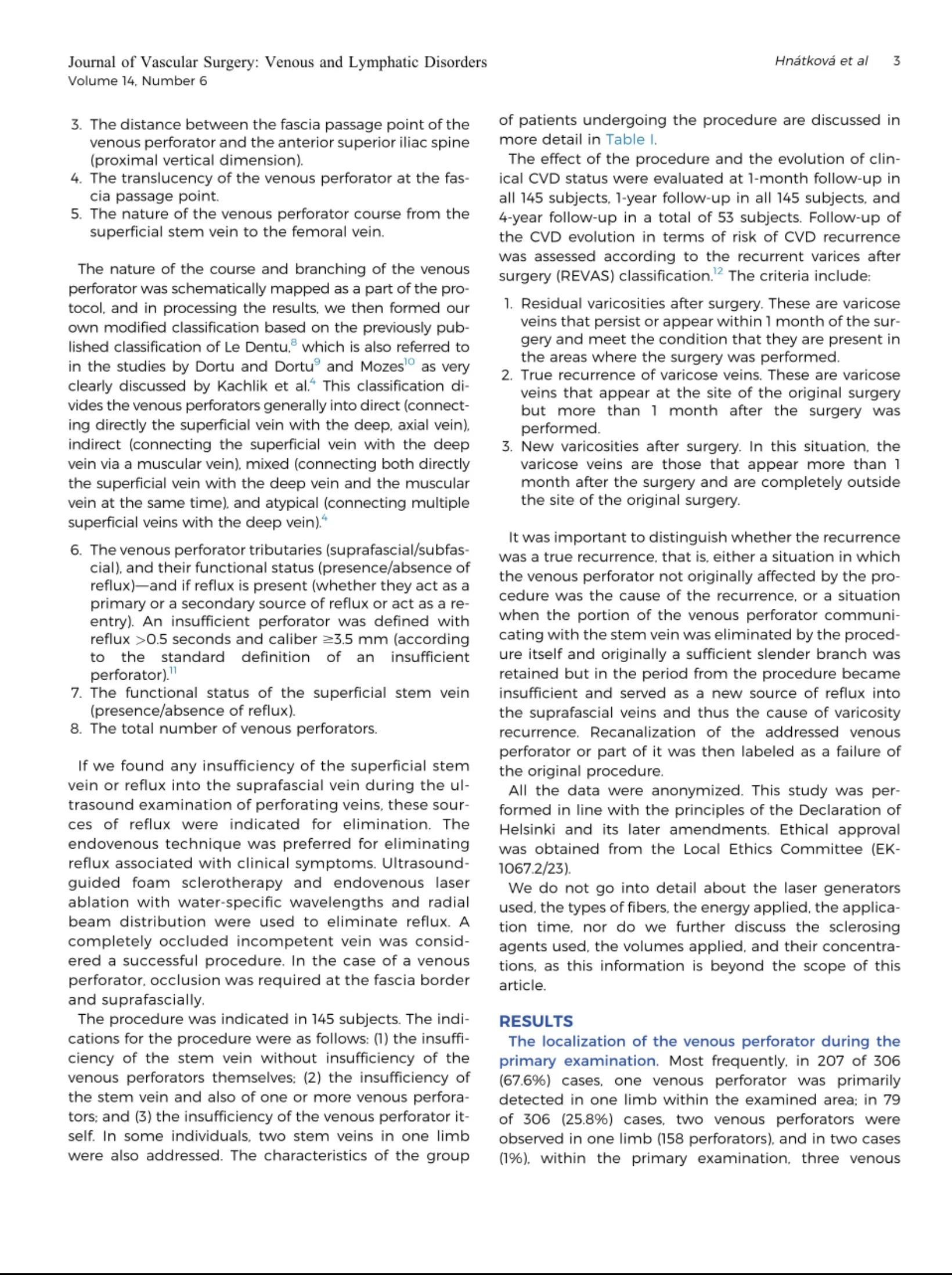
“The Anatomical Conditions of the Venous Perforators of the Medial Side of the Thigh in Progressive Chronic Venous Disease. JVSVL 2026;
A few take-home points that I think are particularly relevant to those of us performing venous duplex ultrasound:
Medial thigh perforators are easy to underestimate
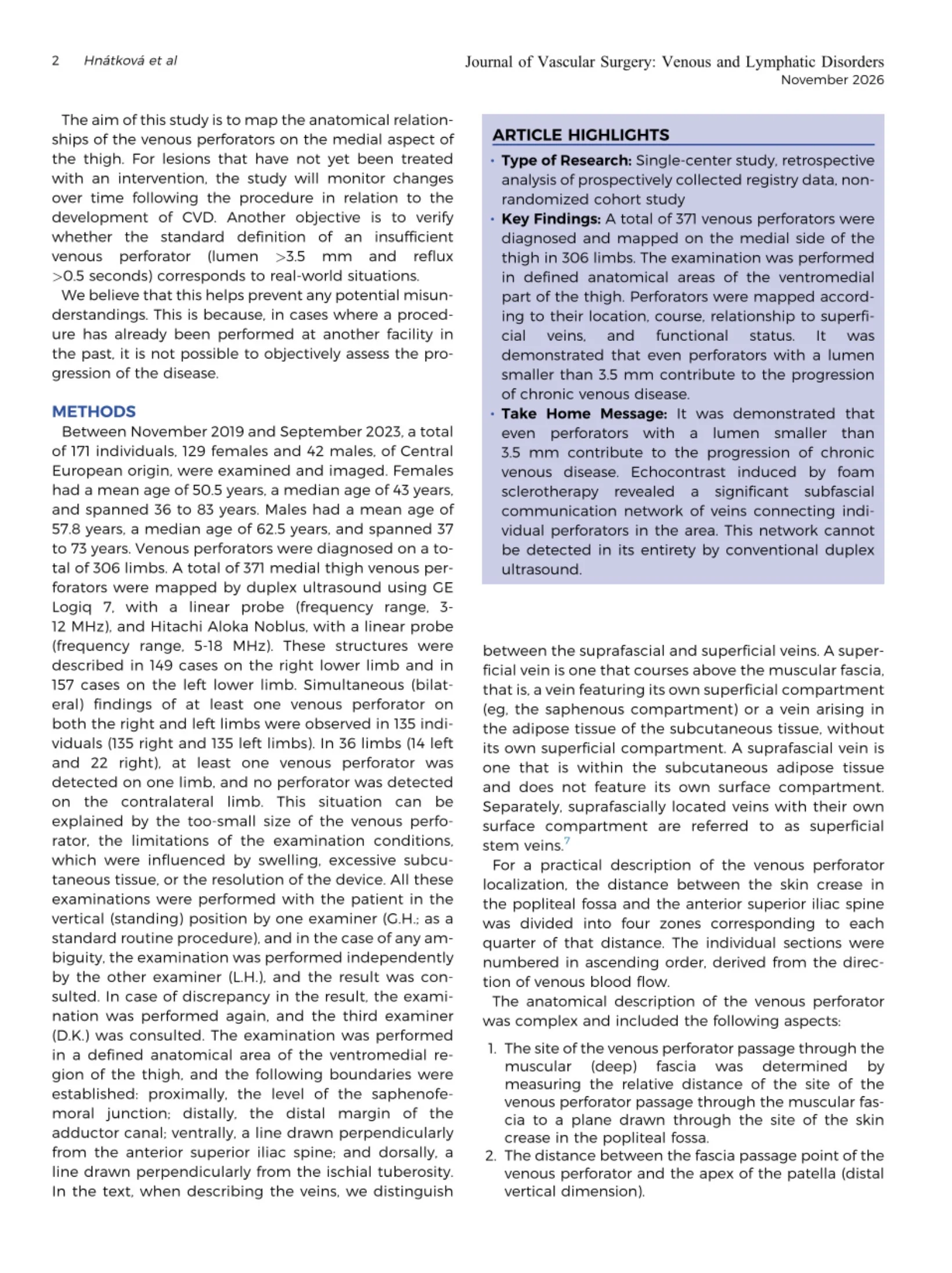
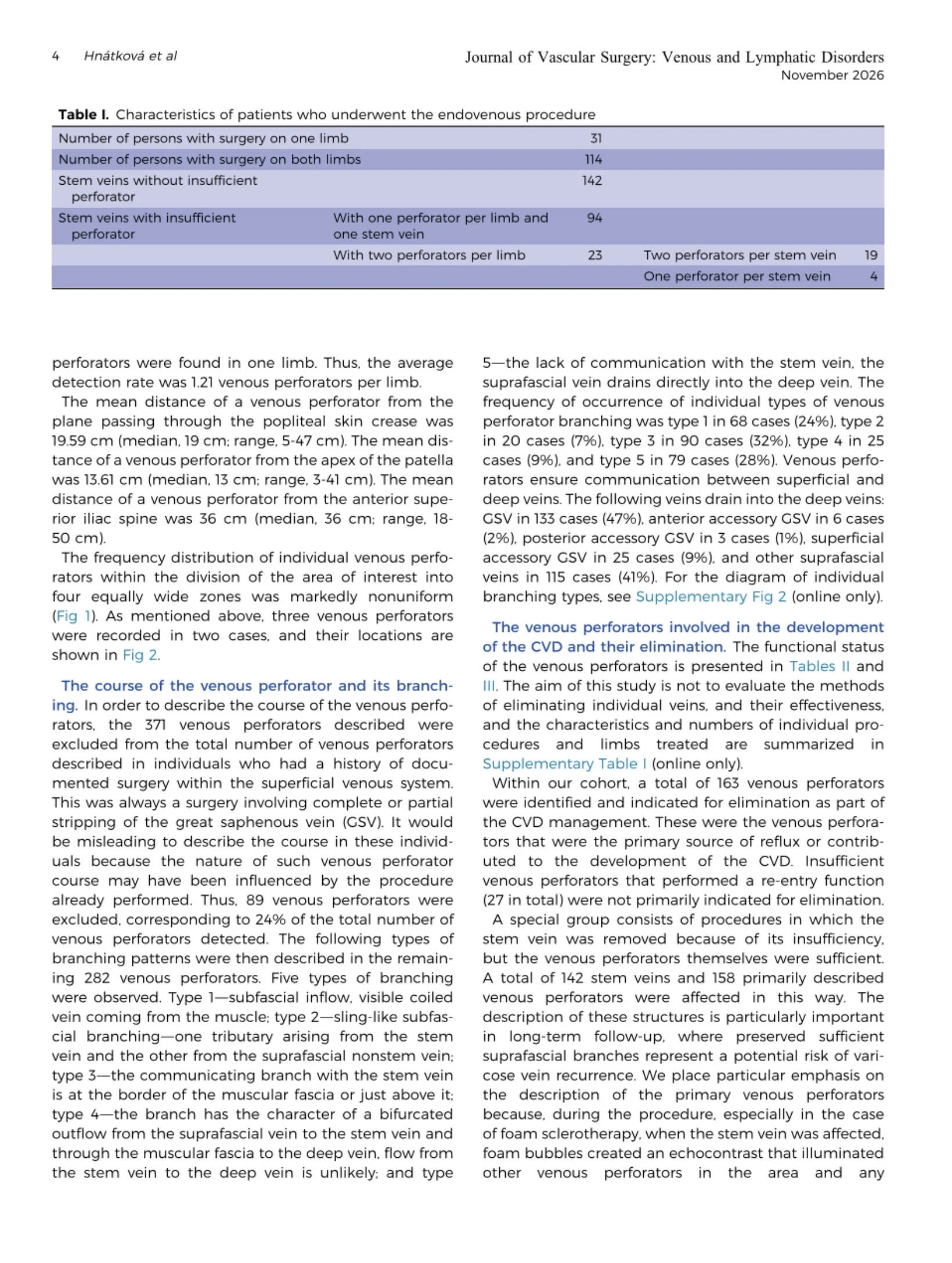
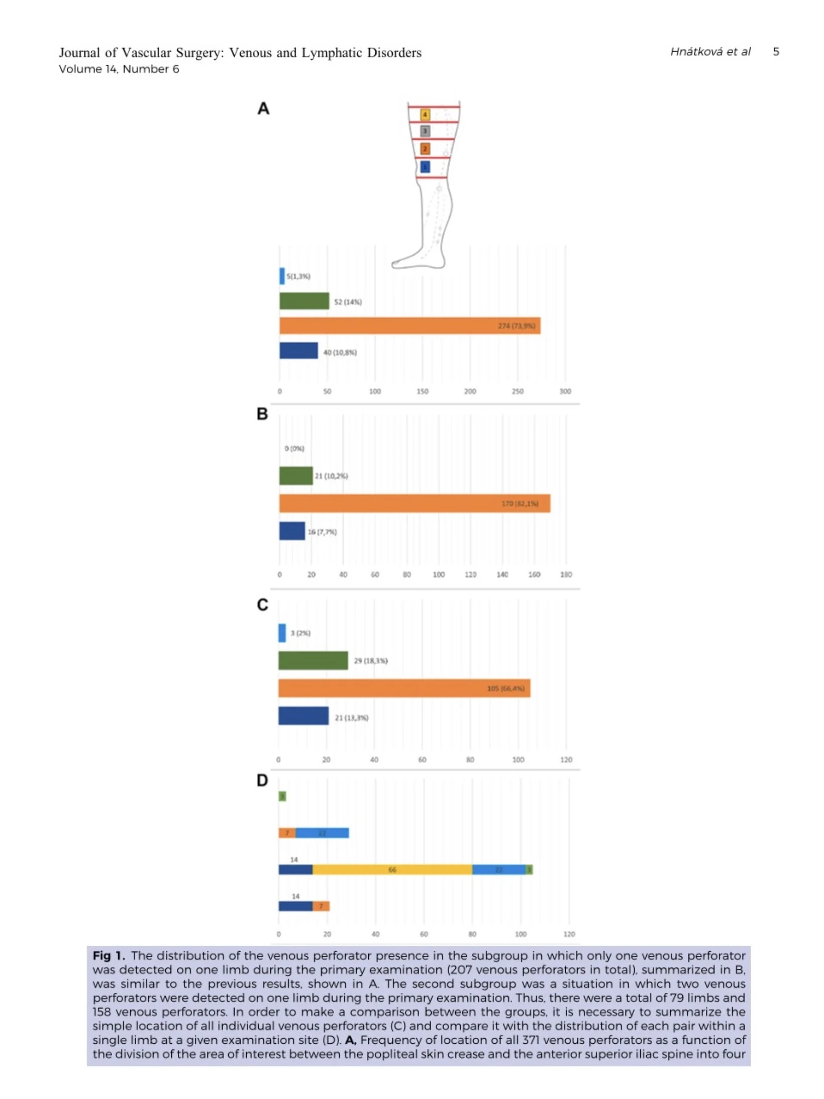
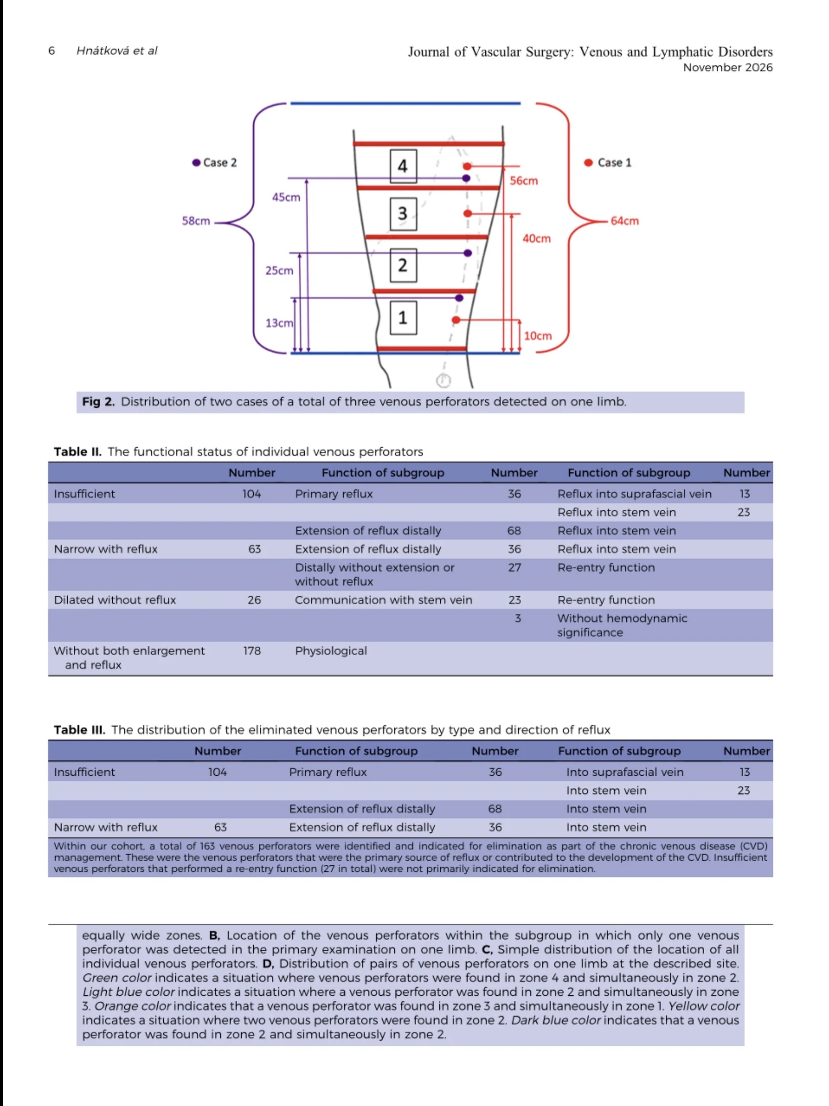
The study identified 371 perforators in 306 limbs, with almost 74% located in the second of four anatomical zones.
PDiameter alone may not tell the whole story
The conventional definition of an insufficient perforator is a diameter greater than or equal to 3.5 mm with reflux greater than 0.5 seconds.
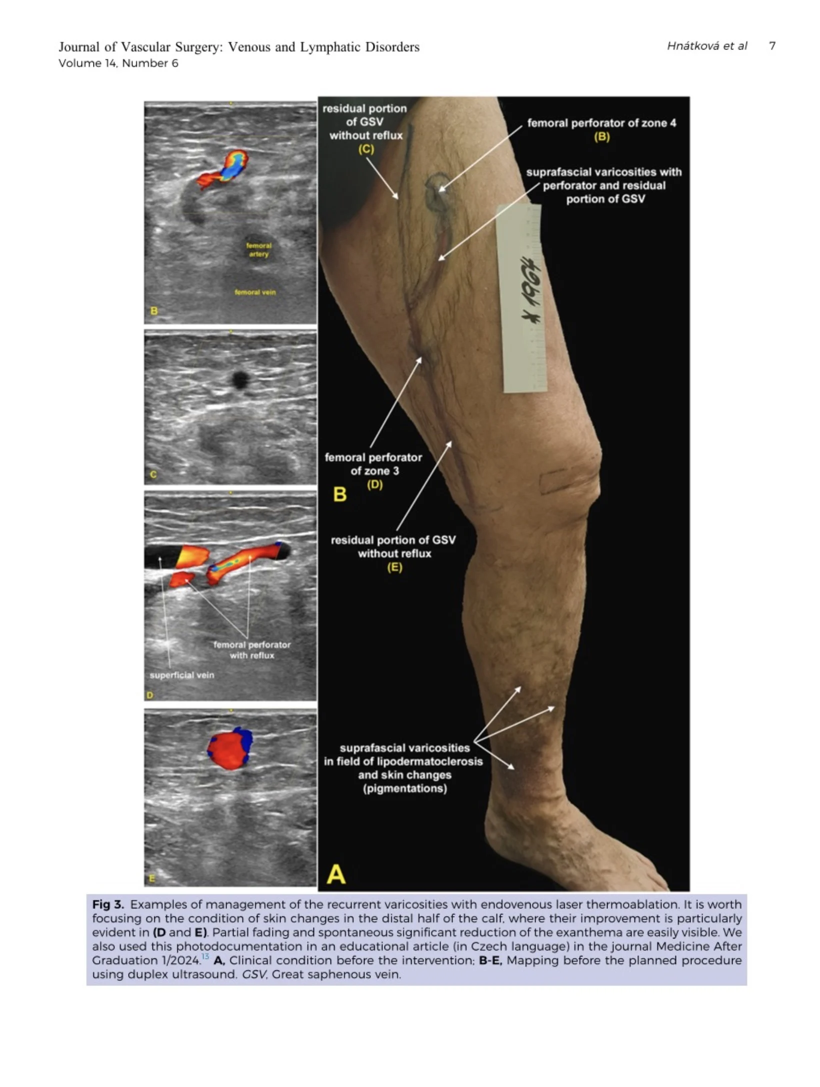
However, the authors identified smaller perforators with prolonged reflux that could contribute to superficial venous reflux.
This raises an important ultrasound point: don’t use diameter as the only determinant of relevance.
Look beyond the individual perforator
The study demonstrated a subfascial network of communicating veins between the perforators and the superficial/deep venous systems.
Conventional duplex may not visualize this network in its entirety.
Foam echocontrast can reveal additional anatomy
In 65 limbs, foam echocontrast demonstrated additional perforators and subfascial communications, increasing detection to 197 perforators, approximately 3 per limb.
Importantly, the authors note that echocontrast demonstrates communication but does not determine the functional status of the perforator.
Location and connections matter
The authors emphasize documenting the anatomical course, connections, reflux and relationship to fixed landmarks rather than relying only on traditional perforator names.
For me, the practical ultrasound message is simple:
When mapping venous disease, particularly recurrent disease, look carefully at the medial thigh perforators, assess their reflux and connections, and don’t let diameter be the only consideration.
Subscribe to the only newsletter built for vascular pros.”
Title: The Anatomical Conditions of the Venous Perforators of the Medial Side of the Thigh in Progressive Chronic Venous Disease
Authors: Gabriela Hnátková, Lukas Hnátek, David Kachlík
Stay updated on all scientific advances with Hemostasis Today.
-
Oct 5, 2026, 13:01Nawsherwan Mohammad: Recognizing TTP Before the ADAMTS13 Result Arrives
-
Oct 5, 2026, 12:51Bruno Pougault: The Hidden Challenges Behind Anti Xa Testing
-
Oct 5, 2026, 12:41Syed Huzaifa Khan: Rethinking CTRP12 as a Biomarker for In Stent Restenosis
-
Oct 5, 2026, 12:29Mirvat Alasnag: Creating a Space for Women in Cardiovascular Medicine
-
Oct 5, 2026, 11:59Khaoula Mazouzi: The Potential of Haemoglobin Vesicles as an Oxygen Carrier
-
Oct 5, 2026, 11:56Muhammet Özbilen: What Does New Evidence Tell Us About IV Iron in Pregnancy?
-
Oct 5, 2026, 11:42Fonkou Steve: A Moment to Celebrate the Journey Ahead
-
Oct 5, 2026, 11:33Abeeha Faisal: Beyond the Textbooks – A Day With Children Living With Thalassemia
-
Oct 5, 2026, 11:26Jan Zlamal: Honoured with the Fritz Schiff Young Investigator Award by the DGTI