


Hereditary Hemorrhagic Telangiectasia: The Molecular Connection Between Vascular Malformations and Bleeding
Hereditary hemorrhagic telangiectasia (HHT), or Osler-Weber-Rendu syndrome, is an autosomal-dominant vascular disease characterized by recurrent mucocutaneous bleeding
and arteriovenous malformations (AVMs).
Its central molecular problem is impaired TGF-β/BMP signaling, which disrupts normal endothelial-cell function and capillary formation, leading to fragile direct connections between arteries and veins.
What is HHT?
HHT is characterized by small mucocutaneous vascular lesions—telangiectasias—and larger visceral AVMs, most often in the lungs, liver, and brain.
Telangiectasias are especially common on the nose, lips, tongue, oral mucosa, gastrointestinal tract, and fingertips; they can bleed spontaneously or after minimal trauma.
The prevalence is estimated at up to 1 in 5,000 people.
Recurrent epistaxis (nosebleeds) is the most frequent manifestation, often beginning around age 12, while gastrointestinal bleeding and iron-deficiency anemia may become more prominent later in life․
Molecular basis
Most HHT cases arise from loss-of-function variants in genes belonging to the transforming growth factor-beta (TGF-β) and bone morphogenetic protein (BMP) signaling pathway.
These pathways normally regulate endothelial-cell behavior, vascular remodeling, vessel-wall integrity, and the formation of stable capillary networks.
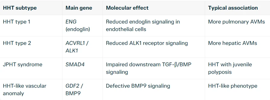
ENG and ACVRL1 account for the vast majority of genetically confirmed cases; SMAD4 and GDF2 variants are less common.
From gene to malformation
In a normal microcirculation, arteries and veins are connected through capillaries, which slow blood flow and allow exchange of oxygen, nutrients, and waste products.
In HHT, impaired BMP9/BMP10–ENG–ALK1–SMAD4 signaling disrupts endothelial homeostasis: postcapillary venules dilate, capillaries are progressively bypassed, and direct artery-to-vein connections form.
The result is a telangiectasia or AVM: a low-resistance, structurally weak vessel with abnormal endothelium, disorganized basement membrane, and insufficient contractile elements in the vessel wall.
These features make the vessels prone to rupture, while the direct high-flow connection can make bleeding brisk and difficult to control.
A “second hit” model helps explain why lesions are focal rather than universal: local injury, inflammation, hemodynamic stress, or an acquired somatic mutation may trigger malformation formation in an already genetically susceptible vascular bed.
Why bleeding occurs
Bleeding in HHT is primarily structural rather than caused by a conventional clotting-factor deficiency.
Thin-walled, superficial telangiectasias rupture easily, particularly in the nasal and gastrointestinal mucosa.
Epistaxis: Recurrent nosebleeds are the most common symptom and can range from minor to severe and transfusion-requiring.
Gastrointestinal bleeding: Telangiectasias in the GI tract may cause chronic occult blood loss or overt bleeding, commonly contributing to iron-deficiency anemia.
Anemia: Repeated mucosal bleeding can deplete iron stores, leading to fatigue, dyspnea, pallor, and reduced exercise tolerance.
Visceral hemorrhage: Less commonly, AVMs in the lungs or brain can bleed; pulmonary AVMs may also cause hemoptysis.
Why AVMs are dangerous
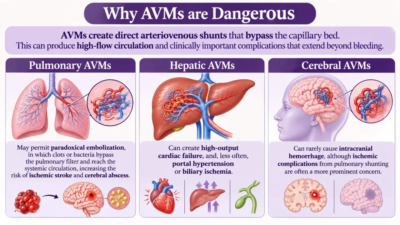
AVMs create direct arteriovenous shunts that bypass the capillary bed. This can produce high-flow circulation and clinically important complications that extend beyond bleeding.
Pulmonary AVMs may permit paradoxical embolization, in which clots or bacteria bypass the pulmonary filter and reach the systemic circulation, increasing the risk of ischemic stroke and cerebral abscess.
Hepatic AVMs can create high-output cardiac failure and, less often, portal hypertension or biliary ischemia.
Cerebral AVMs can rarely cause intracranial hemorrhage, although ischemic complications from pulmonary shunting are often a more prominent concern.
Bleeding and thrombosis: a dual risk
Although HHT is often thought of as a bleeding disorder, the abnormal shunts and altered endothelial biology can also increase thrombotic risk.
A clinical study of 24 patients found bleeding in 100% and thrombotic events in 41.7%; however, such findings require cautious interpretation because of the small sample size.
Elevated von Willebrand factor activity has been observed in HHT patients, consistent with endothelial activation, but it does not by itself prove a primary prothrombotic mechanism.
Anticoagulant or antiplatelet therapy may be necessary in selected patients with thrombosis, yet must be individualized because bleeding risk can be substantial.
Diagnosis
Diagnosis is usually clinical, supported by the Curaçao criteria:
- Spontaneous, recurrent epistaxis
- Mucocutaneous telangiectasias at typical sites
- Visceral AVMs or telangiectasias
- A first-degree relative with HHT
A diagnosis is generally considered definite when at least three criteria are present. Genetic testing for pathogenic variants in ENG, ACVRL1, or SMAD4 can confirm the molecular diagnosis and guide family screening.
Because pulmonary AVMs may be clinically silent but carry embolic risk, screening with transthoracic contrast echocardiography is commonly used; positive findings may require further imaging and specialist management.
Management principles
Treatment focuses on controlling bleeding, correcting iron deficiency, preventing embolic complications, and treating high-risk AVMs. There is currently no therapy that reverses the underlying genetic defect.
Iron replacement and transfusion: Oral or intravenous iron is used for iron-deficiency anemia; transfusion is reserved for severe or acute blood loss.
Nosebleed control: Saline humidification, topical measures, laser or cauterization for accessible lesions, and, in refractory cases, systemic therapies may be considered.
AVM surveillance and intervention: Pulmonary AVMs often require embolization when clinically significant, while brain and liver lesions need multidisciplinary evaluation.
Targeted therapies: Drugs affecting angiogenic pathways, including VEGF-pathway inhibitors such as bevacizumab, have been used in selected patients with severe bleeding or anemia, but treatment should be individualized.
Thrombosis management: Antithrombotic therapy demands careful risk-benefit assessment because HHT patients may have both thrombotic and hemorrhagic vulnerability.
Conclusion
HHT illustrates how a single molecular pathway can link vascular structure to clinical hemostasis. Loss of ENG–ALK1–SMAD4 signaling destabilizes endothelial-cell regulation and capillary formation, producing fragile direct artery-vein connections that bleed easily; in larger organs, the same malformations can create dangerous shunts, embolic risk, and cardiac complications.
Understanding this molecular connection is essential not only for explaining recurrent epistaxis and anemia, but also for developing therapies that target the vascular malformations themselves rather than merely managing their consequences.
10 FAQs
1. What is HHT?
A hereditary vascular disorder that causes fragile blood vessels, telangiectasias, and arteriovenous malformations (AVMs).
2. How is HHT inherited?
It is usually autosomal dominant; a child of an affected parent has about a 50% chance of inheriting it.
3. What are the main symptoms?
Recurrent nosebleeds, small red vascular spots on the skin or mouth, and sometimes gastrointestinal bleeding or anemia.
4. Why does it cause bleeding?
The blood vessels are abnormally thin and fragile, so they rupture easily, especially in the nose and digestive tract.
5. What genes cause HHT?
Most cases involve ENG or ACVRL1; less commonly, SMAD4 or GDF2.
6. What are pulmonary AVMs?
Abnormal direct connections between arteries and veins in the lungs that can allow clots or bacteria to bypass the lungs and reach the brain.
7. How is HHT diagnosed?
Doctors use the Curaçao criteria and may confirm it with genetic testing.
8. What is the Curaçao criteria?
Four features: recurrent nosebleeds, telangiectasias, internal AVMs, and a first-degree relative with HHT. Three or more suggest definite HHT.
9. Is there a cure?
No, but bleeding, anemia, and dangerous AVMs can often be managed with treatment.
10. Can people with HHT live normally?
Yes, especially with regular screening, treatment of anemia, and management of high-risk AVMs.
Written by Anna Stepanyan, MD
Stay updated with Hemostasis Today.
-
Nov 17, 2026, 14:54Cindy Neunert: Beyond Platelet Counts – Addressing Fatigue and Whole-Person Care in ITP
-
Nov 17, 2026, 14:44Strengthening Haemophilia Diagnosis Through Hands-On Laboratory Training – IAHAD
-
Nov 17, 2026, 14:40Platelet Membrane Protrusions Beyond Their Role in Hemostasis – JTH
-
Nov 17, 2026, 14:10Wolfgang Miesbach: Aspirin vs Anticoagulants – Who Needs More Than Aspirin?
-
Nov 17, 2026, 14:08Chaza Alaji: Soluble Transferrin Receptor – A Key to Uncovering Hidden Iron Deficiency
-
Nov 17, 2026, 14:03Shadi Tabibian: Fibrinogen – From Molecular Structure to Clinical Decision Making
-
Nov 17, 2026, 13:54Gabrielle Sarlon: Breaking the Silence on Menstrual Bleeding During Anticoagulant Therapy
-
Nov 17, 2026, 13:47Ksenia Guznova Kats: Advanced Venous Stenting in May–Thurner Syndrome
-
Nov 17, 2026, 13:37Abdlemewla Ibrahim: Prasugrel vs Ticagrelor in ACS Patients Undergoing PCI