

Factor V Deficiency: Diagnostic Challenges and the Role of Combined Factor V and VIII Deficiency
Factor V (FV) deficiency is a rare inherited bleeding disorder that can present with a wide range of clinical manifestations, from mild mucocutaneous bleeding to severe, potentially life-threatening hemorrhage.
Its diagnosis may be challenging because routine coagulation tests are not specific to FV deficiency, and the measured FV activity does not always reflect the severity of the bleeding phenotype.
Recent molecular research has further highlighted the genetic heterogeneity of the condition and the importance of distinguishing isolated FV deficiency from combined factor V and factor VIII deficiency.
Factor V: A Dual Role in Hemostasis
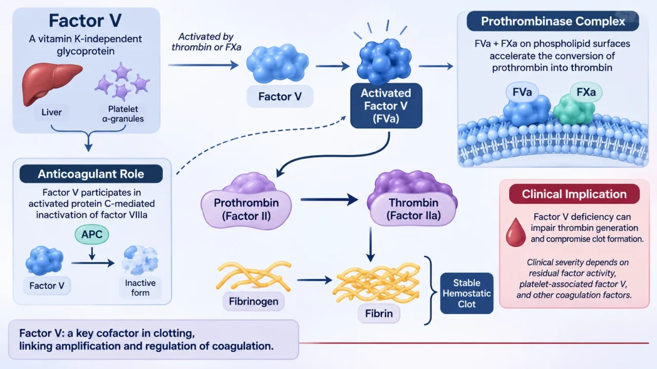
Factor V is a large, vitamin K-independent glycoprotein that circulates in plasma and is also stored in platelet alpha granules. It is synthesized primarily in the liver and is released into the circulation as an inactive precursor. Following proteolytic activation by thrombin or factor Xa, FV is converted into activated factor V (FVa), which serves as an essential cofactor in the prothrombinase complex.
FVa binds activated factor X (FXa) on negatively charged phospholipid surfaces, particularly those provided by activated platelets. This interaction markedly accelerates the conversion of prothrombin (factor II) into thrombin (factor IIa), promoting fibrin formation and the stabilization of the hemostatic plug. A deficiency or functional abnormality of FV therefore compromises thrombin generation and may result in an impaired coagulation response.
Importantly, FV also participates in anticoagulant pathways. Certain molecular forms of FV act as cofactors for activated protein C (APC) in the inactivation of factor VIIIa, while other forms interact with tissue factor pathway inhibitor alpha (TFPIα). This dual procoagulant and anticoagulant activity makes FV more than simply a component of the common coagulation pathway: it is also involved in maintaining the balance between clot formation and inhibition.
Congenital Factor V Deficiency: Clinical and Genetic Characteristics
Congenital FV deficiency, also known as parahemophilia or Owren disease, is a rare autosomal recessive bleeding disorder caused by pathogenic variants in the F5 gene, located on chromosome 1q24.2. The condition is characterized by reduced FV activity, which may result from impaired protein synthesis, secretion, stability, or function. The disorder is exceptionally rare, with an estimated prevalence of approximately one case per million individuals.
Clinical manifestations are highly variable. Patients may experience easy bruising, epistaxis, gingival bleeding, prolonged bleeding following dental extraction or surgery, and heavy menstrual bleeding. More severe cases may present with gastrointestinal bleeding, hematuria, postpartum hemorrhage, or intracranial hemorrhage. Unlike hemophilia A and B, spontaneous hemarthroses are not typically the predominant clinical feature.
FV activity is commonly used to classify disease severity. However, the relationship between residual FV activity and clinical bleeding is not consistently linear. Some individuals with very low FV activity may have relatively mild symptoms, whereas others experience clinically significant bleeding despite higher residual activity. The presence of platelet-derived FV, which contributes to local thrombin generation at sites of vascular injury, may partly explain this variability.
Recent Research: Expanding the Molecular Spectrum of F5 Variants
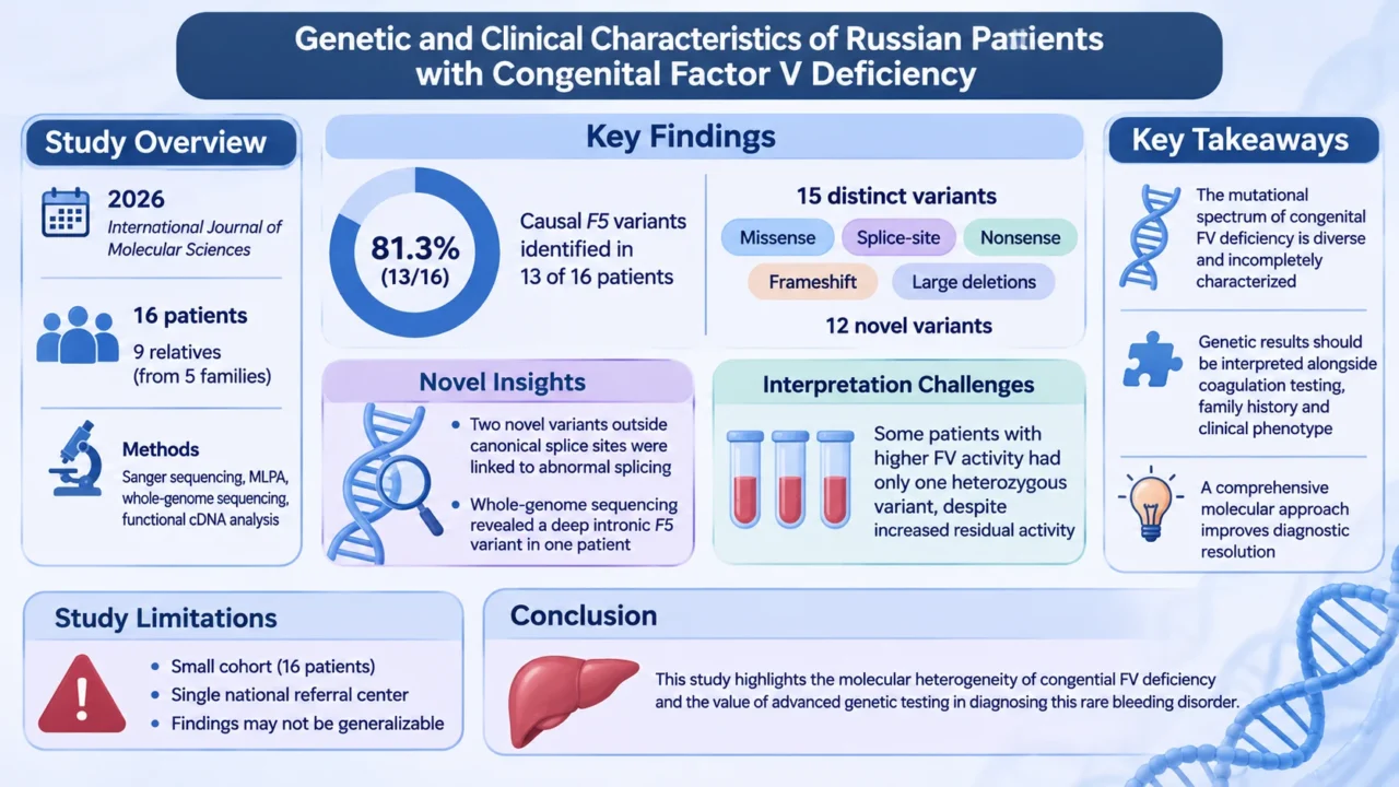
A 2026 study published in the International Journal of Molecular Sciences, titled “Genetic and Clinical Characteristics of Russian Patients with Congenital Factor V Deficiency,” provides additional insight into the molecular heterogeneity of congenital FV deficiency. The study, led by Olesya Pshenichnikova and colleagues, aimed to characterize F5 variants in a Russian patient cohort and examine their clinical and molecular significance.
The researchers analyzed 16 unrelated patients with congenital FV deficiency and nine relatives from five families. Their diagnostic approach combined Sanger sequencing of functionally relevant regions of F5 with multiplex ligation-dependent probe amplification (MLPA) to identify large deletions or duplications. Whole-genome sequencing and functional cDNA analysis were also performed in selected cases, allowing the investigators to explore variants that might not be detected through conventional sequencing approaches.
Causal F5 variants were identified in 13 of the 16 unrelated patients (81.3%). In total, 15 distinct variants were reported, including missense variants, splice-site alterations, nonsense variants, frameshift variants, and large deletions. Twelve variants were novel, illustrating how the mutational spectrum of this rare disorder remains incompletely characterized.
One particularly relevant finding was the identification of two previously unreported variants outside the canonical splice-site dinucleotides that were associated with abnormal splicing.
In one patient, conventional Sanger sequencing and MLPA did not identify a causative variant; whole-genome sequencing subsequently revealed a deep intronic F5 variant. This finding demonstrates how non-canonical splice-altering and deep intronic variants may contribute to cases in which routine molecular testing is inconclusive.
The study also identified an important limitation in interpreting genetic results. Although all five patients with FV activity below 5% carried biallelic F5 variants, several patients with higher residual activity had only one identified heterozygous variant. The authors therefore considered the possibility of additional genetic or non-genetic modifiers. These findings reinforce that molecular results should be interpreted alongside coagulation testing, family history, and the patient’s clinical phenotype.
Despite its contribution, the study has limitations. The cohort was relatively small and drawn from a national referral center, meaning that the findings cannot necessarily be generalized to all populations with congenital FV deficiency. Nevertheless, the combination of sequencing, structural-variant analysis, and functional investigation provides a useful example of how a comprehensive molecular approach can improve diagnostic resolution in rare bleeding disorders.
Diagnostic Challenges and Laboratory Evaluation
The initial laboratory evaluation of suspected FV deficiency usually reveals prolongation of both prothrombin time (PT) and activated partial thromboplastin time (aPTT), reflecting the involvement of FV in the common pathway of coagulation.
Thrombin time (TT) is generally normal because the conversion of fibrinogen to fibrin is not directly dependent on FV. However, this pattern is not specific and may also occur in deficiencies of other common pathway factors or in acquired coagulation abnormalities.
A mixing study is an important step in the investigation. Patient plasma is mixed with normal pooled plasma, and PT and aPTT are reassessed. Correction of the prolonged clotting times supports a factor deficiency, whereas failure to correct raises suspicion for an inhibitor, including an acquired FV inhibitor. Interpretation should take into account the limitations of mixing studies, including the possibility of partial correction and the effects of anticoagulant drugs or lupus anticoagulants.
The diagnosis is confirmed by measuring FV activity using a specific coagulation factor assay, usually a one-stage clotting assay based on PT correction in FV-deficient plasma. Other common pathway factors, particularly factors II and X, should also be assessed when indicated.
Measuring FV antigen may help distinguish quantitative deficiency, in which both antigen and activity are reduced, from qualitative deficiency, in which FV antigen is relatively preserved despite reduced functional activity.
A comprehensive evaluation should also include factor VIII activity. This is particularly important because reduced FV activity may occur as part of combined factor V and VIII deficiency rather than as an isolated abnormality.
Depending on the clinical presentation, testing for von Willebrand factor antigen and activity may also be required to investigate alternative explanations for reduced FVIII activity.
The differential diagnosis includes other common pathway factor deficiencies, liver disease, vitamin K deficiency, disseminated intravascular coagulation, and acquired FV inhibitors. A sudden onset of bleeding in a patient without a previous bleeding history should particularly raise concern for an acquired inhibitor. In such cases, a specific inhibitor assay, such as a modified Bethesda assay, may be required to establish the diagnosis.
Combined Factor V and Factor VIII Deficiency: A Distinct Diagnostic Entity
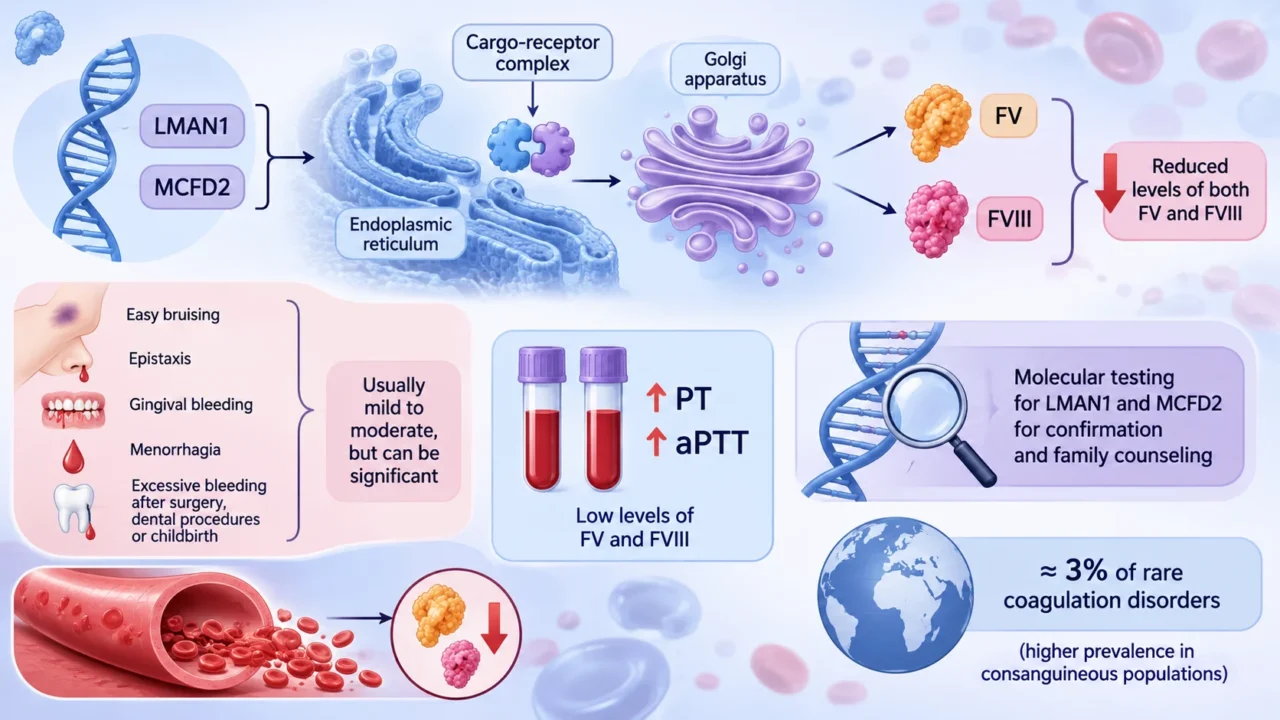
Combined deficiency of factors V and VIII (F5F8D) is a rare inherited bleeding disorder characterized by reduced plasma levels of both coagulation factors. It is distinct from isolated FV deficiency because the underlying molecular mechanism involves intracellular protein transport rather than pathogenic variants in the F5 gene itself. The disorder is usually inherited in an autosomal recessive manner and is associated with pathogenic variants in either LMAN1 or MCFD2.
LMAN1 and MCFD2 encode proteins that form a cargo-receptor complex involved in the transport of selected proteins from the endoplasmic reticulum to the Golgi apparatus. Both FV and FVIII depend on this pathway for efficient intracellular transport and secretion. Defects in either component of the complex can therefore reduce the circulating levels of both coagulation factors.
Patients with combined FV and FVIII deficiency may present with easy bruising, epistaxis, gingival bleeding, menorrhagia, and excessive bleeding after surgery, dental procedures, or childbirth. The clinical phenotype is often mild to moderate, although significant bleeding can occur.
The disorder is estimated to account for approximately 3% of rare coagulation disorders, with a higher prevalence reported in populations where consanguineous marriage is more common.
The laboratory profile typically demonstrates prolongation of both PT and aPTT, reflecting the reduced activity of FV and FVIII. The diagnosis requires measurement of both factors: identifying a low FV level alone is insufficient to distinguish isolated FV deficiency from F5F8D. Molecular testing for LMAN1 and MCFD2 can then help establish the genetic diagnosis and support family counseling.
Why Distinguishing the Two Disorders Matters
Although isolated FV deficiency and combined FV and FVIII deficiency can produce similar screening coagulation results, distinguishing them has important diagnostic and clinical implications. In a patient with prolonged PT and aPTT and reduced FV activity, FVIII activity should not be overlooked. A simultaneous reduction in both factors should prompt consideration of F5F8D and further evaluation of the LMAN1 and MCFD2 genes.
The distinction is also relevant when interpreting the relationship between factor levels and bleeding. Research in F5F8D has shown that the balance between FV and FVIII may influence the clinical phenotype in ways that are not explained by either factor level alone.
In particular, a relatively low FV level may reduce the procoagulant drive and partially modify the bleeding tendency associated with reduced FVIII. This observation illustrates the complex interaction between coagulation factors and cautions against interpreting an isolated laboratory value without considering the broader hemostatic context.
Management and Hemostatic Support
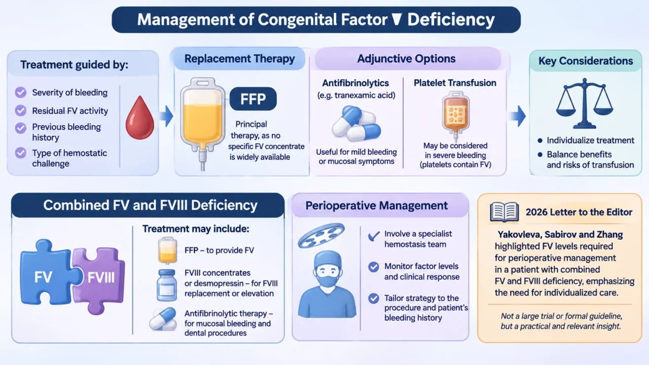
Management of congenital FV deficiency is guided by the severity of bleeding, residual FV activity, previous bleeding history, and the type of hemostatic challenge. There is currently no widely available specific FV concentrate. Fresh frozen plasma (FFP) is therefore the principal replacement therapy for clinically significant bleeding and for selected invasive procedures, as it provides a source of FV.
For patients with mild bleeding or mucosal symptoms, antifibrinolytic agents such as tranexamic acid may be useful, either alone or as an adjunct to replacement therapy. Platelet transfusion may also be considered in selected severe bleeding situations because platelets contain FV in their alpha granules, providing a local pool that may contribute to hemostasis. Treatment decisions should be individualized, taking into account the clinical situation and the risks associated with transfusion.
In combined FV and FVIII deficiency, treatment may require a strategy that addresses both factor deficiencies. FFP can provide FV, while FVIII concentrates or desmopressin may be considered for FVIII replacement or elevation in selected patients, depending on the clinical context and demonstrated response.
Antifibrinolytic therapy may also be useful for mucosal bleeding and dental procedures. Perioperative planning should involve a specialist hemostasis team, with factor levels and clinical response monitored according to the procedure and the patient’s bleeding history.
A 2026 letter to the editor by Yakovleva, Sabirov, and Zhang specifically addressed FV levels required for perioperative management in a patient with combined FV and FVIII deficiency. Although this publication is not a large clinical trial or a formal guideline, it highlights the practical difficulty of planning surgical hemostasis in patients with this rare combined disorder and the need for individualized management.
Looking Beyond Factor Levels
Congenital FV deficiency remains a diagnostic challenge because its clinical presentation is heterogeneous, its laboratory abnormalities are not disease-specific, and the relationship between FV activity and bleeding severity is imperfect.
Recent molecular studies have expanded the known spectrum of F5 variants and demonstrated the value of combining conventional sequencing with structural-variant analysis, whole-genome sequencing, and functional studies when routine testing does not provide a clear explanation.
Equally important is the recognition of combined FV and FVIII deficiency as a separate inherited disorder involving intracellular protein transport. Measuring FVIII activity in patients with reduced FV is therefore an essential part of a comprehensive diagnostic evaluation.
Ultimately, accurate diagnosis requires the integration of clinical history, coagulation testing, factor activity measurements, inhibitor assessment, and molecular investigation to guide appropriate management and improve care for patients with rare bleeding disorders.
FAQ
1. What is congenital factor V deficiency?
Congenital factor V deficiency is a rare autosomal recessive bleeding disorder caused by pathogenic variants in the F5 gene, resulting in reduced factor V activity.
2. Why are both PT and aPTT prolonged in factor V deficiency?
Factor V participates in the common coagulation pathway. Its deficiency can therefore impair both the extrinsic and intrinsic pathway-based clotting assays.
3. What is the difference between factor V deficiency and combined factor V and VIII deficiency?
Isolated factor V deficiency involves reduced FV activity, whereas combined deficiency involves simultaneous reductions in FV and FVIII due to abnormalities in their shared intracellular transport mechanism.
4. Which genes are associated with combined FV and FVIII deficiency?
The principal genes are LMAN1 and MCFD2, which encode proteins involved in the intracellular transport of coagulation factors.
5. Can factor V deficiency occur with normal platelet counts?
Yes. Factor V deficiency primarily affects coagulation factor activity and does not necessarily cause thrombocytopenia.
6. Why is fresh frozen plasma used to treat factor V deficiency?
Fresh frozen plasma contains factor V and can provide replacement in patients with clinically significant bleeding or before invasive procedures.
7. Can factor V levels predict bleeding severity?
Not reliably. Clinical manifestations vary considerably, and plasma factor V activity does not consistently correlate with bleeding severity.
8. Why is genetic testing important in factor V deficiency?
Genetic testing can identify pathogenic F5 variants, clarify inheritance, support family assessment, and investigate cases in which conventional laboratory findings are suggestive but molecular diagnosis remains uncertain.
9. Why should factor VIII be measured in patients with suspected factor V deficiency?
Measuring factor VIII helps distinguish isolated factor V deficiency from combined FV/FVIII deficiency and other conditions associated with reduced factor VIII activity.
10. What is the main treatment difference between isolated and combined FV/FVIII deficiency?
Both conditions may require fresh frozen plasma for factor V replacement. However, combined deficiency additionally requires assessment and, when necessary, replacement of factor VIII.
Written by Hermine Sayiyan, MD.
Stay updated with Hemostasis Today.
-
Nov 18, 2026, 14:29Maxime Dely: When Patient Encounters Become Life Lessons
-
Nov 18, 2026, 14:23Basanta Khatiwada: Advancing Haematology Through Research and Innovation at HAEMATOCON 2026
-
Nov 18, 2026, 14:13Alvin Katumba: Sickle Cell Disease Research Recognised With a 2026 ASH Abstract Achievement Award
-
Nov 18, 2026, 13:34Vincenzo De Angelis: Strengthening Plasma Management and Quality Control in Eastern Europe
-
Nov 18, 2026, 13:34Standardizing Bleeding Outcome Assessment in Hemophilia Clinical Trials – JTH
-
Nov 18, 2026, 13:34Nicolas Gendron: Towards Sustainable Anticoagulation
-
Nov 18, 2026, 13:33Breaking the Silence on Women and Girls with Bleeding Disorders – EHC
-
Nov 18, 2026, 13:32Maria Elisa Mancuso: Next-Generation Protein Therapies in Hemophilia
-
Nov 18, 2026, 13:25Maia Meier: Advocating for Women and Girls with Bleeding Disorders