


Jan Sloves: Why Pelvic Venous Disorders Remain Underrecognized
Jan Sloves, President and Consultant at Vascular Imaging Professionals LLC, shared a post on LinkedIn about a recent article by Francine Freitas Fernandes et al., published in Ultrasonography, adding:
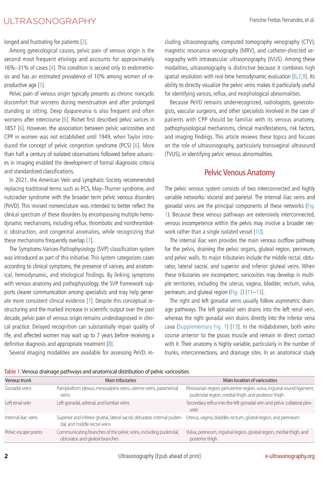
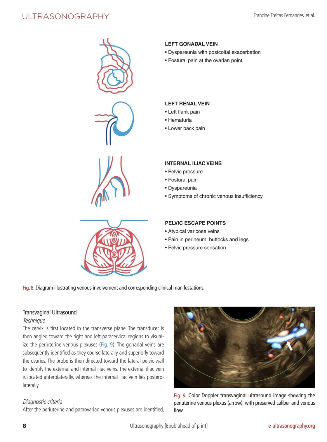
“Pelvic pain of venous origin remains underrecognized, despite being a major contributor to chronic pelvic pain in women and a frequent driver of gynecologic consultation.
This New Ultrasonography Review by Francine Freitas Fernandes and Colleagues is Important Because it Reframes How Vascular Specialists Should Think About Both Terminology and Imaging Strategy at a High Academic Level.
My Key Take home Points
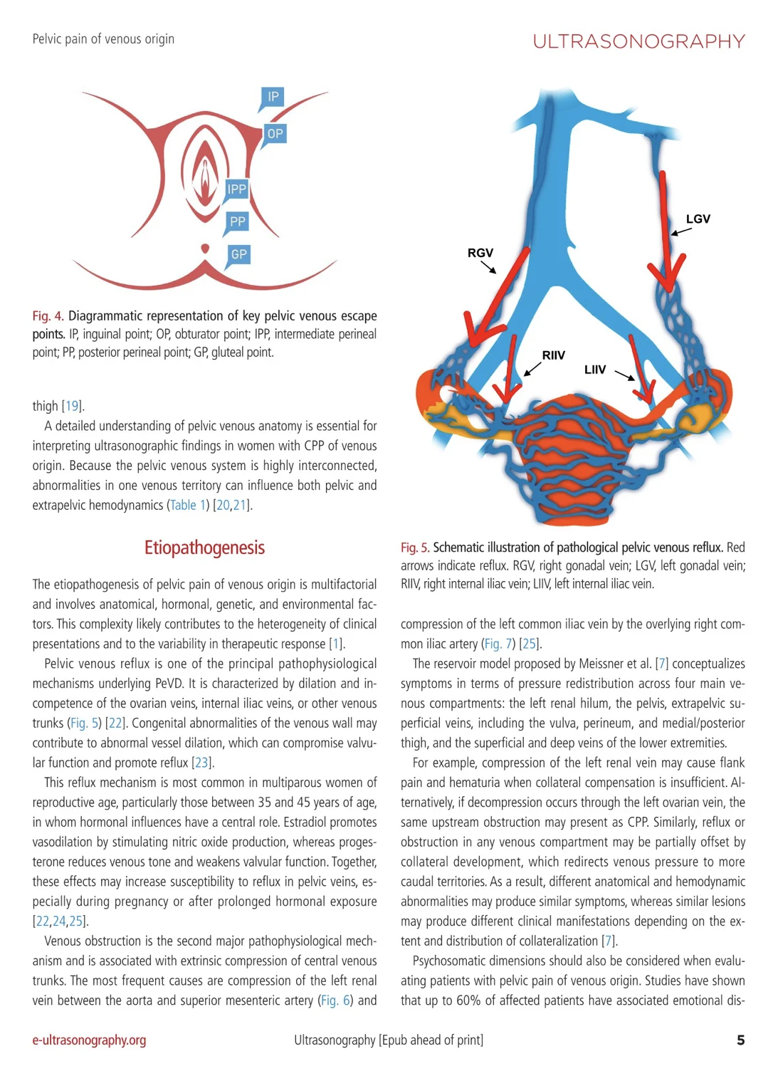
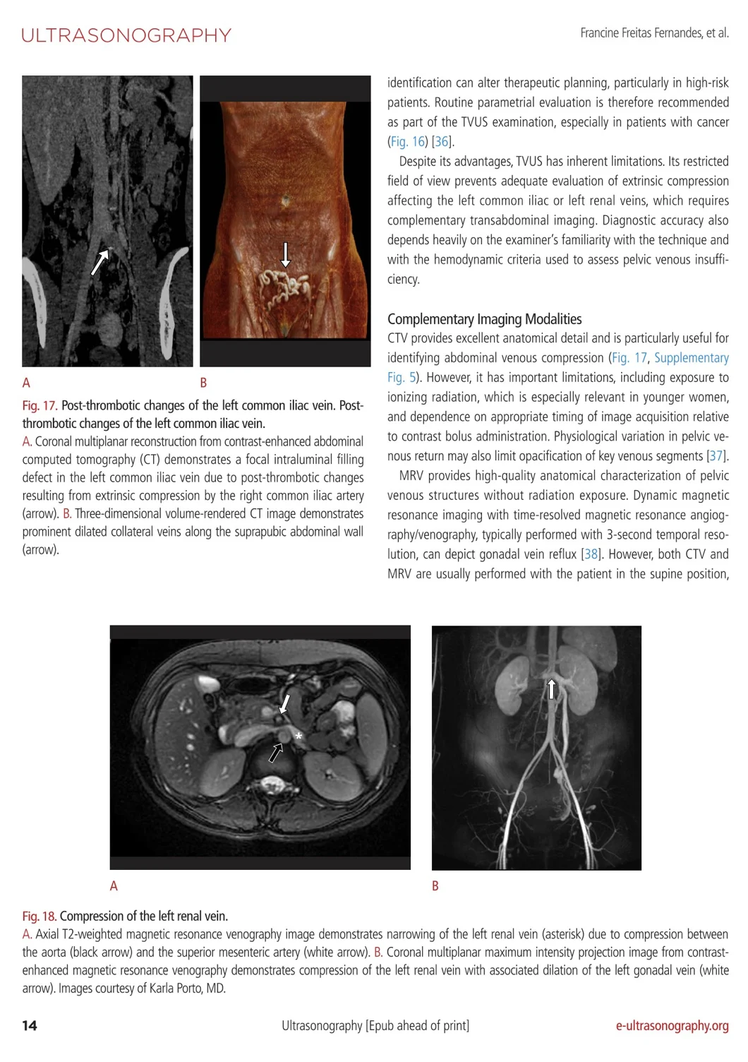
From ‘PCS’ to Pelvic Venous Disorders (PeVD): The move away from narrow labels such as pelvic congestion syndrome acknowledges that reflux, thrombotic and nonthrombotic obstruction and congenital anomalies frequently coexist rather than present as isolated entities
Symptom–varix–pathophysiology linkage: The SVP framework connects symptoms, varices, and anatomic/hemodynamic findings, creating a more rigorous language between gynecology, vascular surgery, and radiology
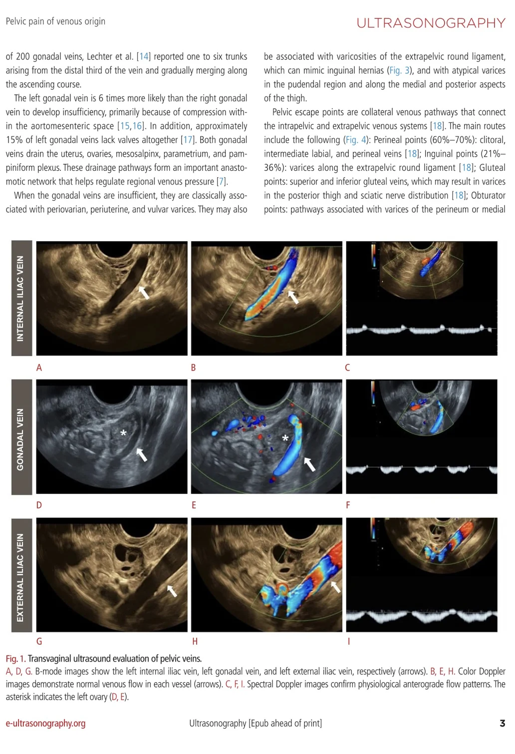
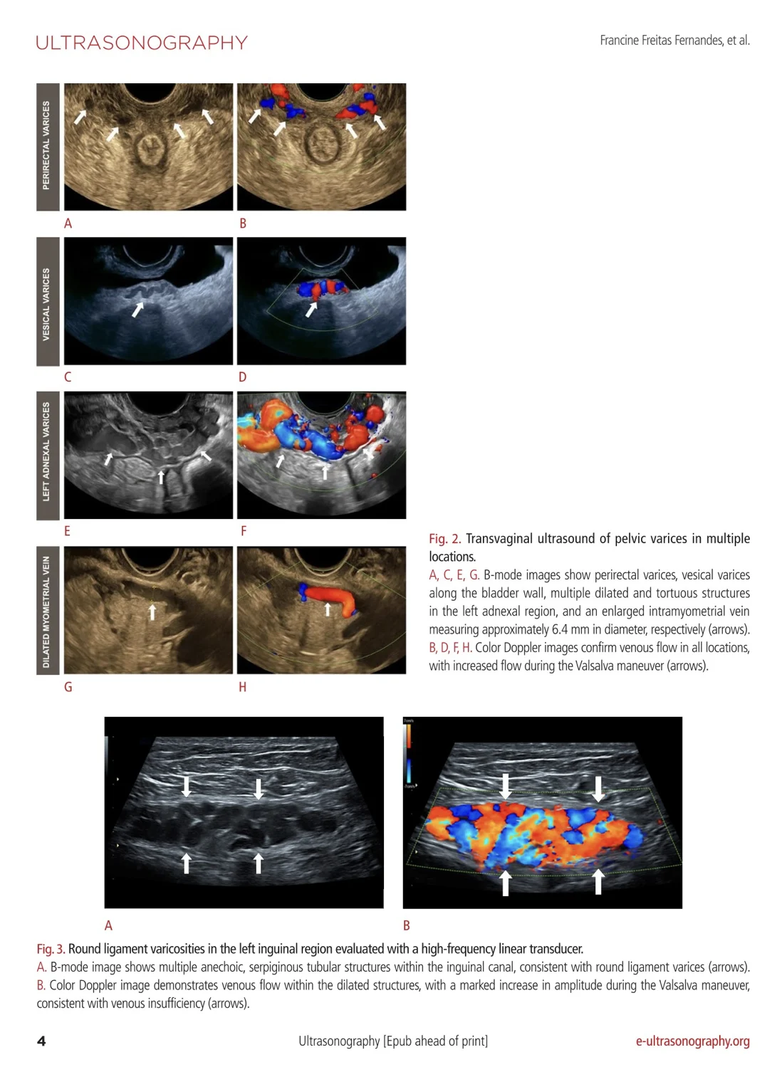
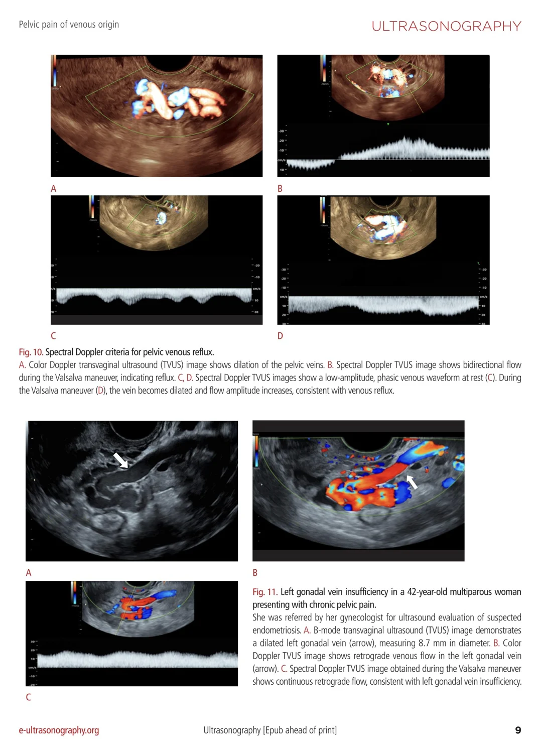
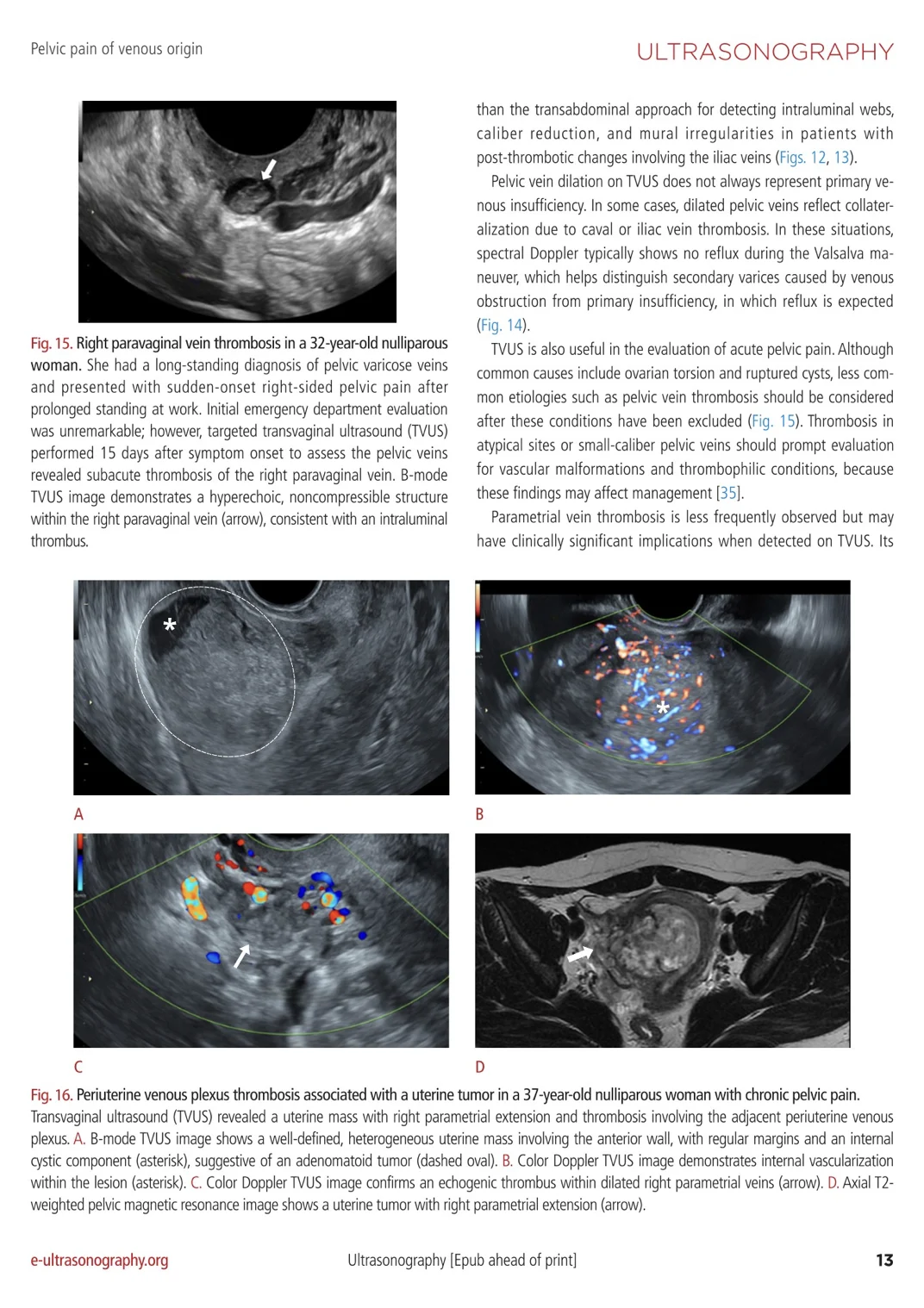
Transvaginal ultrasound as a hemodynamic examination: When performed with appropriate positioning, venous filling and Doppler interrogation during Valsalva, TVUS can define ovarian and internal iliac venous insufficiency with a degree of functional detail that is often under appreciated
Diameter alone is insufficient: Venous caliber thresholds are useful, but spectral evidence of reflux and overall hemodynamic context remain essential for distinguishing clinically meaningful disease from incidental dilation.
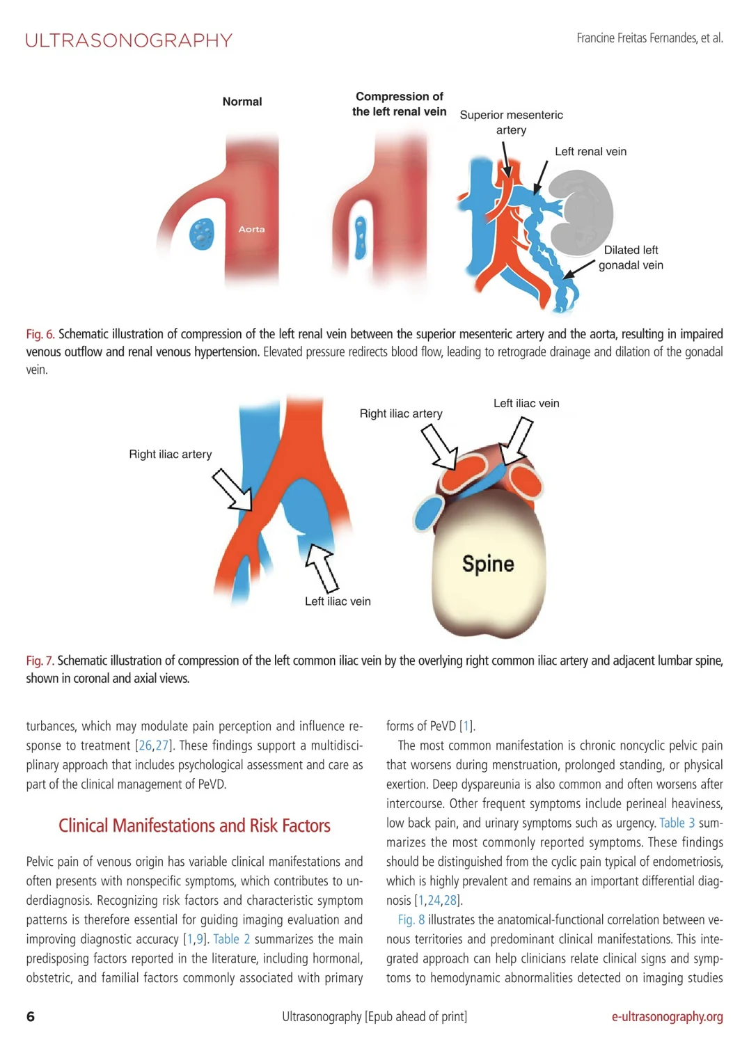
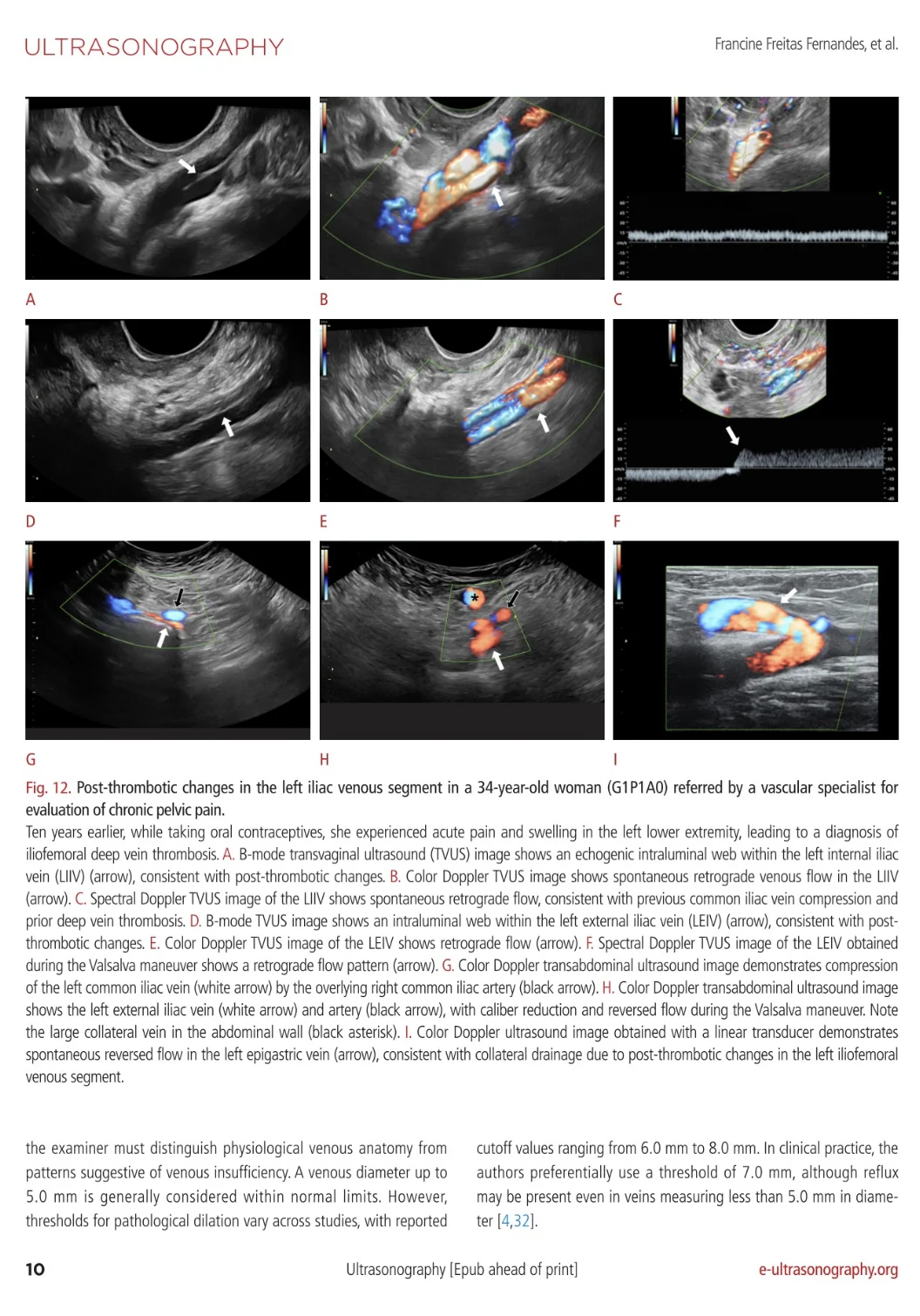
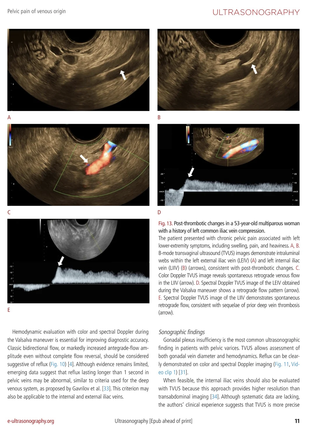
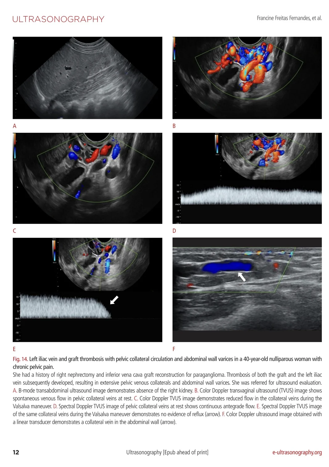
Reflux versus collateralization: Dilated pelvic veins may represent primary venous insufficiency or collateral pathways secondary to iliac or caval obstruction, making Doppler behavior central to interpretation
Diagnostic delay matters: The paper emphasizes that these patients may wait years for definitive diagnosis and failure to recognize a venous etiology contributes directly to delayed care and inappropriate management
Protocol integration is overdue: A structured four-stage ultrasound approach offers a credible framework for routine practice and in my view, this is the level of pelvic venous assessment our labs should be delivering consistently.”
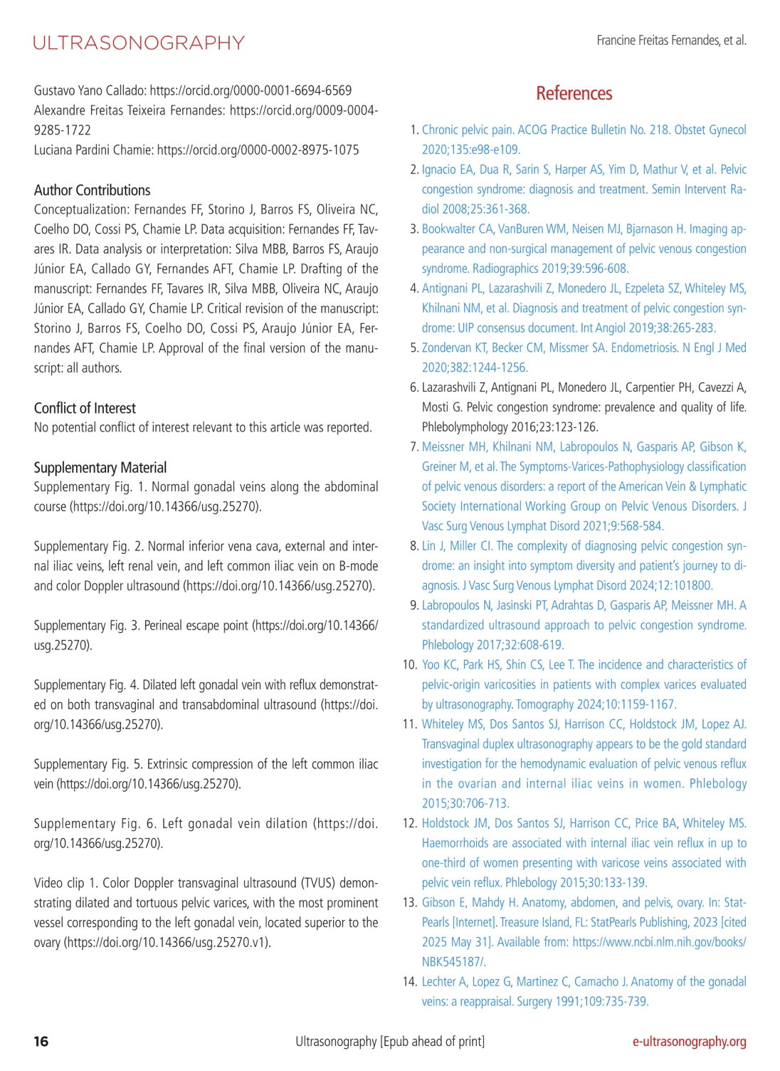
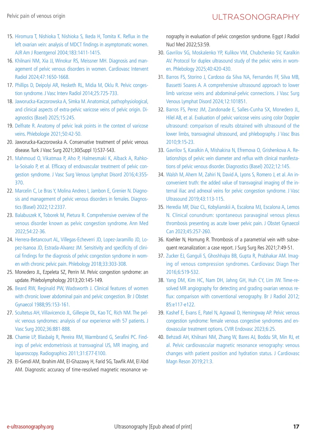
Title: Pelvic pain of venous origin: diagnostic insights from transvaginal ultrasonography
Authors: Francine Freitas Fernandes, Isabela Rodrigues Tavares, Manuella Brandão Barreto Silva, Joana Storino, Fanilda Souto Barros, Nathalia Cardoso Oliveira, Dionésio Oliveira Coelho, Paulo Sérgio Cossi, Edward Araujo Júnior, Gustavo Yano Callado, Alexandre Freitas Teixeira Fernandes, Luciana Pardini Chamie
Stay updated on all scientific advances with Hemostasis Today.
-
Oct 2, 2026, 13:50Arnav Garcha: New Insights Into Platelet Transport in Arterial Flow
-
Oct 2, 2026, 13:49Alessandro Lucchesi: RiseMPN 2026 – Shaping the Future of MPN Treatment
-
Oct 2, 2026, 13:43Ronald Mahomane: Bangladesh Moves Toward Sustainable Hemophilia Treatment Access
-
Oct 2, 2026, 13:38Sathya-Prasad Burjonrappa: Pediatric Trauma and Deep Venous Thrombosis
-
Oct 2, 2026, 12:50Ravi C Dara: ADHAR 2026 – Red Cell Exchange in Sickle Cell Disease
-
Oct 2, 2026, 12:37David McIntosh: WeDonate Partners with Dr. Martens to Reward UK Blood Donors
-
Oct 2, 2026, 12:31Ahmed Salah El-Deen: HELLP Syndrome – An Obstetric Emergency
-
Oct 2, 2026, 12:28Swapnil Hiremath: Transitioning from Idiopathic Thrombocytopenia to Antiphospholipid Syndrome in Recurrent Severe Preeclampsia
-
Oct 2, 2026, 12:08Austin Camp: Learn How to Treat HIT from an ICU Pharmacist in 60 Seconds!