

Glanzmann Thrombasthenia Beyond Platelet Transfusion
Glanzmann thrombasthenia (GT) is becoming a model for a broader change in the treatment of inherited bleeding disorders: moving from replacing defective blood components toward bypassing the underlying hemostatic defect and, ultimately, correcting it.
GT is caused by quantitative or qualitative abnormalities of platelet integrin αIIbβ3.
Despite a normal or near-normal platelet count, patients have severely impaired platelet aggregation.
Defective αIIbβ3 prevents effective fibrinogen-mediated platelet–platelet bridging and compromises clot retraction, resulting in impaired primary hemostasis.
Bleeding is predominantly mucocutaneous but can be severe, with recurrent epistaxis, gastrointestinal bleeding, heavy menstrual bleeding, postpartum hemorrhage, postoperative bleeding, and trauma-related bleeding.
The long-term burden may also include iron-deficiency anemia, hospitalization, transfusion exposure, reproductive complications and limitations in daily activities.
For decades, treatment has relied on local hemostatic measures, antifibrinolytics, platelet transfusion, and recombinant activated factor VII (rFVIIa). These remain important therapies.
However, most are used in response to bleeding or around high-risk situations.
The emerging therapeutic pipeline is addressing a different question:
Can hemostasis be restored without supplying donor platelets and without requiring αIIbβ3 to function normally?
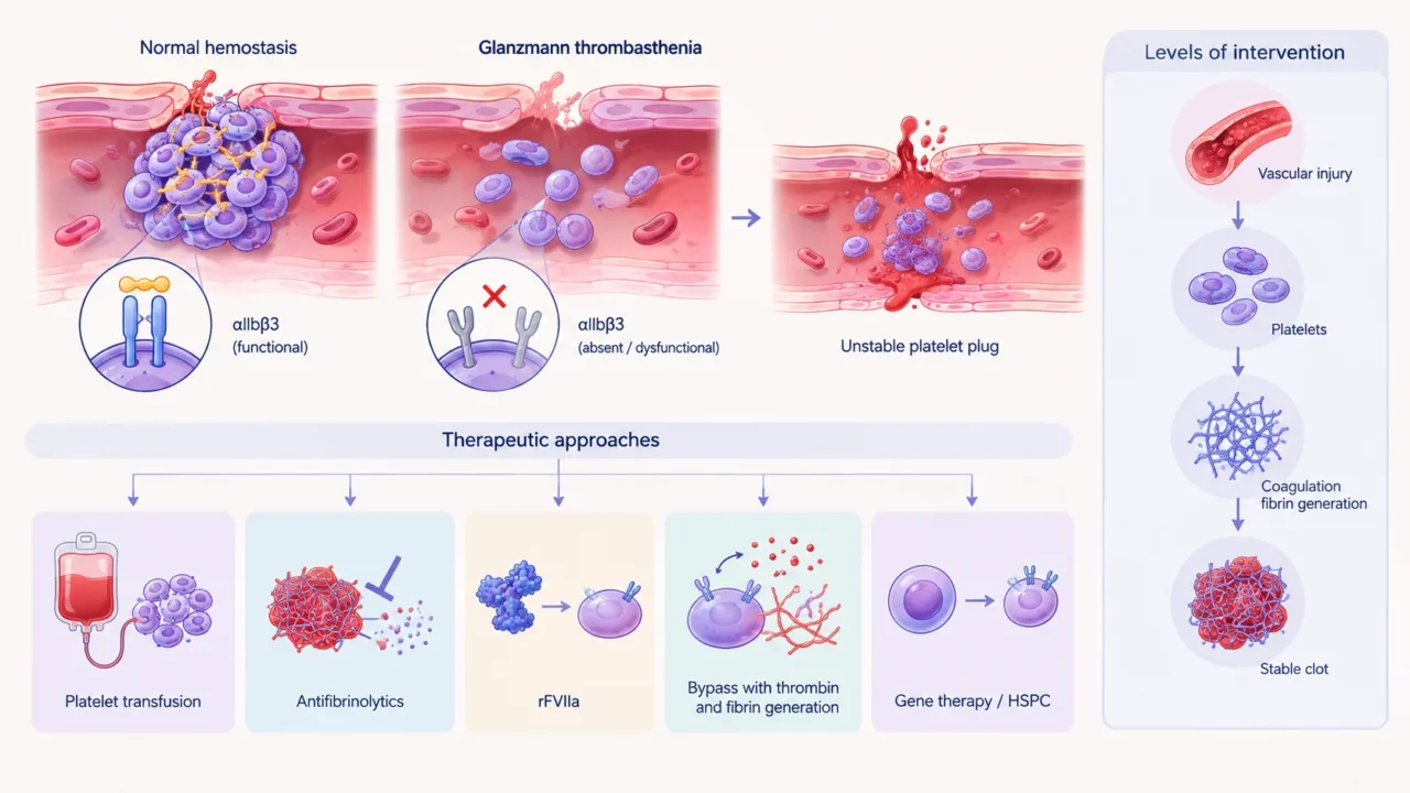
The defect is platelet function.
The central problem in GT is not platelet production but platelet interaction.
Platelets can circulate and adhere to sites of vascular injury, but they cannot efficiently aggregate because αIIbβ3 is absent, reduced or dysfunctional.
Consequently, fibrinogen cannot effectively bridge activated platelets, leaving the developing platelet plug unstable.
This creates several therapeutic possibilities.
Functional platelets can be supplied through transfusion, defective aggregation can be bypassed by increasing thrombin and fibrin generation, coagulation factors can be pharmacologically recruited to activated platelets or the underlying genetic defect can potentially be corrected in hematopoietic stem and progenitor cells.
These strategies act at different levels of the hemostatic system.
From Transfusion to Pathway Bypass
Platelet transfusion remains an important treatment for major bleeding and high-risk procedures.
It provides platelets containing functional αIIbβ3 and can rapidly restore platelet aggregation.
The major limitation is repeated exposure to donor antigens. Patients may develop antibodies against HLA or platelet-specific antigens, leading to platelet refractoriness and making subsequent bleeding increasingly difficult to manage.
This is particularly relevant in patients requiring repeated transfusions, women with pregnancy-related transfusion exposure and individuals who may later require major surgery or transplantation.
rFVIIa introduced an important transfusion-sparing approach.
Rather than correcting the αIIbβ3 defect, it enhances thrombin generation at the site of injury, promoting fibrin formation and stabilization of the hemostatic plug.
It can be particularly useful in patients with platelet antibodies or refractoriness, or when compatible platelets are unavailable.
Consensus recommendations commonly use approximately 80–120 μg/kg every 2–3 hours during bleeding or around procedures, although treatment should be individualized.
Its limitations are equally important. rFVIIa is administered intravenously, often requires repeated dosing, and is primarily used during active bleeding or around invasive procedures. This makes it difficult to use as routine long-term prophylaxis.
The next generation of therapies is attempting to retain the bypass principle while moving toward prevention.
Sutacimig and the Move Toward Prophylaxis
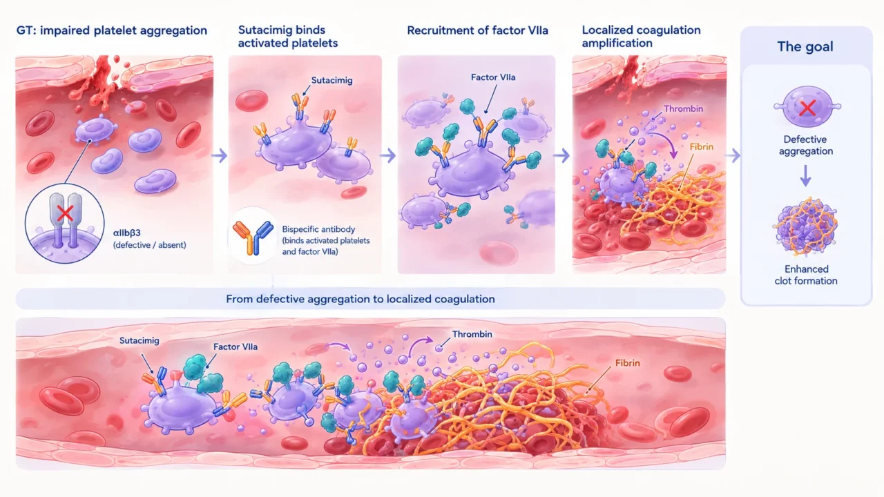
Sutacimig is a bispecific antibody being developed as a potential prophylactic treatment for GT.
Rather than replacing αIIbβ3-deficient platelets, sutacimig is designed to bind activated platelets and recruit or localize factor VIIa at sites of vascular injury.
The resulting local amplification of coagulation is intended to increase thrombin and fibrin generation around defective platelets.
The concept is straightforward:
Defective platelet aggregation to localized coagulation amplification
The therapeutic goal is therefore not to restore platelet aggregation itself, but to make the aggregation defect less consequential for clot formation.
What the Clinical Data Show?
Sutacimig has been evaluated in the Phase 1/2 HMB-001-CL101 study.
At the 2025 ASH meeting, the sponsor reported an approximately 50% reduction in mean annualized treated bleeding across evaluated dose groups, supporting further development toward a pivotal Phase 3 study.
Updated long-term-extension data presented at ISTH 2026 included 34 patients treated for a median of 6.9 months, with exposure extending to 15.9 months.
Hemab reported sustained reductions in bleeding, including a 62% mean reduction in high-intensity annualized treated bleeding rate. Three procedures performed during the extension period were reported to have successful hemostatic outcomes.
These findings are encouraging in a rare disorder in which large comparative trials are challenging. They suggest the possibility of reducing bleeding between acute episodes rather than relying exclusively on rescue treatment.
However, the evidence remains preliminary. The available data come from a small, non-comparative clinical program and include sponsor-reported analyses. They should therefore be interpreted as an encouraging signal rather than definitive evidence of clinical benefit.
As of September 2026, the FDA has reportedly endorsed progression to a pivotal Phase 3 study using a planned dose of 0.2 mg/kg once weekly, with initiation anticipated during the second half of 2026.
Sutacimig also received FDA Breakthrough Therapy Designation in March 2026.
Breakthrough Therapy Designation facilitates regulatory interaction and development but does not constitute approval or establish efficacy.
What Phase 3 needs to answer?
The pivotal study will need to establish whether weekly sutacimig can provide consistent protection across the heterogeneous GT population.
Important questions include whether it reduces annualized treated bleeding compared with individualized standard care, whether benefit is maintained in patients with severe bleeding phenotypes, and whether it remains effective in patients with platelet alloimmunization or refractoriness.
Its role during surgery, dental procedures, childbirth, and trauma will also be important.
Equally relevant will be the incidence of thrombosis and other coagulation-related complications, as well as potential reductions in hospitalization, platelet exposure, rFVIIa use, and treatment burden.
The most clinically meaningful benefit may not simply be fewer mucosal bleeding episodes.
Prevention of hospital-level hemorrhage, postpartum bleeding, procedure-related bleeding, and episodes requiring intensive platelet or rFVIIa support could have greater implications for patients.
Gene Therapy: Correcting the Megakaryocyte
A different strategy is aimed at the underlying cellular defect.
GT results from pathogenic variants in ITGA2B or ITGB3, which encode the αIIb and β3 subunits of αIIbβ3.
Because mature platelets are anucleate, gene therapy would need to target hematopoietic progenitors and the megakaryocyte lineage rather than circulating platelets.
An autologous hematopoietic stem-cell approach could involve collection of CD34⁺ cells, introduction of a functional copy of ITGA2B or ITGB3, and reinfusion after conditioning.
If successful, the corrected cells could generate megakaryocytes and platelets expressing functional αIIbβ3.
The potential advantage is fundamental: instead of repeatedly compensating for defective platelets, treatment could provide a durable source of autologous functional platelets.
This could reduce dependence on donor platelet transfusion and potentially decrease the risk of alloimmunization.
However, gene therapy for GT remains experimental. Conditioning toxicity, engraftment, vector-related risks, immune responses, manufacturing complexity, and long-term durability remain major considerations.
Genetic heterogeneity adds another layer of complexity.
Variants in ITGA2B and ITGB3 can affect protein expression, folding, intracellular trafficking, surface expression, or receptor activation. Future approaches may therefore need to consider the specific molecular defect rather than treating all GT genotypes identically.
Gene Editing and Precision Disease Modeling
Gene editing provides another route to understanding and potentially correcting GT.
CRISPR-based approaches have been used to introduce disease-causing ITGA2B and ITGB3 variants into CD34⁺ cell-derived megakaryocytes. These models allow investigators to examine how individual mutations affect integrin synthesis, trafficking, surface expression, platelet production, and receptor function.
They also provide a platform for testing gene replacement and editing strategies and for determining which mutations may be amenable to molecular rescue.
At present, the most realistic role of gene editing is likely to remain within research and therapeutic development. Clinical translation will require rigorous assessment of off-target editing, chromosomal abnormalities, clonal selection, and long-term hematopoietic safety.
Measuring More than Bleeding
The impact of a new GT therapy should not be judged solely by annualized bleeding rates.
Reducing emergency visits and hospitalizations, limiting exposure to platelets and rFVIIa, preventing alloimmunization, improving management of heavy menstrual bleeding and pregnancy, and facilitating safer surgery could all represent meaningful clinical benefits.
Quality of life and treatment burden are also important.
A therapy that reduces bleeding while decreasing repeated intravenous treatment and allowing greater participation in education, employment, and physical activity could substantially change life with GT.
Conversely, a treatment that reduces bleeding but requires frequent administration, intensive monitoring, or carries significant thrombotic risk may offer limited benefit for some patients.
A New Treatment Architecture
The evolving therapeutic landscape can be viewed as a progression from replacement toward increasingly disease-directed approaches.
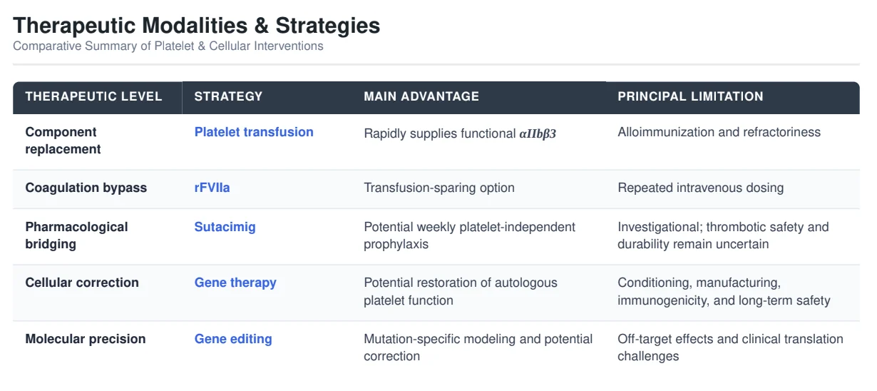
GT therefore provides a unique model of how hemostasis can potentially be restored at multiple levels: the platelet, the coagulation cascade, the megakaryocyte, or the hematopoietic stem cell.
Questions that will define the field
Several questions remain central to the development of these approaches.
- Can pharmacological bypass provide consistent protection across different GT phenotypes?
- Can increased coagulation activity maintain an adequate margin between hemostasis and thrombosis, particularly during surgery, pregnancy, inflammation, or other prothrombotic states?
- For gene therapy, what proportion of corrected megakaryocytes and platelets is required to achieve clinically meaningful protection?
- And ultimately, how should treatment be selected?
The future is unlikely to involve a single replacement for platelet transfusion. Treatment may instead be individualized according to bleeding phenotype, genotype, alloantibody status, reproductive plans, procedural risk, venous access, and patient preference.
A New Direction for Glanzmann Thrombasthenia
Glanzmann thrombasthenia illustrates a broader change in inherited platelet disorder management.
For decades, treatment has largely focused on controlling bleeding when it occurs. New approaches are beginning to address the defect from a different direction: bypassing impaired platelet aggregation or, eventually, correcting the cellular and genetic cause.
Sutacimig is currently the most advanced clinical example of this strategy, with Phase 2 and long-term-extension data supporting progression toward a pivotal Phase 3 study.
It remains investigational, however, and transfusion-independent prophylaxis cannot yet be considered established care.
Gene therapy and gene editing offer a more definitive possibility, but remain at earlier stages of development.
The future of GT may therefore be a layered approach rather than a single therapy: platelet transfusion and antifibrinolytics for selected acute settings, rFVIIa or pharmacological bypass for transfusion-sparing treatment, and potentially cellular or genetic correction for selected patients.
The goal is no longer simply to replace what is missing.
It is to make the platelet defect less relevant – or eventually correct it at its source.
FAQ
1. What causes Glanzmann thrombasthenia?
Biallelic pathogenic variants in ITGA2B or ITGB3 cause absent, reduced, or dysfunctional platelet integrin αIIbβ3, resulting in severe platelet aggregation defects.
2. Why do patients bleed despite a normal platelet count?
The problem is platelet function, not platelet number. Defective αIIbβ3 prevents fibrinogen-mediated platelet–platelet bridging and effective aggregation.
3. What are the typical bleeding manifestations?
GT mainly causes mucocutaneous bleeding, including epistaxis, gingival bleeding, heavy menstrual bleeding, and easy bruising. Severe patients may develop gastrointestinal, postpartum, postoperative or trauma-related bleeding.
4. What is the current treatment for GT?
Treatment includes local hemostatic measures, antifibrinolytics, platelet transfusion, and rFVIIa. Platelets and rFVIIa are particularly important for major bleeding and procedures.
5. Why is rFVIIa used in a platelet function disorder?
rFVIIa bypasses the defective platelet aggregation pathway by increasing local thrombin generation and fibrin formation. It strengthens clot formation without correcting αIIbβ3.
6. What is the main limitation of repeated platelet transfusion?
Repeated transfusions can cause HLA or platelet-specific alloimmunization, leading to platelet refractoriness and making future transfusions less effective.
7. What is sutacimig?
Sutacimig is an investigational bispecific antibody designed to bind activated platelets and localize FVIIa activity, thereby amplifying coagulation at sites of vascular injury.
8. How could sutacimig change GT treatment?
Unlike rescue therapies, sutacimig is being developed as prophylaxis, with the potential to reduce bleeding through regular dosing without requiring donor platelets.
9. Can gene therapy correct GT?
Potentially. Gene therapy aims to modify hematopoietic stem/progenitor cells so that megakaryocytes produce platelets with functional αIIbβ3. It remains experimental.
10. What would define a major advance in GT treatment?
The goal is not only fewer bleeding episodes but also fewer hospitalizations, less platelet and rFVIIa exposure, reduced alloimmunization, safer pregnancy and procedures, and better quality of life.
Written by Elen Avetisyan, MD
Stay updated with Hemostasis Today.
-
Sep 15, 2026, 15:29Abdul Muqtadir Abbasi: Recognizing Thalassemia Trait Is Essential for Every Physician
-
Sep 15, 2026, 15:17Shehab Mohamed: A New Regional Consensus on HLH Diagnosis and Management
-
Sep 15, 2026, 15:17Sheharyar Raza: Managing Randomness in Artificial Intelligence for Transfusion Medicine
-
Sep 15, 2026, 15:00Sarah Wachtman: Modeling AAV-Based Gene Therapy for Hemophilia A Using Pediatric Liver Organoids
-
Sep 15, 2026, 15:00Ashenafi Tazebew Amare: From Blood Donation to Innovation and Patient Access
-
Sep 15, 2026, 14:55Christina Pohlman: The Hidden Complexity of Warfarin Therapy in APS
-
Sep 15, 2026, 14:44Consuela A. Albright: Building a More Connected Sickle Cell Care System
-
Sep 15, 2026, 14:43Andrew Jackson: Cyanosis as a Clue to Central Venous Obstruction
-
Sep 15, 2026, 14:43Andy Darlington: Randomized Evidence for Catheter-Based Treatment of Intermediate-High-Risk PE