

Brigitta Nicorici: Pregnancy and Laboratory Medicine
Brigitta Nicorici, Specialist Biochemist at Salvosan Ciobanca Cluj, shared a post on LinkedIn:
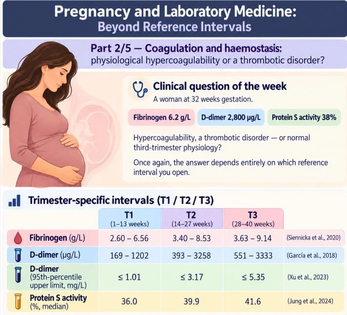
”Pregnancy and Laboratory Medicine: Beyond Reference Intervals
Part 2/5 — Coagulation and haemostasis
Clinical question of the week
A woman at 32 weeks gestation. Fibrinogen 6.2 g/L. D-dimer 2,800 µg/L. Protein S activity 38 percent.
Hypercoagulability, a thrombotic disorder — or normal third-trimester physiology?
It depends on which interval you open.
Trimester-specific intervals (T1/T2/T3)
Fibrinogen (g/L): T1 2.60–6.56; T2 3.40–8.53; T3 3.63–9.14 (Siennicka et al., 2020)
D-dimer (µg/L): T1 169–1202; T2 393–3258; T3 551–3333 (García et al., 2018)
D-dimer (95th percentile, mg/L): T1 up to 1.01; T2 up to 3.17; T3 up to 5.35 (Xu et al., 2023)
Protein S activity (percent, median): T1 36.0; T2 39.9; T3 41.6 (Jung et al., 2024)
Against non-pregnant cut-offs, every value looks alarming: D-dimer exceeds 500 µg/L in 99% of third-trimester women (García et al., 2018) and in 99.33 percent of low-risk women in the third trimester (Sun et al., 2021); fibrinogen exceeds 4 g/L in 63.93% (Sun et al., 2021). Against pregnancy-specific intervals, all three values are unremarkable (Jung et al., 2024).
Why does this happen?
Pregnancy induces a hypercoagulable state to protect against haemorrhage at delivery (Li et al., 2025). PT, APTT and TT shorten, and fibrinogen rises significantly versus non-pregnant controls, with p less than 0.05 (Li et al., 2025). FIBTEM MCF rises from the second to the third trimester: 13 mm, with an interquartile range of 11 to 16 mm, in controls versus 19 mm, with an interquartile range of 17 to 23 mm, in the third trimester, with p less than 0.001 (Carrabin et al., 2009). Fibrinolysis decreases in late pregnancy (Karlsson et al., 2012).
D-dimer and fibrinogen rise with each trimester and correlate positively in the second trimester, with r equal to 0.475 and p less than 0.0001 (Siennicka et al., 2020). Protein S activity falls substantially (Jørgensen et al., 2010), to a mean of about 46 percent in the first half of pregnancy (Ignjatović et al., 2010). Protein S antigen and activity fall throughout pregnancy, and specific activity drops from the second trimester (Takeuchi et al., 2020), possibly aided by plasma-volume expansion (Oruç et al., 2000).
Take-home
- Never read late-pregnancy coagulation tests against non-pregnant intervals.
- A D-dimer above 500 µg/L has no rule-out value in the third trimester (García et al., 2018); trimester-specific thresholds are needed (Kline et al., 2005). A negative result retains its sensitivity and negative predictive value (Favresse et al., 2018).
- Do not diagnose protein S deficiency in pregnancy. Defer testing until at least six weeks postpartum (Oruç et al., 2000), or at least 40 days postpartum (Johnston et al., 2007), and confirm positive results postpartum (Kate and Meer, 2008). Protein S assay specificity is low in pregnancy (Kate et al., 2010).
- Fibrinogen and D-dimer intervals must be trimester-specific and assay-specific. A value above the population cut-off is not automatically pathological (Siennicka et al., 2020).
A single value rarely answers the question.
The trend does.”
Stay updated with Hemostasis Today.
-
Oct 29, 2026, 15:22Ugo Clinton։ Tranexamic Acid From Discovery to a Life-Saving Treatment
-
Oct 29, 2026, 15:01Owen McCarty: Targeting FXI and FXII to Reduce Device-Associated Thrombosis and Inflammation
-
Oct 29, 2026, 14:47Ingeborg Hers: Exploring New Frontiers in Platelet Biology and Thrombosis Research
-
Oct 29, 2026, 14:36Theodore Iwashyna: Could Hemoglobin Thresholds Lead to Unnecessary Blood Transfusions in ICU Patients?
-
Oct 29, 2026, 14:24Aryabhatta Sadhu: Contributing to Nepal’s First CAR-T Cell Therapy Case
-
Oct 29, 2026, 13:50Lainie Kazan’s Story and the Clinical Risks of Post-Injury Immobilization
-
Oct 29, 2026, 12:17Bridget King: IBM Employees Support Blood Donation Efforts
-
Oct 29, 2026, 12:08Georgina Pearson: A Promising Blood Donation Partnership With Missed Potential
-
Oct 29, 2026, 11:59Dianne E. van der Wal: Cryopreserved Platelets in Transfusion Medicine