


Amir Hussain: The Science Behind Blood Clot Prevention in Healthy Circulation
Amir Hussain, Medical Laboratory Supervisor at Natural Medical and Health Research Centre Islamabad, shared a post on LinkedIn:
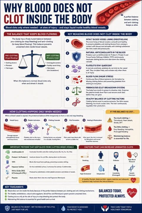
“Blood does not clot inside healthy blood vessels because the body maintains a delicate balance between pro-clotting (procoagulant) and anti-clotting (anticoagulant) mechanisms.
Clotting is activated only when a blood vessel is injured.
1. Healthy Endothelium (Blood Vessel Lining)
The innermost lining of blood vessels is called the endothelium.
It acts as a natural anti-clotting surface.
Functions of the endothelium:
- Prevents platelets from sticking to the vessel wall.
- Produces Nitric Oxide (NO), which relaxes blood vessels and inhibits platelet activation.
- Produces Prostacyclin (PGI₂), which prevents platelets from aggregating.
- Expresses Heparan sulfate, which enhances the activity of antithrombin.
2. Platelets Remain Inactive
Platelets circulate freely in the bloodstream but do not become activated unless they encounter:
- Damaged blood vessel walls
- Exposed collagen
- Tissue factor
In healthy vessels, these activating signals are hidden beneath the intact endothelium.
3. Natural Anticoagulant System
Several proteins continuously prevent unwanted clot formation.
Anticoagulant Function
- Antithrombin III Inactivates thrombin and Factors IXa, Xa, XIa, XIIa
- Protein C Inactivates Factors Va and VIIIa
- Protein S Cofactor for Protein C
- TFPI (Tissue Factor Pathway Inhibitor) Blocks the tissue factor pathway
These proteins keep the coagulation cascade under control.
4. Continuous Blood Flow
Normal blood flow:
- Dilutes activated clotting factors.
- Carries them away from the vessel wall.
- Prevents platelets from accumulating.
Stagnant or slow blood flow increases the risk of thrombosis.
5. Fibrinolytic System
The body also has a clot-removal system called fibrinolysis.
What happens when a blood vessel is injured?
When a vessel is damaged, the protective balance shifts temporarily toward clot formation.
Step 1: Vasoconstriction
The injured vessel narrows to reduce blood loss.
Step 2: Platelet Adhesion
Platelets attach to exposed collagen with the help of von Willebrand factor (vWF).
Step 3: Platelet Activation
Activated platelets release:
- ADP
- Thromboxane A₂ (TXA₂)
- Serotonin
These substances recruit additional platelets.
Step 4: Platelet Aggregation
Platelets bind together through fibrinogen bridges to form a temporary platelet plug.
Step 5: Coagulation Cascade
Clotting factors activate sequentially, producing thrombin, which converts fibrinogen – fibrin.
Fibrin forms a stable mesh that strengthens the clot.
Step 6: Healing
After the vessel heals, plasmin breaks down the fibrin clot, restoring normal blood flow.
Unwanted clots usually develop when one or more components of Virchow’s triad are present:
- Endothelial injury – trauma, surgery, inflammation.
- Abnormal blood flow – stasis or turbulence (e.g., prolonged bed rest, atrial fibrillation).
- Hypercoagulability – inherited or acquired conditions that increase clotting tendency (e.g., Factor V Leiden mutation, cancer, pregnancy).”
Stay updated with Hemostasis Today.
-
Sep 21, 2026, 03:49Jessica Lee: Walking Together for a Stronger Bleeding Disorders Community
-
Sep 21, 2026, 03:36Syed Asif Ali Shah: From Pakistan to Australia – A Clinical Haematology Milestone
-
Sep 21, 2026, 03:22Katie Baca-Motes: Estradiol Fluctuations May Help Explain Menstrual Migraine
-
Sep 21, 2026, 03:00Family History Can Reveal Hidden Risk of Aortic Dissection – The Marfan Foundation
-
Sep 21, 2026, 02:47Transitioning from Paediatric to Adult Haemophilia Care – Haemophilia Foundation Australia
-
Sep 21, 2026, 02:29Diana Castro Paupério: A New Era for Patient Blood Management
-
Sep 21, 2026, 00:29Iheanyichukwu Ike: Childhood Anaemia in Nigeria Through an Analysis of the 2018 NDHS
-
Sep 21, 2026, 00:27Nicole De Vita: The Global Burden of Iron Deficiency and Anemia in Women at SABM
-
Sep 21, 2026, 00:25Giuseppe Panuccio: IVUS and OCT Guidance Reduce MACE and Stent Thrombosis in Calcified Coronary Lesions