


Saiyed raza Abbas: Understanding White Blood Cell Development and Maturation in Peripheral Blood Smears
Saiyed raza Abbas, Laboratory Operations at PathCare Labs Private Limited, shared a post on LinkedIn:
“WBC Development in Blood Smear Understanding Leukopoiesis
Leukopoiesis is the process of formation and maturation of White Blood Cells (WBCs), primarily occurring in the bone marrow.
WBCs develop through different maturation stages, and identifying these stages during Peripheral Blood Smear (PBS) examination is an essential skill in hematology.
There are five major WBC lineages:
Neutrophilic Lineage
Myeloblast to Promyelocyte to Myelocyte to Metamyelocyte to Band Cell to Segmented Neutrophil
- Myeloblast: Large cell with a high N:C ratio and prominent nucleoli.
- Promyelocyte: Primary (azurophilic) granules appear.
- Myelocyte: Specific granules develop; last stage capable of mitosis.
- Metamyelocyte: Kidney shaped or indented nucleus.
- Band Cell: Horseshoe-shaped nucleus.
- Segmented Neutrophil: Mature cell with 2 to 5 nuclear lobes.
Function: Phagocytosis and defense against bacterial infections.
Eosinophilic Lineage
Myeloblast to Promyelocyte to Eosinophilic Myelocyte to Eosinophilic Metamyelocyte to Band Cell to Mature Eosinophil
- Characterized by prominent orange red granules.
- Mature eosinophils typically have a bilobed nucleus.
Function: Important in parasitic infections and allergic responses.
Basophilic Lineage
Myeloblast to Promyelocyte to Basophilic Myelocyte to Basophilic Metamyelocyte to Band Cell to Mature Basophil
- Contains coarse, dark blue purple granules.
- Granules may partially obscure the nucleus.
Function: Releases histamine and heparin and participates in allergic and inflammatory reactions.
Monocytic Lineage
Monoblast to Promonocyte to Monocyte
- Monoblast: Large cell with prominent nucleoli.
- Promonocyte: Irregular or folded nucleus.
- Monocyte: Large circulating WBC with abundant gray blue cytoplasm and kidney shaped nucleus.
Function: Phagocytosis and differentiation into tissue macrophages.
Lymphocytic Lineage
Lymphoblast to Prolymphocyte to Mature Lymphocyte
- * Lymphoblast: Large cell with fine chromatin and prominent nucleoli.
- * Prolymphocyte: More condensed chromatin.
- * Mature lymphocyte: Dense nucleus with a scant rim of cytoplasm.
Types: B lymphocytes, T lymphocytes and Natural Killer (NK) cells.
Clinical Importance in Peripheral Blood Smear Examination
Understanding WBC maturation helps laboratory professionals to:
- Identify immature and abnormal cells.
- Recognize left shift and increased immature granulocytes.
- Detect circulating blasts.
- Support the investigation of leukemia and other hematological disorders.
- Correlate automated analyzer flags with microscopic findings.
Remember: Accurate identification of WBC maturation stages is fundamental to peripheral blood smear interpretation and hematological diagnosis.
Knowledge of morphology is the foundation of hematology!”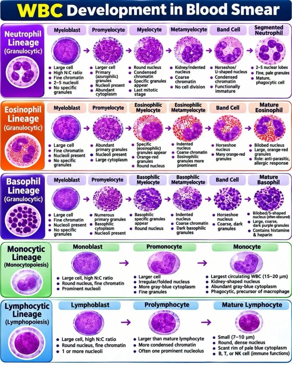
Stay updated with Hemostasis Today.
-
Nov 22, 2026, 12:46Rui Providência: Anticoagulation After AF Ablation A Timely Discussion
-
Nov 22, 2026, 12:41Teresa Gerardi: KIT Inhibition with Avapritinib and Its Potential Impact on Mastocytosis-Related Osteoporosis
-
Nov 22, 2026, 12:37Rahul Bhargava: 2026 Haematology’s Shift From Chemotherapy to Immunotherapy
-
Nov 22, 2026, 12:27Saleh Shamse Basha: Can a Platelet Count Be Trusted After Accidental Centrifugation of a Citrate Tube?
-
Nov 22, 2026, 12:19Sophia Delicou: Transforming Haemoglobin Disorders Through Innovation and Patient-Centred Care
-
Nov 22, 2026, 12:15Toby Richards: Adenomyosis and Its Impact on Female Fertility
-
Nov 22, 2026, 11:11George Kumar: Early Gastric Cancer Detection Through Red Blood Cell DNA Profiling
-
Nov 22, 2026, 10:50Kemal Karaagac: A Phenotype-Guided Approach to Stroke Prevention
-
Nov 22, 2026, 10:32Bianca Rocca: Thromboinflammation and Microvascular Injury in Shock-Associated Limb Ischaemia