


Tareq Abadl: Extravascular Hemolysis The High-Yield Guide
Tareq Abadl, Medical Lab Specialist, shared a post on LinkedIn:
“Extravascular Hemolysis
Where are the RBCs destroyed?
In extravascular hemolysis, RBCs are removed from circulation by macrophages, mainly in the spleen and liver.
What happens in the Lab?
- High Indirect Bilirubin
- Hemoglobin is broken down within macrophages, increasing bilirubin production.
- High Reticulocytes
- The bone marrow responds to increased RBC destruction by increasing RBC production.
- High LDH
- LDH may be elevated as a marker of hemolysis, but it is not specific for extravascular hemolysis.
- Low Haptoglobin
- Haptoglobin can decrease in hemolysis, but this finding occurs in both intravascular and extravascular hemolysis and should not be used alone to distinguish them.
Peripheral blood smear
Spherocytes may be seen, particularly in:
- Hereditary spherocytosis
- Warm autoimmune hemolytic anemia
Hemoglobinuria or Hemosiderinuria
These findings are more characteristic of intravascular hemolysis and are generally absent in typical extravascular hemolysis.
Common Examples
- Warm autoimmune hemolytic anemia
- Hereditary spherocytosis
- Hypersplenism
- Lab memory tip
Extravascular occurs in the spleen and liver
RBCs are broken down by macrophages, leading to heme breakdown and increased indirect bilirubin.
Remember:
Not every hemolysis marker tells you where the RBC was destroyed.
The mechanism plus peripheral smear plus urine findings should be interpreted together.”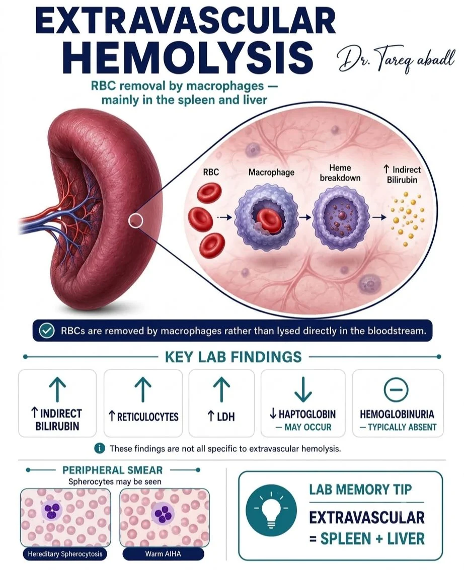
Stay updated with Hemostasis Today.
-
Oct 19, 2026, 19:44Where You Live Should not Determine Whether You Receive Care – World Federation of Hemophilia
-
Oct 19, 2026, 19:42Belen Bianco: Prevention, Early Suspicion and an Interdisciplinary Approach Save Lives
-
Oct 19, 2026, 19:40Christina Chrysohoou: Catheter-Based LAAC vs OAC for Stroke Prevention in Atrial Fibrillation
-
Oct 19, 2026, 19:38Matthew Flick: A Two-Day Research Meeting on Fibrinogen, Hemostasis and Thrombosis
-
Oct 19, 2026, 19:37Hayat Saeed: Excited to Share Our Publication on the FGL1–Hepcidin Axis in Iron Homeostasis and Disease
-
Oct 19, 2026, 19:08IPAW 2026 Comes to a Close, but the Work Continues – PPTA
-
Oct 19, 2026, 17:44Paul Riley: Moderating a Webinar on Anticoagulation Monitoring
-
Oct 19, 2026, 16:36Christopher Pittman: Blood Clot Prevention Cannot End at Hospital Discharge
-
Oct 19, 2026, 16:13Javier M. Floren: When Immune Cells Move With Their Environment