


Jalen Hurts Sidelined by a Superficial Blood Clot: What to Know
Philadelphia Eagles quarterback Jalen Hurts will miss the team’s October 18 game against the Carolina Panthers after a follow-up ultrasound revealed movement in a previously identified superficial blood clot in his calf. The clot was initially detected in London ahead of Philadelphia’s October 11 game against the Jacksonville Jaguars.
Hurts had sustained a calf injury during the October 4 game against the Los Angeles Rams after being struck by a cleat. Although an initial ultrasound identified a superficial blood clot, medical staff cleared him to play after assessing the findings. A subsequent ultrasound prompted the team to reconsider his participation, highlighting why superficial venous thrombosis can require careful monitoring.
What Happened to Jalen Hurts?
According to the Eagles’ official statement, Hurts first sought treatment for his calf on October 8, four days after the Rams game. He practiced fully on Wednesday and Thursday, October 7–8, and continued practicing on Friday, when he was added to the injury report.
An ultrasound performed in London on Saturday, October 10, revealed a superficial blood clot. After reviewing the results, the team’s medical staff and physicians in London determined that he could safely play the following day. They recommended a follow-up ultrasound within 72 hours of the game and the return flight to Philadelphia.
Hurts played against Jacksonville on October 11 without reported limitations and received treatment on Monday and Tuesday. However, a follow-up ultrasound on Wednesday night, October 14, revealed movement in the clot. The Eagles subsequently ruled him out of their October 18 game while determining the appropriate course of treatment.
The team has not publicly specified the exact vein involved, the clot’s dimensions, or its proximity to the deep venous system. It has also not confirmed whether the clot extended toward a deep vein or whether any pulmonary embolism occurred. These details are important because the clinical significance of superficial venous thrombosis depends on its anatomical location, extent, and potential for progression.
What Is Superficial Venous Thrombosis?
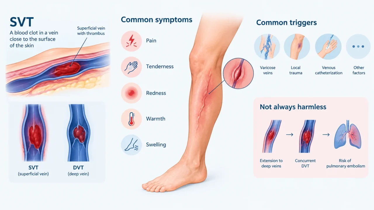
Superficial venous thrombosis (SVT) is the formation of a blood clot in a vein located close to the surface of the skin. When accompanied by inflammation of the affected vein, it is often referred to as superficial thrombophlebitis. Common symptoms include localized pain, tenderness, redness, warmth, and swelling, although some cases may present with relatively mild symptoms.
In the lower extremities, SVT frequently develops in association with varicose veins. However, it can also occur following local trauma, venous catheterization, or other provoking factors. The clinical presentation depends on the affected vein and the extent of the thrombus.
Although isolated SVT is generally less dangerous than deep vein thrombosis (DVT), it should not automatically be considered harmless. Some cases are associated with extension into the deep venous system, concurrent DVT, or an increased risk of pulmonary embolism. Consequently, identifying a clot as superficial is only the beginning of the clinical assessment.
Why Can a Superficial Clot Be Clinically Significant?
Superficial and deep veins are connected through perforating veins and major venous junctions. These anatomical connections allow a thrombus originating in the superficial system to extend into the deep veins. The risk is particularly important when the clot is extensive or located close to a junction between the superficial and deep venous systems.
SVT may also coexist with DVT at the time of diagnosis. Because DVT can lead to pulmonary embolism (PE), assessing the deep venous system is an important component of the evaluation of lower-limb SVT. However, a superficial clot does not mean that a deep clot or pulmonary embolism is necessarily present.
The location and length of the thrombus, proximity to the deep venous system, symptom severity, and individual risk factors help determine the likelihood of complications. This is why the clinical management of SVT should be guided by imaging findings and an individualized assessment rather than symptoms alone.
Can a Sports Injury Trigger a Blood Clot?
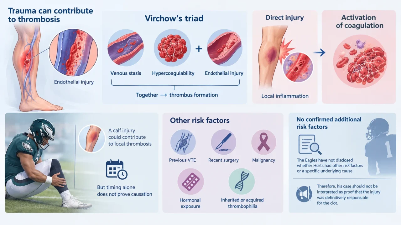
Trauma may contribute to thrombosis through endothelial injury, one component of Virchow’s triad. The other components are venous stasis and hypercoagulability, which together describe the principal mechanisms that promote thrombus formation. Direct injury to tissues or a blood vessel may initiate local inflammation and activate coagulation.
A blow to the calf could plausibly contribute to local thrombosis, but the timing of Hurts’ injury and subsequent diagnosis does not establish a direct causal relationship. Other factors, including previous venous thromboembolism, recent surgery, malignancy, hormonal exposure, and inherited or acquired thrombophilia, can also influence thrombotic risk.
The Eagles have not publicly disclosed whether Hurts had additional risk factors or whether investigations identified a specific underlying cause. Therefore, his case should not be interpreted as evidence that a direct sports injury was definitively responsible for the clot.
The Role of Ultrasound in Detecting and Monitoring SVT
Duplex ultrasonography is a key investigation for suspected lower-extremity SVT. It helps establish the location and extent of the thrombus, assess its proximity to the deep venous system, and identify associated DVT. These findings are important for determining the need for anticoagulation and the appropriate duration of treatment.
Follow-up imaging may be appropriate when clinicians need to reassess a thrombus or investigate possible progression. The decision depends on the initial findings, the patient’s symptoms, the treatment plan, and the suspected risk of extension. Repeat ultrasound can provide information that was not available or has changed since the initial assessment.
In Hurts’ case, the follow-up scan reportedly revealed movement in the clot. However, the team has not clarified whether this represented extension along the vein, movement toward a deep-vein junction, or another imaging finding. Without further information, the precise clinical significance of this change cannot be determined.
How Is Superficial Venous Thrombosis Treated?
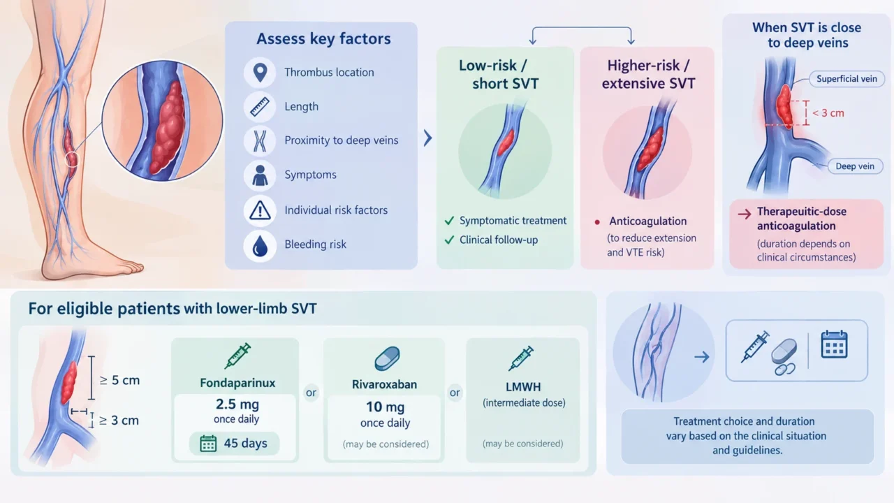
Treatment depends on the thrombus’s location, length, proximity to the deep venous system, associated symptoms, individual risk factors, and bleeding risk. Selected patients with short, low-risk SVT may receive symptomatic treatment and clinical follow-up. Other patients may require anticoagulation to reduce the risk of thrombus extension and venous thromboembolic complications.
For eligible patients with lower-limb SVT at least 5 cm long and at least 3 cm from the junction with the deep veins, European Society for Vascular Surgery (ESVS) guidelines recommend fondaparinux 2.5 mg once daily for 45 days. Rivaroxaban 10 mg once daily or an intermediate dose of low-molecular-weight heparin may be considered in appropriate circumstances.
When SVT extends to within 3 cm of a deep-vein junction, therapeutic-dose anticoagulation is generally recommended, although the regimen and duration depend on the clinical circumstances and applicable guidelines. These recommendations describe general management principles and do not indicate which treatment, if any, was selected for Hurts.
Why Might an Athlete Be Sidelined Despite Feeling Well?
Symptoms alone cannot establish whether a venous thrombus is stable or progressing. An athlete may retain full strength, mobility, and exercise tolerance while an imaging abnormality still requires further assessment. Preserved physical performance therefore does not exclude a clinically significant thrombotic process.
Return-to-play decisions must consider more than the athlete’s ability to perform. In contact sports, further trauma may increase bleeding concerns, particularly if anticoagulant treatment is required. The potential consequences of a thrombotic complication must also be weighed against the risks of continued participation.
Temporarily withholding an athlete from competition can therefore be a precautionary clinical decision while the medical team clarifies the diagnosis and determines the appropriate management strategy. In Hurts’ case, the Eagles stated that his health and safety remained their highest priority.
Warning Signs That Should Not Be Ignored
Persistent or worsening calf pain, localized tenderness, warmth, redness, or unilateral swelling should prompt medical evaluation, particularly when symptoms develop after trauma or in the presence of other venous thromboembolism risk factors. These findings are not specific to thrombosis, but they warrant appropriate assessment when a clot is suspected.
Sudden shortness of breath, chest pain that worsens with breathing, coughing up blood, or collapse may indicate pulmonary embolism and require emergency medical attention. Such symptoms should never be dismissed as routine consequences of exercise or a sports injury.
Although not every superficial clot progresses or causes complications, recognizing warning signs and obtaining appropriate diagnostic testing can help clinicians identify higher-risk cases. The severity of symptoms alone is not a reliable measure of the extent of venous thrombosis.
The Takeaway
Jalen Hurts’ case illustrates why superficial venous thrombosis warrants careful clinical assessment, even in an athlete who remains physically active. Although SVT is distinct from DVT, its anatomical location, extent, and potential for progression can influence the risk of complications and the need for anticoagulation.
Duplex ultrasound provides essential information for diagnosis and risk stratification, while repeat imaging may help clarify a changing clinical picture. Decisions about treatment and return to sport should be individualized according to the imaging findings, clinical assessment, and potential risks.
Until additional details are released, Hurts’ precise thrombotic anatomy and treatment plan remain unknown. His case nevertheless provides an opportunity to highlight an important clinical message: a superficial blood clot should not automatically be dismissed as harmless.
FAQ
1. What type of blood clot was diagnosed in Jalen Hurts?
The Philadelphia Eagles reported a superficial blood clot in his calf. The team has not publicly identified the exact vein involved or provided detailed measurements of the thrombus.
2. Is a superficial blood clot the same as DVT?
No. SVT affects veins close to the skin, whereas DVT affects deeper veins. However, SVT can extend into the deep venous system or coexist with DVT.
3. Can a direct blow to the calf cause a blood clot?
Trauma can contribute to thrombosis by injuring the vessel wall and triggering local inflammation. However, the reported sequence of events alone cannot establish that Hurts’ injury caused his clot.
4. Why was Hurts initially cleared to play?
According to the Eagles, the medical staff and physicians in London reviewed the initial ultrasound and determined that he could safely participate. The follow-up ultrasound later revealed movement in the clot, prompting a change in the team’s decision.
5. Does movement of a blood clot mean it has become a pulmonary embolism?
No. The team reported movement in the clot but did not announce a pulmonary embolism. Further clinical assessment and imaging details would be needed to establish whether the thrombus had extended or another complication had occurred.
6. How is superficial venous thrombosis diagnosed?
Clinical assessment and duplex ultrasonography are central to diagnosis. Ultrasound helps identify the thrombus’s location and extent and assess the deep veins for associated thrombosis.
7. Does every superficial blood clot require anticoagulation?
No. Treatment depends on clot length, location, proximity to the deep venous system, symptoms, risk factors, and bleeding risk. Some low-risk cases can be managed symptomatically, whereas others require anticoagulant therapy.
8. How long is anticoagulation usually given for SVT?
For eligible patients with lower-limb SVT at least 5 cm long and at least 3 cm from a deep-vein junction, guidelines recommend 45 days of anticoagulation. Thrombi close to the deep venous system or cases with high-risk features may require a different regimen or longer treatment.
9. Can an athlete have a blood clot without obvious symptoms?
Yes. Symptoms vary, and preserved mobility or exercise performance does not exclude venous thrombosis. Persistent or unexplained calf symptoms warrant appropriate clinical assessment.
10. What symptoms may indicate a serious complication?
New or worsening unilateral leg swelling, pain, warmth, or redness requires prompt medical evaluation. Sudden breathlessness, pleuritic chest pain, coughing up blood, or collapse may indicate pulmonary embolism and require emergency care.
Written by Hermine Sayiyan, MD
Stay updated with Hemostasis Today.
-
Oct 17, 2026, 09:33Robert Lookstein: Mechanical Thrombectomy Shows Functional Benefits in Intermediate-High-Risk PE
-
Oct 17, 2026, 09:18Sarra Hmidi: Tunisia Highlights Thrombosis Prevention on World Thrombosis Day
-
Oct 17, 2026, 09:05Addressing the Hidden Burden of Bleeding Disorders in Women – Hemophilia Society Mumbai Chapter
-
Oct 17, 2026, 08:49Maria Magnusson: Karolinska Experts Explore Gene Therapy in Sickle Cell Disease and Hemophilia
-
Oct 17, 2026, 04:07Weihao Li: Global HEPATIC Registry Findings on Post-Transplant Arterial Thrombosis
-
Oct 17, 2026, 03:56Ghaith Abu-Zeinah: ASH Publishes 2026 Guidelines for Myelofibrosis Management in Blood Advances
-
Oct 17, 2026, 03:48Tristan Siokos: Why Vascular Occlusion Is More Than Just Clots – Breakdown of the 8 Pathways
-
Oct 17, 2026, 02:36Archil Jaliashvili: Oxygen Transport in Capillaries
-
Oct 17, 2026, 02:35Carly Newton: Understanding Access to Red Blood Cell Exchange for People Living With SCD