

Jan Sloves: Pelvic Venous Insufficiency in Atypical and Recurrent Varicose Veins
Jan Sloves, President and Consultant at Vascular Imaging Professionals LLC, shared a post on LinkedIn about a recent article by Joana Storino et al, adding:
”In day‑to‑day venous lab practice, I still see pelvic venous insufficiency being under‑recognized in women who present with ‘atypical’ or recurrent varicose veins.
The issue is not the technology; it’s that our mapping remains too focused on the saphenous trunks and junctions and not enough on how pelvic reflux actually reaches the leg.
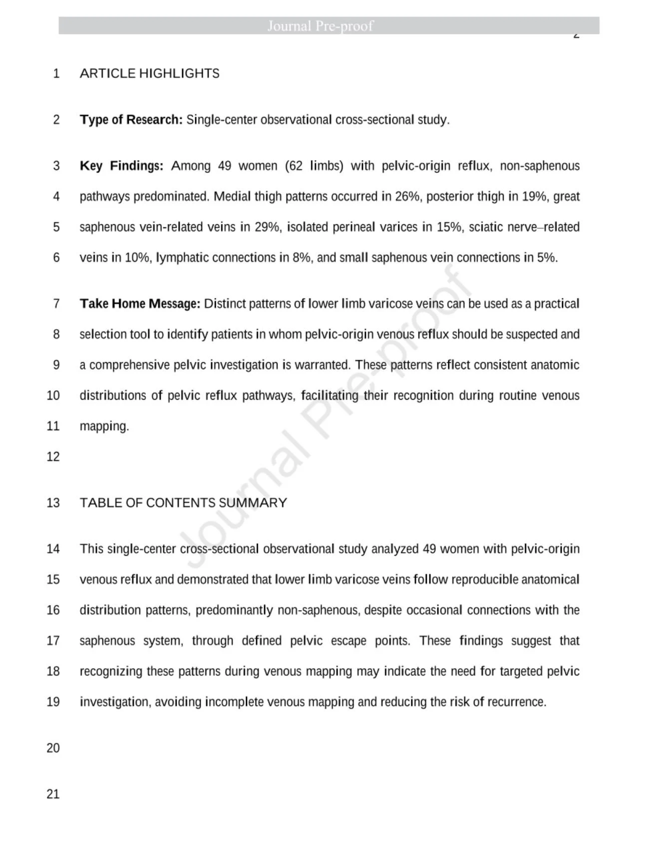
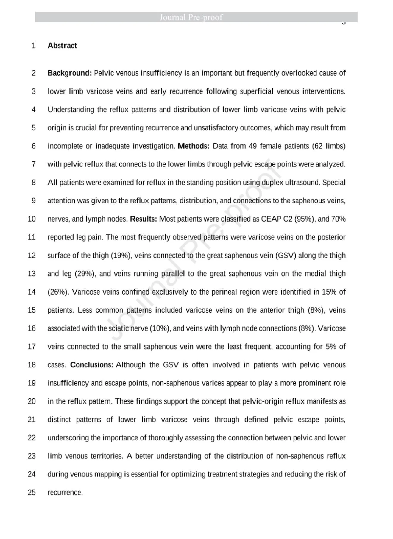
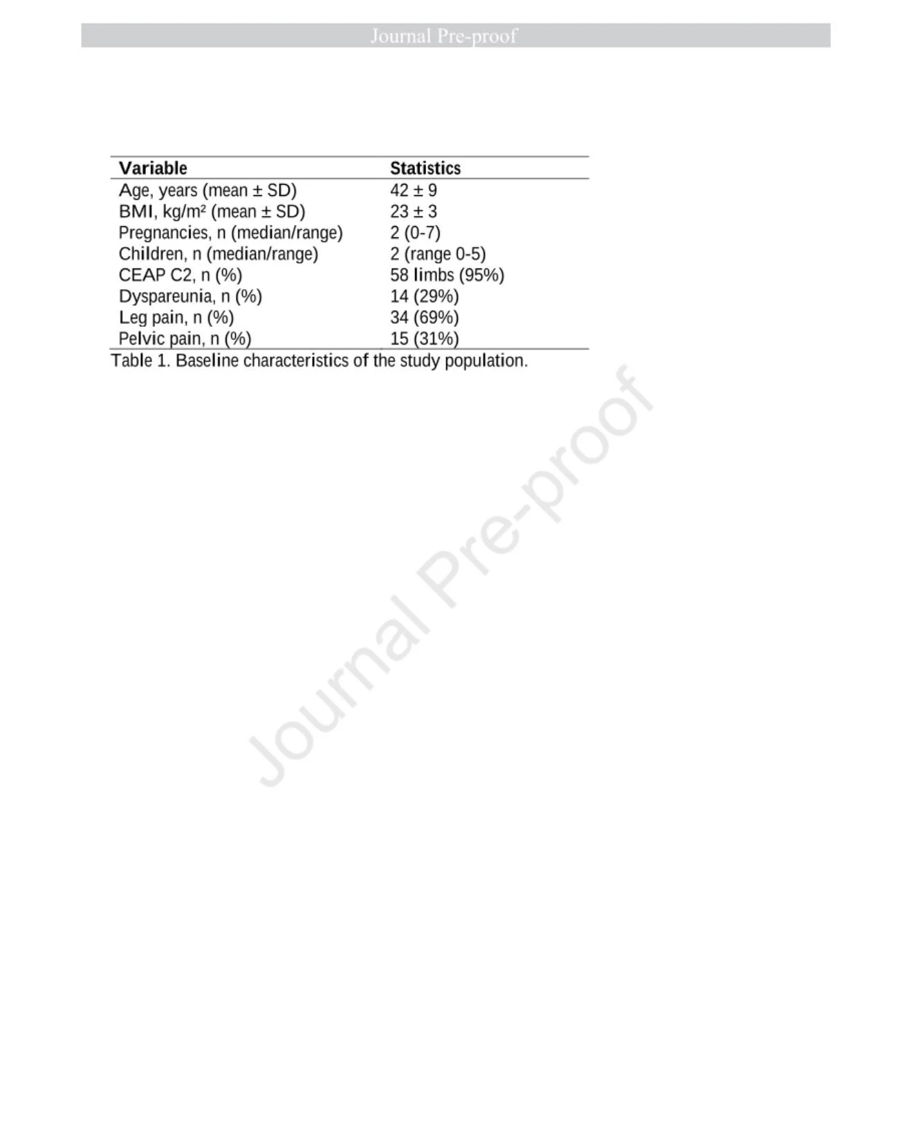
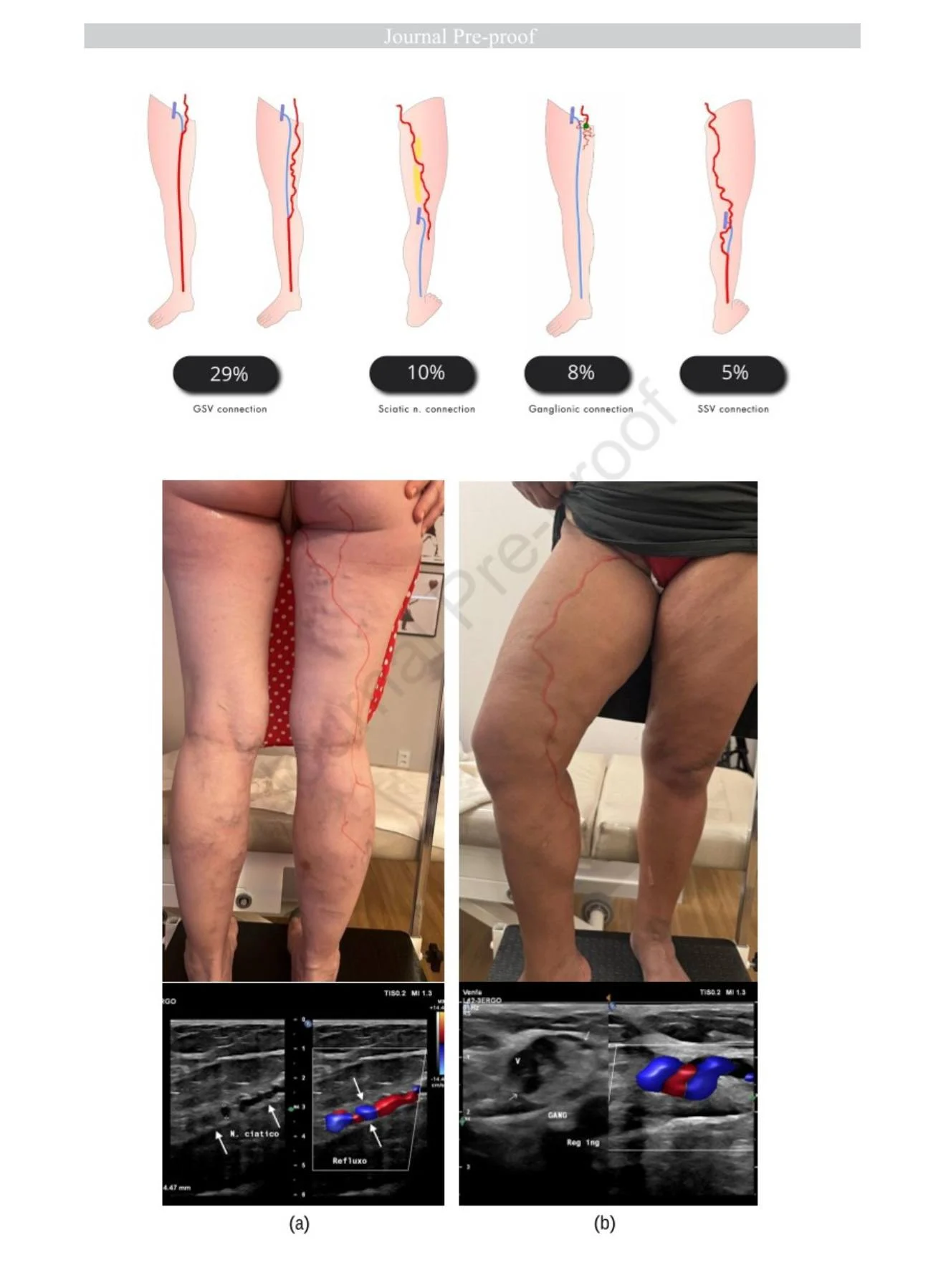
In a new paper in JVSVL, ‘Anatomical Patterns of Pelvic Venous Reflux to the Lower Limbs’ by Joana Storino, Fanilda Barros, and Nathalia Cardoso, the authors studied 49 women (62 limbs) with duplex confirmed pelvic reflux and escape points into the lower limbs. What struck me is how often the patterns we label as ‘weird’ or ‘recurrent’ are actually reproducible anatomic signatures of pelvic‑origin reflux.
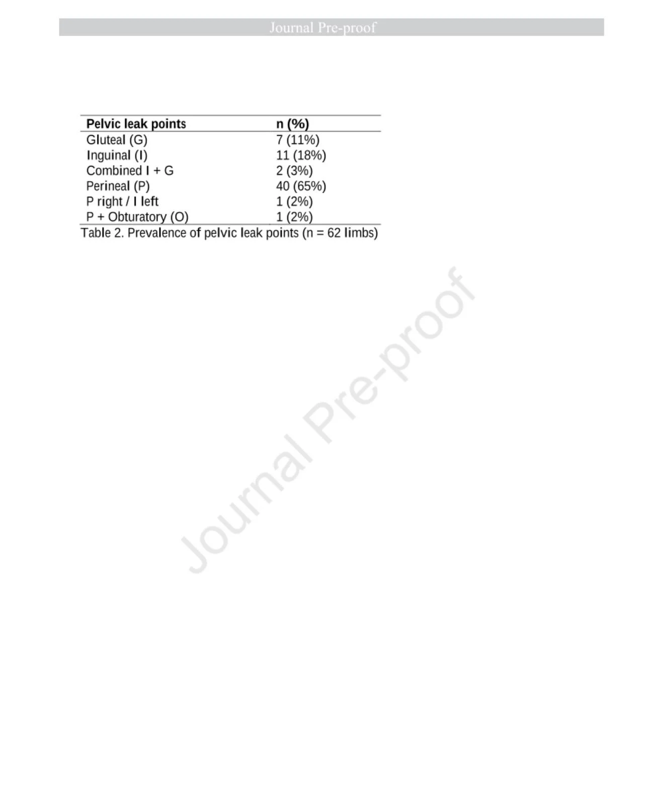
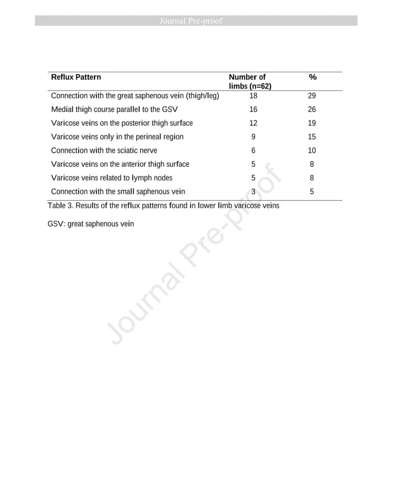
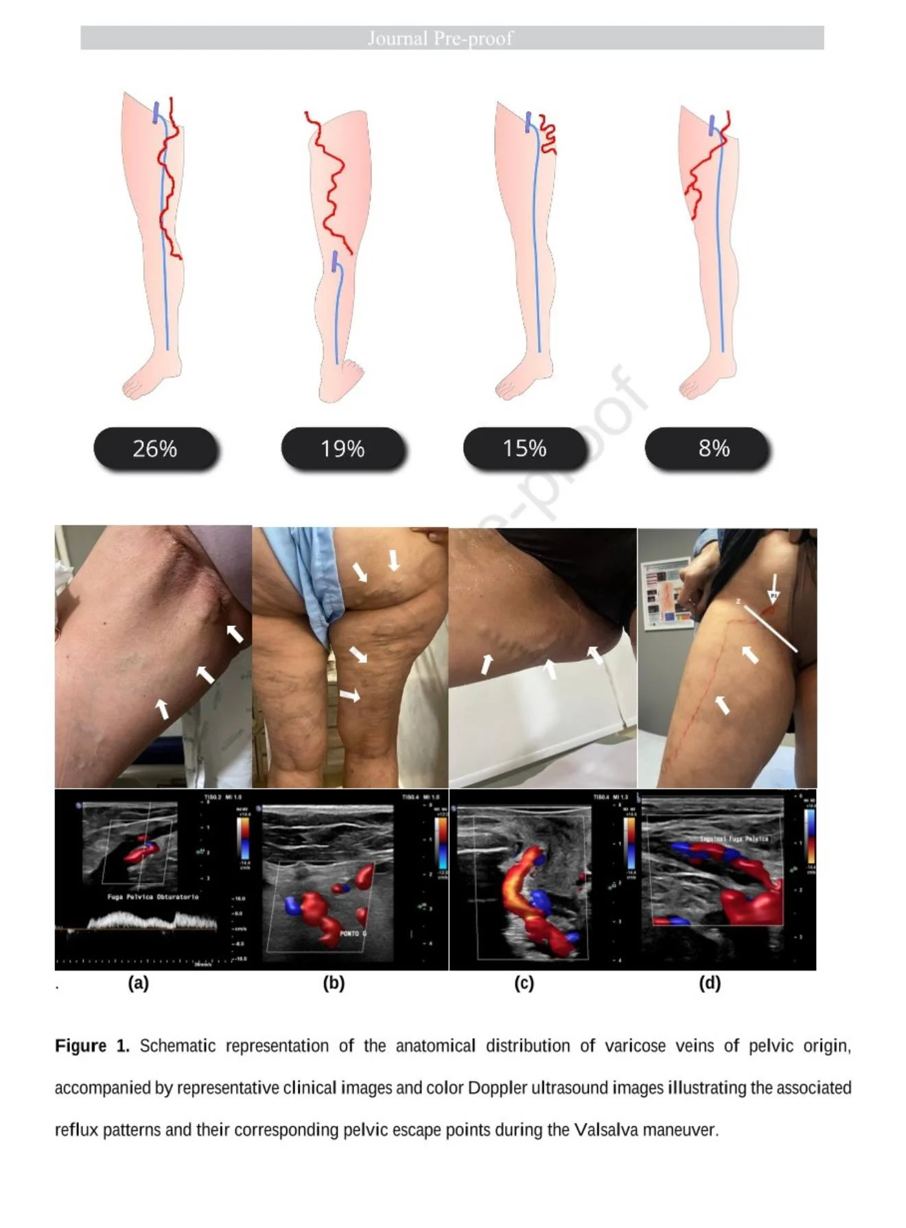
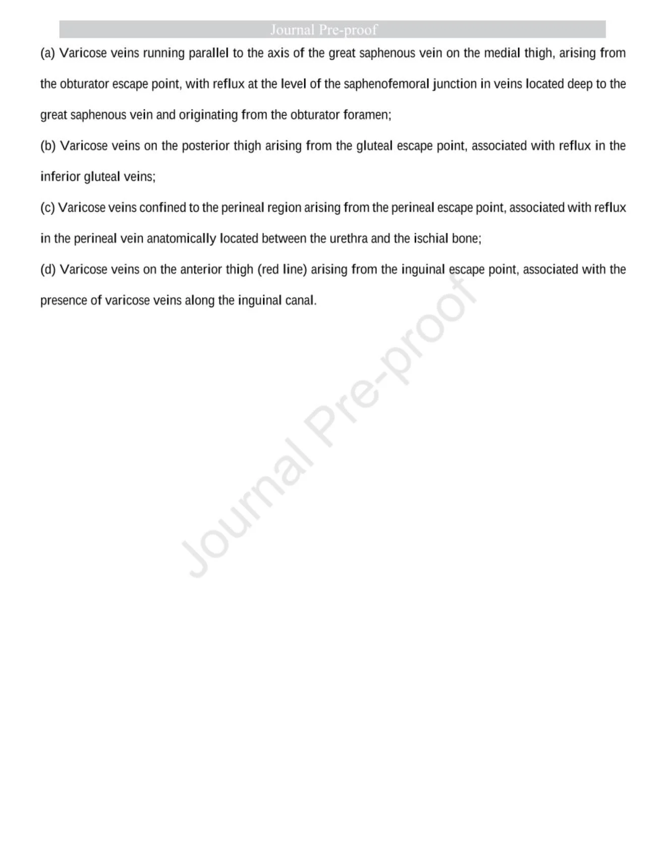
They describe seven main patterns:
- Medial thigh veins tracking parallel to the GSV
- True GSV‑related connections along thigh/leg
- Posterior thigh varices
- Isolated perineal varices
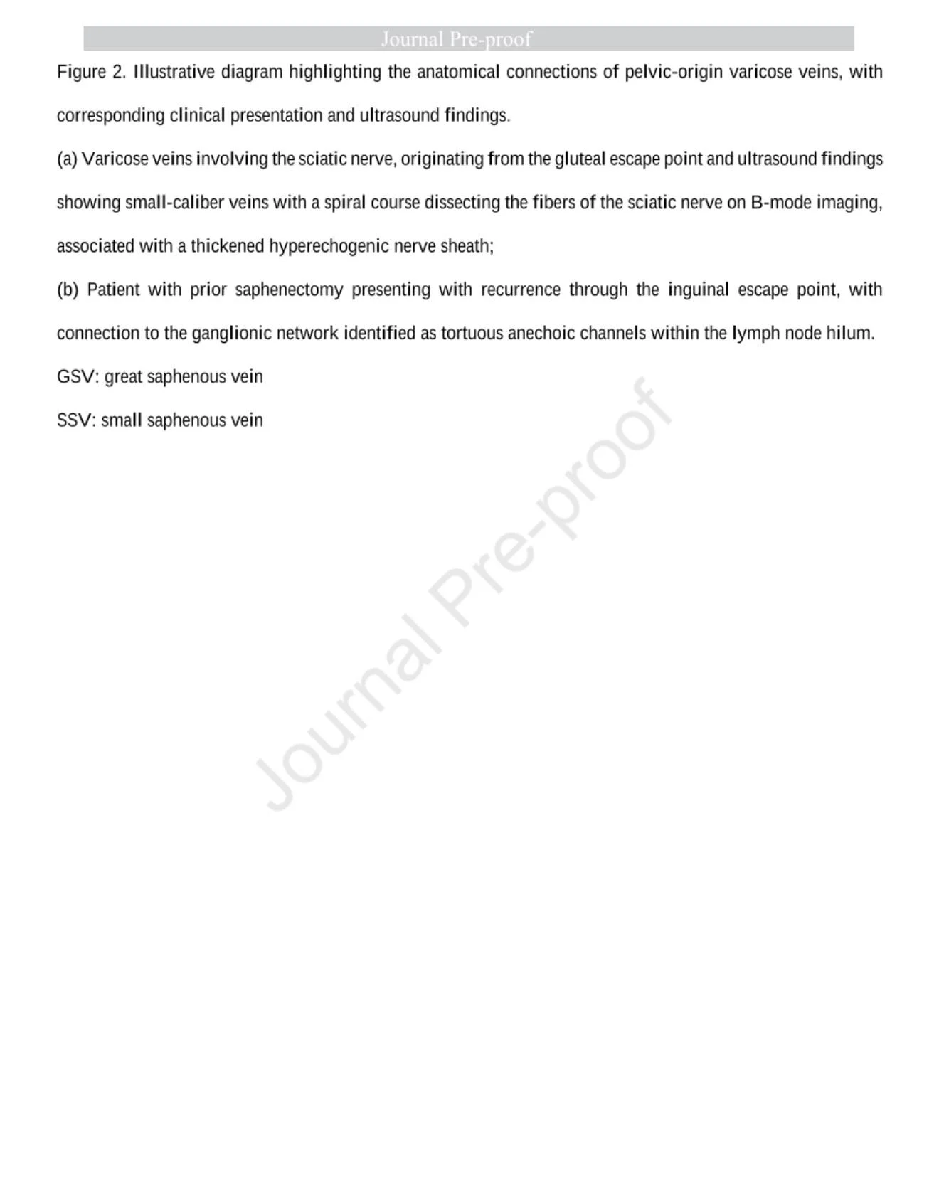
- Sciatic nerve – related veins
- Lymph node – related channels in the groin
- Less frequent SSV connections
Most of these are non‑saphenous pathways that may only touch the truncal system secondarily. Several correlate consistently with specific pelvic escape points (perineal, gluteal, inguinal, obturator), which means the limb is often just the projection screen for pelvic disease.
For me, the practical message is simple: when I see medial‑thigh ‘GSV‑parallel’ veins, posterior‑thigh tracks, perineal‑only clusters, or sciatic/lymphatic‑axis varices on standing duplex, I pause the truncal script and deliberately search for a pelvic connection.
We don’t need to send every patient for pelvic imaging. But if we want to meaningfully reduce recurrence in women, we do need to train ourselves and our labs to read these patterns as potential pelvic reflux, not just as odd variants of limb disease.
Subscribe to the only newsletter built for vascular pros.”
Title: Anatomical patterns of pelvic venous reflux to the lower limbs
Authors: Joana Storino, Fanilda Barros, Nathalia Cardoso Oliveira

Other articles featuring Jan Sloves on Hemostasis Today.
-
Jul 7, 2026, 06:04Brian A Beh: An Open Letter to Lisa Murphy Honouring the 30 Years of the Stroke Foundation’s Dedication
-
Jul 7, 2026, 05:55Alan Nurden: A Landmark Review Comparing Hemophilia and Hereditary Angioedema
-
Jul 7, 2026, 05:40Andrew Agwunobi: UConn Health is Proud to Advance Stroke Rehabilitation for Veterans
-
Jul 6, 2026, 22:54Louise St. Germain Bannon: I Wanted to Recognize the Incredible Staff Behind ISTH 2026
-
Jul 6, 2026, 21:46Armghan Ans: UPMC Washington Stroke Program Earns AHA Gold Plus Award for 16th Consecutive Year
-
Jul 6, 2026, 20:31Manik Madaan: Master Transfusion Reactions for USMLE In Less Than 60 Seconds
-
Jul 6, 2026, 19:25Ambreen Kashif: Highlighting the Impact of Temporary Donor Deferrals on Donor Retention
-
Jul 6, 2026, 19:23Rob Mac Sweeney: Anticoagulation Strategies for Cardiopulmonary Bypass in Heparin-Induced Thrombocytopenia
-
Jul 6, 2026, 19:22Nthabiseng Bapela: Interpreting INR in Patients on Warfarin – A Practical Clinical Framework