


Samwel Mikaye: Why Early Recognition of Pulmonary Embolism Matters
Samwel Mikaye, Medical Doctor at MSK, shared a post on LinkedIn:
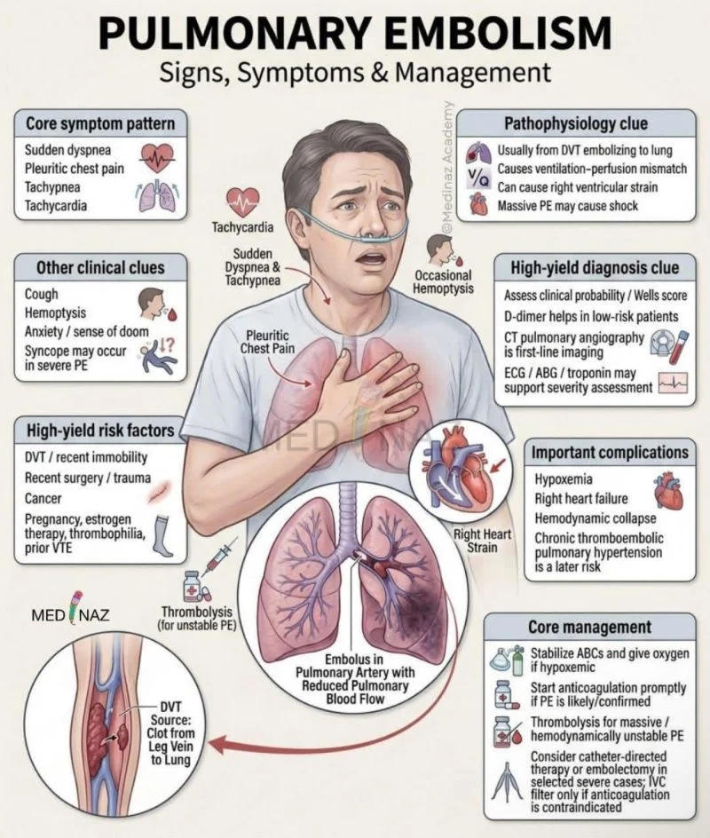
“Pulmonary Embolism (PE)
Definition
Pulmonary embolism is the obstruction of one or more pulmonary arteries by a thrombus, usually originating from a deep vein thrombosis (DVT) of the lower limbs or pelvis.
Risk Factors
Virchow’s Triad
1. Venous stasis
- Prolonged immobilization
- Surgery
- Long-distance travel
- Heart failure
2. Hypercoagulability
- Pregnancy and puerperium
- Malignancy
- Thrombophilia
- Oral contraceptives
3. Endothelial injury
- Trauma
- Surgery
- Central venous catheters
Clinical Features
Symptoms
- Sudden onset dyspnea
- Pleuritic chest pain
- Cough
- Hemoptysis
- Syncope (massive PE)
- Anxiety
Signs
- Tachypnea
- Tachycardia
- Hypoxia
- Fever (low-grade)
- Hypotension (massive PE)
- Raised jugular venous pressure
Signs of DVT:
- Unilateral leg swelling
- Calf tenderness
- Warmth and erythema
Diagnosis
Initial Tests
- Pulse oximetry
- ECG:
- Sinus tachycardia (most common)
- S1Q3T3 pattern
- Right heart strain
Laboratory
- D-dimer (elevated)
- Arterial blood gas:
- Hypoxemia
- Respiratory alkalosis
Imaging
- CT pulmonary angiography (CTPA) – gold standard in stable patients
- Ventilation-perfusion (V/Q) scan if CTPA contraindicated
- Doppler ultrasound of lower limbs for DVT
- Echocardiography in unstable patients
Management
Initial Stabilization
- Oxygen therapy
- IV access and monitoring
- Hemodynamic support if required
Anticoagulation
- Enoxaparin
- Heparin
- Direct oral anticoagulants (DOACs)
- Warfarin
Massive PE (Hemodynamic Instability)
- Thrombolysis with Alteplase
- Surgical or catheter-directed embolectomy if thrombolysis is contraindicated or unsuccessful
Prevention
- Early mobilization
- Compression stockings
- Prophylactic anticoagulation in high-risk patients
Complications
- Shock
- Right ventricular failure
- Cardiac arrest
- Recurrent PE
- Chronic thromboembolic pulmonary hypertension
Most PEs originate from deep vein thrombosis of the lower limbs.
Classic triad: dyspnea, pleuritic chest pain, and hemoptysis (not always present).
CT pulmonary angiography (CTPA) is the diagnostic imaging test of choice in stable patients.
D-dimer is useful for excluding PE in low-risk patients.
Massive PE may present with hypotension, shock, and syncope and requires urgent thrombolysis.”
Other posts featuring Samwel Mikaye on Hemostasis Today.
-
Sep 25, 2026, 10:49Shefina S: From Presenting to Hosting at TRANSCON 2026
-
Sep 25, 2026, 10:37Michelle Leona Cecil: Hemophilia Advocacy and the Power of Community
-
Sep 25, 2026, 10:13Michael Baria: Platelet Concentration and PRP Outcomes in Partial Rotator Cuff Tears
-
Sep 25, 2026, 09:49Haley Coleman: Volunteering to Support Hemophilia Awareness
-
Sep 25, 2026, 08:55Hitarth Mehta: BHMS Students Gain Practical Exposure to Haemophilia Diagnosis
-
Sep 25, 2026, 07:25Varun Capoor: How Therapeutic Apheresis and Transfusion Medicine Can Complement Clinical Management in SCD
-
Sep 25, 2026, 07:14Manshi Kesharwani: SAK-GQDs Modulate Thrombus Burden and Endothelial Function in Deep Vein Thrombosis
-
Sep 25, 2026, 06:20Farjah Hassan Algahtani: How Can We Better Identify Patients at Risk of Blood Clots after Metabolic and Bariatric Surgery?
-
Sep 25, 2026, 05:59Carla Lewis: Kids Conquering Sickle Disease Joins Nemours Children’s Hospital for 2nd Annual Awareness Event