


Lady Joan Branson’s Story – The Hidden Danger of Hospitalization
For Lady Joan Branson, the wife of British entrepreneur Sir Richard Branson, a fall that initially appeared to be an ordinary accident ultimately became a fatal medical event. At 80 years of age, she sustained fractures to her pelvis and right hip after falling at Necker Island in the British Virgin Islands.
Weeks later, on November 17, 2025, she was admitted to the Lister Hospital in Chelsea, London, where she remained at high risk of developing venous thromboembolism (VTE).
Her hospitalization ended tragically. On November 24, 2025, Lady Joan died following a cardiac arrest caused by a pulmonary embolism (PE) originating from deep vein thrombosis (DVT).
A subsequent inquest in September 2026 concluded that the absence of prophylactic anticoagulation had probably contributed to her death. Her case brought renewed attention to a frequently underestimated complication of hospitalization: hospital-associated thrombosis.
From an Accidental Fall to a Fatal Pulmonary Embolism
Lady Joan’s medical history, as presented during the inquest, began with a fall on Necker Island several weeks before her hospital admission. She reportedly described the accident to her general practitioner as tripping over slippers near her bed. The fall resulted in fractures involving her pelvis and right hip, significantly limiting her mobility.
On November 17, she was admitted to the private Lister Hospital. According to evidence presented at Westminster Coroner’s Court, she was considered at extremely high risk of developing blood clots, with her fracture and restricted mobility identified as the major contributing factors.
However, Lady Joan did not receive pharmacological VTE prophylaxis during her hospitalization. The inquest also heard that an immediate VTE risk assessment had not been completed and that she had not received thromboembolism-deterrent stockings during the first 48 hours after admission. Her admitting doctor had also failed to complete a bleeding risk assessment on the day of admission.
Haematologist Dr George Adams, who provided evidence at the inquest but was not involved in her care, explained that appropriate prophylactic anticoagulation would probably have reduced the likelihood of a fatal pulmonary embolism.
Senior Coroner Professor Fiona Wilcox concluded that the lack of anticoagulation had probably caused or contributed to Lady Joan’s death.
She also clarified that the case was not considered one of neglect or an absence of medical treatment. Nevertheless, the findings highlighted how failures in risk assessment, preventive treatment and communication between healthcare professionals can have devastating consequences.
Understanding Hospital-Associated Thrombosis
Hospital-associated thrombosis refers to venous thromboembolic events that develop during hospitalization or in the period following discharge. It encompasses both DVT and PE and can occur in patients admitted for medical conditions, surgery, trauma or other causes of acute illness.
Hospitalization often brings together several prothrombotic factors, including reduced mobility, tissue injury, systemic inflammation, advanced age, malignancy and surgical procedures. Consequently, even patients without a previous history of thrombosis may develop VTE during or after a hospital stay.
Hospital-associated VTE is particularly concerning because its clinical manifestations may be nonspecific or initially absent. A thrombus can develop silently in the deep veins and subsequently present as a potentially fatal pulmonary embolism. This makes prevention and systematic risk assessment essential components of inpatient care.
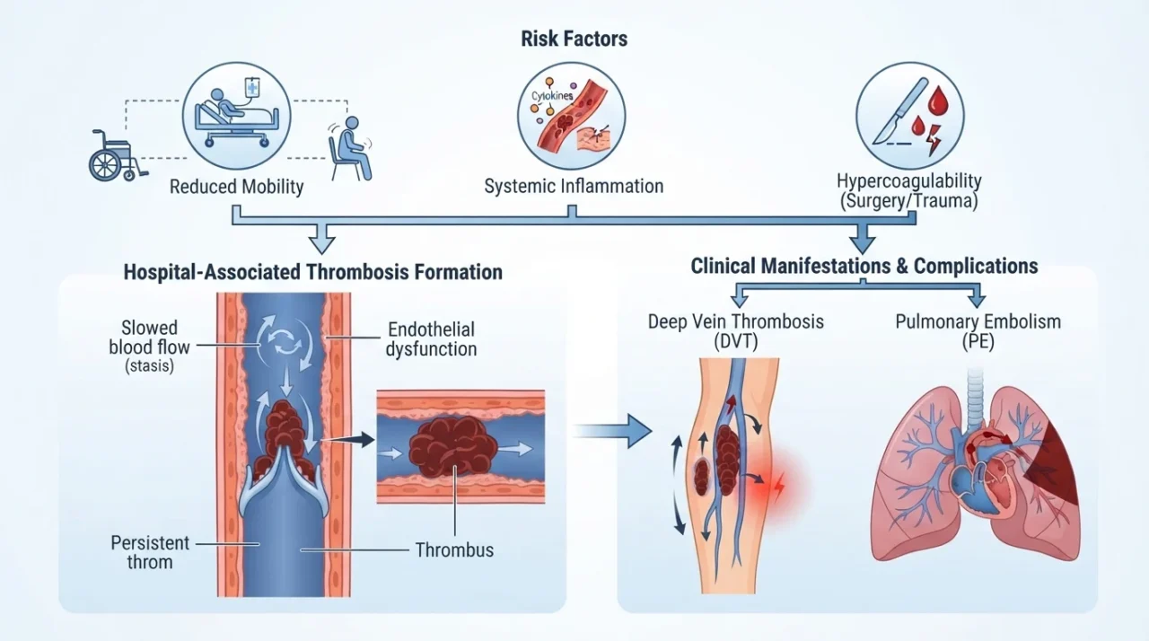
The Pathophysiology Behind Hospital-Associated Thrombosis
The development of venous thrombosis is traditionally explained by Virchow’s triad, which identifies three interconnected mechanisms: venous stasis, endothelial injury and hypercoagulability. Hospitalized patients frequently experience several of these mechanisms simultaneously, creating an environment that favors thrombus formation.
1. Venous stasis
Prolonged bed rest, reduced walking and immobilization following fractures or surgery decrease the activity of the calf-muscle pump. Under normal circumstances, contraction of the calf muscles compresses the deep veins and facilitates venous return toward the heart. When mobility is restricted, blood flow slows, particularly in the deep veins of the lower extremities.
Reduced blood flow also contributes to local accumulation of activated coagulation factors and decreases their clearance. These changes facilitate thrombin generation and fibrin formation, promoting the development of venous thrombi.
In Lady Joan’s case, pelvic and hip fractures, together with substantial immobility, represented major contributors to venous stasis.
2. Endothelial injury and tissue trauma
Fractures and traumatic injuries can damage vascular structures and surrounding tissues. Exposure of tissue factor and activation of inflammatory pathways promote the initiation of coagulation. Tissue factor binds factor VII/VIIa, activating the coagulation cascade and ultimately generating thrombin.
Thrombin converts fibrinogen into fibrin and activates platelets and other coagulation factors, contributing to the formation and stabilization of a thrombus. In addition, inflammatory mediators released after tissue injury can promote endothelial activation and a prothrombotic vascular environment.
3. Hypercoagulability and systemic inflammation
Acute illness, trauma and inflammation can shift the hemostatic balance toward coagulation. Inflammatory cytokines stimulate tissue factor expression and activate endothelial cells, monocytes and platelets. At the same time, natural anticoagulant pathways and fibrinolytic activity may become relatively impaired.
The resulting imbalance favors thrombin generation and fibrin deposition. Advanced age and other underlying conditions may further increase susceptibility to thrombosis, particularly when combined with immobility and tissue injury.
These mechanisms are not independent. In a patient with a major fracture, reduced mobility and an inflammatory response may occur together, producing a particularly unfavorable prothrombotic environment. This combination helps explain why patients with hip and pelvic fractures require careful VTE risk assessment.
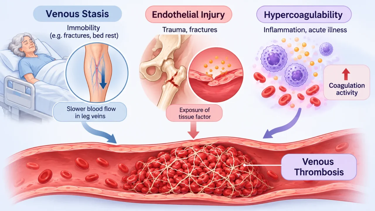
Why Hospitalized Patients Are at Increased Risk
Hospital-associated VTE is not limited to patients undergoing major surgery. Both medical and surgical inpatients may develop thrombosis, although the underlying risk factors and appropriate preventive strategies differ.
Important risk factors include:
- Advanced age.
- Previous VTE or known thrombophilia.
- Active malignancy.
- Major trauma and fractures, particularly involving the pelvis or hip.
- Recent surgery.
- Prolonged immobilization or bed rest.
- Acute infection and systemic inflammation.
- Obesity and certain chronic medical conditions.
- Pregnancy and estrogen-containing medications in relevant patient populations.
The presence of several risk factors simultaneously can substantially increase thrombotic risk.
In older patients, fractures involving the hip or pelvis are particularly important because they frequently result in prolonged immobility. Pain, impaired weight-bearing and the need for assistance with movement can further reduce venous return. The combination of trauma, age and immobility makes systematic thromboprophylaxis assessment especially important in this population.
From Deep Vein Thrombosis to Pulmonary Embolism
Most clinically significant venous thrombi originate in the deep veins of the lower extremities or pelvis. When part of a thrombus detaches, it travels through the inferior vena cava to the right atrium and right ventricle before entering the pulmonary arterial circulation.
Once lodged in the pulmonary arteries, the embolus obstructs blood flow and increases pulmonary vascular resistance. This creates an acute pressure load on the right ventricle, potentially resulting in right ventricular dilation, impaired contractility and reduced cardiac output.
In severe cases, acute right ventricular failure can lead to systemic hypotension, obstructive shock and cardiac arrest. The severity of PE depends on the extent of pulmonary vascular obstruction, the patient’s cardiopulmonary reserve and the degree of right ventricular dysfunction.
Lady Joan’s case illustrates this progression from a thrombotic event associated with lower-extremity and pelvic injury to a fatal pulmonary embolism. Her experience also demonstrates that a patient may remain hospitalized for several days before a clinically catastrophic thromboembolic event becomes apparent.
Recognizing the Clinical Signs of DVT and PE
DVT may present with unilateral leg swelling, pain, tenderness, warmth and skin discoloration. However, clinical manifestations vary, and some patients have no noticeable symptoms. In hospitalized patients, these signs may also be difficult to distinguish from symptoms caused by the original injury, postoperative changes or other medical conditions.
Pulmonary embolism may present with sudden dyspnea, pleuritic chest pain, tachycardia, hemoptysis or syncope. Severe PE can cause hypotension and cardiovascular collapse. Because these symptoms are not specific to PE, clinical suspicion must be informed by the patient’s medical history and thrombotic risk factors.
For suspected DVT, compression ultrasonography is commonly used to identify non-compressible deep veins. Suspected PE is generally evaluated using clinical probability assessment and appropriate diagnostic investigations, including computed tomography pulmonary angiography (CTPA) when indicated.
Preventing Hospital-Associated Thrombosis
VTE prevention begins with systematic assessment of both thrombotic and bleeding risks. According to NICE guideline NG89, all patients admitted to hospital should undergo an assessment of their risk of VTE and bleeding as soon as possible after admission. Risk should also be reassessed when the patient’s clinical condition changes.
Pharmacological thromboprophylaxis commonly involves low-molecular-weight heparin (LMWH), such as enoxaparin, or unfractionated heparin (UFH) in selected patients. These agents enhance antithrombin activity and inhibit key coagulation factors, particularly factor Xa and, depending on the agent, thrombin (factor IIa). By reducing thrombin generation and fibrin formation, they decrease the likelihood of venous thrombus development.
The choice of agent, dose and timing depends on renal function, body weight, the clinical indication and bleeding risk.
Mechanical prophylaxis is another important component of prevention, particularly when pharmacological prophylaxis is contraindicated or when additional protection is appropriate. Intermittent pneumatic compression devices periodically inflate and deflate around the lower extremities, increasing venous flow and reducing stasis. Graduated compression stockings are another option in appropriately selected patients.
Mechanical and pharmacological methods may be combined in patients with particularly high thrombotic risk, depending on their bleeding risk and clinical circumstances.
Early mobilization, when medically appropriate, is also essential. Encouraging patients to resume walking and lower-limb movement as soon as their clinical condition allows can help restore venous return. Adequate hydration and patient education may provide additional support but should not replace indicated pharmacological or mechanical prophylaxis.
For patients with fragility fractures of the pelvis, hip or proximal femur, NICE recommends considering VTE prophylaxis for one month when the thrombotic risk outweighs the bleeding risk. Recommended options include LMWH or fondaparinux, with timing determined by the patient’s surgical status and clinical circumstances. If pharmacological prophylaxis is contraindicated, intermittent pneumatic compression may be considered.
Importantly, anticoagulant prophylaxis is not automatically appropriate for every hospitalized patient. Active bleeding, recent bleeding events, severe thrombocytopenia and other clinical circumstances may influence the decision. The objective is to balance the prevention of potentially fatal VTE against the risk of anticoagulant-associated bleeding.
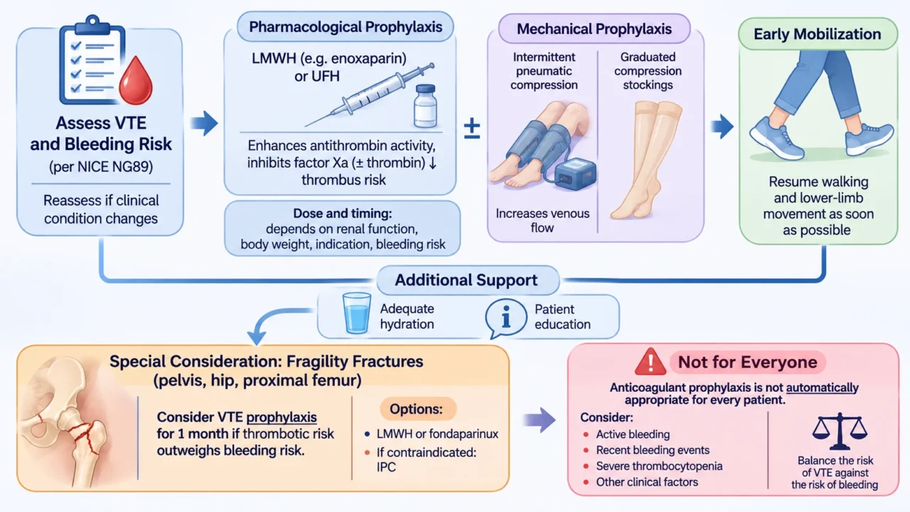
The Importance of Risk Assessment and Communication
Lady Joan’s case highlighted not only the importance of prescribing appropriate prophylaxis but also the need for reliable communication between healthcare professionals. The inquest heard that the initial VTE and bleeding risk assessments had not been completed and that the recommended preventive measures had not been implemented.
These findings emphasize that VTE prevention should be integrated into routine admission procedures, with clear documentation of risk assessment, contraindications, prescribed prophylaxis and reassessment when necessary.
The case also prompted discussion of institutional responsibility and patient safety. During the inquest, the hospital’s medical staff acknowledged shortcomings in the processes surrounding VTE prevention, and the consultant described efforts to strengthen risk assessment and prevention procedures to reduce the likelihood of similar events.
The Clinical Significance of Lady Joan Branson’s Story
Hospital-associated thrombosis is a preventable complication in many patients, yet it remains an important cause of morbidity and mortality. Lady Joan Branson’s death demonstrates how trauma, advanced age and prolonged immobility can combine to create a high-risk thrombotic state.
Her story also reinforces that the absence of visible symptoms does not eliminate the possibility of developing DVT and that preventive measures must be considered before a pulmonary embolism occurs.
For healthcare professionals, the central lesson is that VTE risk assessment is not simply an administrative requirement. It is a fundamental part of inpatient care that requires clinical judgment, timely intervention, appropriate documentation and effective teamwork.
Ultimately, Lady Joan’s case serves as a reminder that preventing thrombosis is often more effective than treating its most severe consequence. Recognizing risk early and implementing appropriate thromboprophylaxis can make a critical difference between recovery and a potentially fatal outcome.
FAQ
1. What is hospital-associated thrombosis?
Hospital-associated thrombosis refers to DVT or PE that develops during hospitalization or following discharge. It is associated with factors such as immobilization, surgery, trauma, acute illness and underlying medical conditions.
2. What happened to Lady Joan Branson?
Lady Joan Branson sustained pelvic and right hip fractures following a fall. After being admitted to the Lister Hospital, she did not receive prophylactic anticoagulation. She subsequently developed DVT complicated by a fatal pulmonary embolism and died on November 24, 2025.
3. Why does immobilization increase the risk of DVT?
Immobilization reduces calf-muscle pump activity, slowing venous blood flow and promoting venous stasis. This facilitates local accumulation of activated coagulation factors and thrombus formation.
4. How do fractures contribute to venous thrombosis?
Fractures can cause tissue and vascular injury, activate inflammatory pathways and promote tissue factor-mediated coagulation. When combined with reduced mobility, these mechanisms substantially increase thrombotic risk.
5. What is Virchow’s triad?
Virchow’s triad describes three major contributors to thrombosis: venous stasis, endothelial injury and hypercoagulability. Hospitalized patients may experience all three simultaneously, particularly following trauma or major surgery.
6. How does DVT lead to pulmonary embolism?
Part of a thrombus originating in the deep veins may detach and travel through the inferior vena cava and right side of the heart into the pulmonary arteries. Obstruction of pulmonary blood flow increases right ventricular afterload and can result in acute right ventricular failure and cardiovascular collapse.
7. Which hospitalized patients are at increased risk of VTE?
Patients with advanced age, previous VTE, active cancer, major trauma, fractures, recent surgery, prolonged immobility or acute inflammatory conditions may have an increased risk. The presence of multiple risk factors further increases concern.
8. How is hospital-associated VTE prevented?
Prevention involves assessing both VTE and bleeding risks, early mobilization when appropriate, mechanical prophylaxis and pharmacological prophylaxis when indicated. LMWH and UFH are commonly used anticoagulants in hospitalized patients.
9. Does every hospitalized patient require anticoagulant prophylaxis?
No. The decision depends on individual thrombotic and bleeding risks. Patients with a high risk of bleeding may require mechanical prophylaxis instead, while others may benefit from pharmacological prophylaxis or a combination of preventive strategies.
10. What is the main clinical lesson from Lady Joan Branson’s case?
Her case highlights the importance of early VTE risk assessment, appropriate thromboprophylaxis and effective communication between healthcare professionals. Timely prevention may reduce the risk of a potentially fatal pulmonary embolism in vulnerable hospitalized patients.
Written by Hermine Sayiyan, MD
Stay updated with Hemostasis Today.
-
Nov 3, 2026, 22:00Ravi C Dara: What’s New in ASFA’s 10th Edition – Practical Updates for the Apheresis Clinician
-
Nov 3, 2026, 21:46Cedric Hermans: Treatment Differentiators for Shared Decision-Making in Haemophilia in 2026
-
Nov 3, 2026, 21:38Arwa Z. Al-Riyami: AI Opportunities and Future Directions in Transfusion Medicine
-
Nov 3, 2026, 21:11Yazan Abou-Ismail: Presenting at American College of Emergency Physicians and MWTCC in the Same Week
-
Nov 3, 2026, 21:05Anirban Sen Gupta: Escalating-Dose Lyophilized SynthoPlate for Emergency Trauma Mitigation
-
Nov 3, 2026, 20:50Mostafa Faisal Mohammed Saleh: Targeting Plasma Cells to Control Persistent ADAMTS13 Autoantibodies in iTTP
-
Nov 3, 2026, 16:57Ahmed Alotaibi: Which Macrophage Receptor ‘eats’ anti-E and Anti-K Coated RBCs?
-
Nov 3, 2026, 16:53Jan Sloves: Key Factors for Achieving Durable Patency in Advanced Venous Stenting
-
Nov 3, 2026, 16:45Nina Weber: Review on Kidney Injury After Stem Cell Transplantation