

Thrombotic Microangiopathies: From Clinical Syndrome to Molecularly Targeted Therapy
Thrombotic microangiopathies (TMAs) are entering a new era in 2026, with the treatment landscape moving beyond simply recognizing and controlling the disease.
New therapeutic strategies are increasingly targeting the mechanisms driving endothelial injury, platelet-rich microvascular thrombosis and complement activation, reshaping the treatment landscape for thrombotic microangiopathies.
TMAs are a heterogeneous group of rare, life-threatening disorders characterized by microangiopathic hemolytic anemia, thrombocytopenia and organ injury caused by microvascular thrombosis.
Despite advances in mechanism-based therapies, TMAs remain a time-critical hematologic emergency in 2026.
Prompt recognition, rapid diagnostic evaluation and accurate subclassification are essential to guide targeted treatment and prevent irreversible renal, neurologic, and other end-organ injury.
What Changed in TMA in 2026?
The TMA landscape is evolving rapidly in 2026, with several developments shifting treatment toward more targeted, mechanism-based approaches.
In immune-mediated TTP, caplacizumab-based strategies continue to reshape acute management, while recombinant ADAMTS13 is strengthening the role of enzyme replacement in congenital TTP.
Recent data from a 2026 tertiary-center cohort of adults with TMA showed that approximately three-quarters of patients achieved a complete response, while in-hospital mortality remained as high as 25.5%.
Non-response to treatment and impaired post-treatment renal function were independently associated with mortality, highlighting the importance of early diagnosis and prompt, mechanism-based treatment.
In complement-mediated TMA, research is moving beyond established C5 inhibition toward earlier components of the complement pathway, including factor B and other proximal targets.
Meanwhile, STEC-HUS remains largely dependent on supportive care, but new toxin-directed therapies are entering late-stage clinical development.
Perhaps most notably among secondary TMAs, transplant-associated TMA has become a major focus of complement-directed research, with narsoplimab emerging as an important therapeutic development and ongoing studies evaluating other complement inhibitors.
Together, these advances highlight a broader shift in TMA care – from treating the clinical syndrome to identifying and targeting the biological mechanism driving disease.
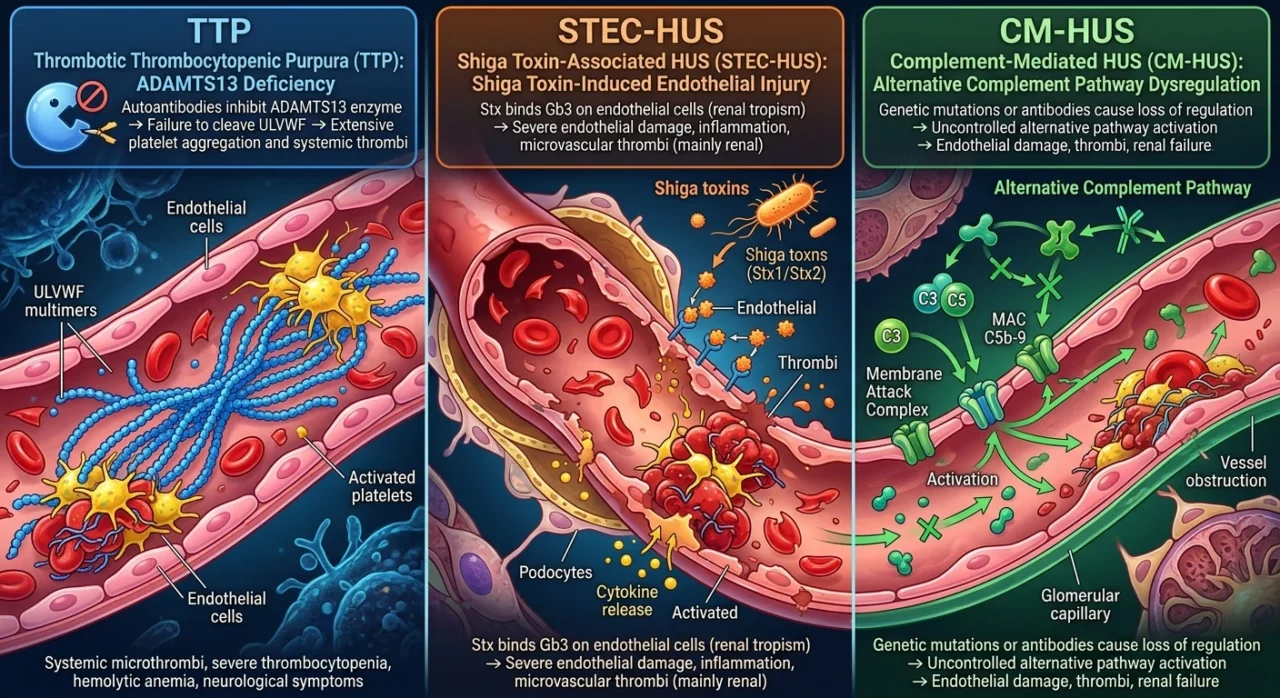
Based on advances in molecular diagnostics and pathophysiology TMAs are classified as primary or secondary.
Primary Thrombotic Microangiopathies:
- Thrombotic Thrombocytopenic Purpura (TTP): ADAMTS13 deficiency
- Shiga toxin-associated hemolytic uremic syndrome (STEC-HUS): Shiga toxin-induced endothelial injury
- Complementmediated hemolytic uremic syndrome (CM-HUS): Alternative complement pathway dysregulation
Thrombotic Thrombocytopenic Purpura (TTP)
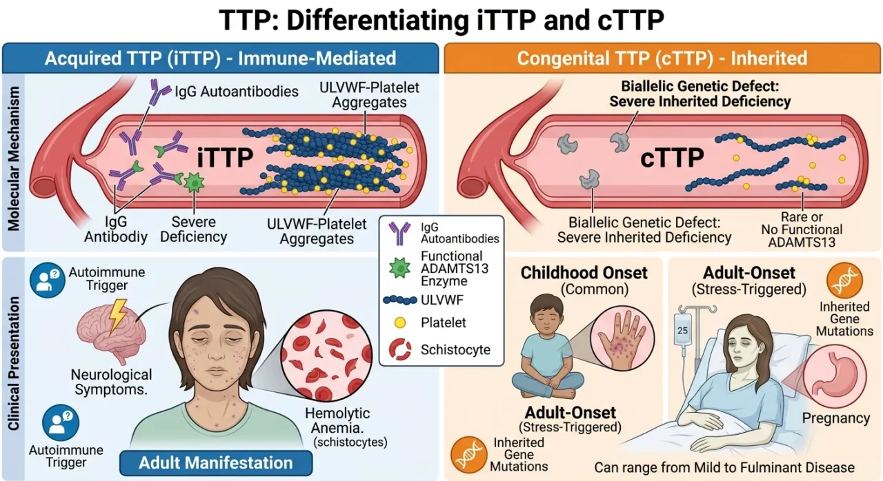
TTP is a rare, potentially life-threatening TMA in which severe thrombocytopenia and microangiopathic hemolytic anemia result from widespread microvascular thrombosis.
TTP arises from a severe deficiency of ADAMTS13 activity, typically below 10% of normal, which prevents the cleavage of ultra-large von Willebrand factor (vWF) multimers.
Their accumulation promotes spontaneous platelet adhesion and the formation of platelet-rich microthrombi within the microvasculature.
TTP can be classified as:
- Acquired TTP or immune-mediated TTP (iTTP) – The predominant form of TTP in adults, severe ADAMTS13 deficiency is caused most commonly by inhibitory autoantibodies, particularly IgG, although IgA and IgM antibodies as well as immune complexes have also been described.
- Congenital TTP (cTTP) – Results from biallelic pathogenic variants in the ADAMTS13 gene, causing a severe inherited deficiency of the enzyme. Although it is often recognized in childhood, cTTP can manifest at any age, with adult-onset episodes – including those triggered by pregnancy or other physiological stressors – sometimes presenting as fulminant disease rather than a mild phenotype.
Hemolytic uremic syndrome (HUS)
Hemolytic uremic syndrome (HUS) is a thrombotic microangiopathy characterized by the triad of microangiopathic hemolytic anemia, thrombocytopenia, and acute kidney injury, resulting from widespread microvascular thrombosis that predominantly affects the renal glomeruli.
HUS can be classified as:
- Shiga toxin-associated (typical HUS) – Arises from infection with Shiga toxin – producing Escherichia coli (STEC), in which Shiga toxin binds to glomerular and systemic endothelial cells, triggering direct endothelial injury and a prothrombotic state.
The resulting microvascular thrombosis leads to mechanical hemolysis, platelet consumption, and acute kidney injury, classically following an episode of bloody diarrhea.
- Complement‑mediated HUS (atypical HUS / C‑TMA) – Arises from dysregulated activation of the complement system on endothelial cells, due to genetic variants or autoantibodies involving complement regulators, which leads to persistent complement‑mediated endothelial injury and a prothrombotic state.
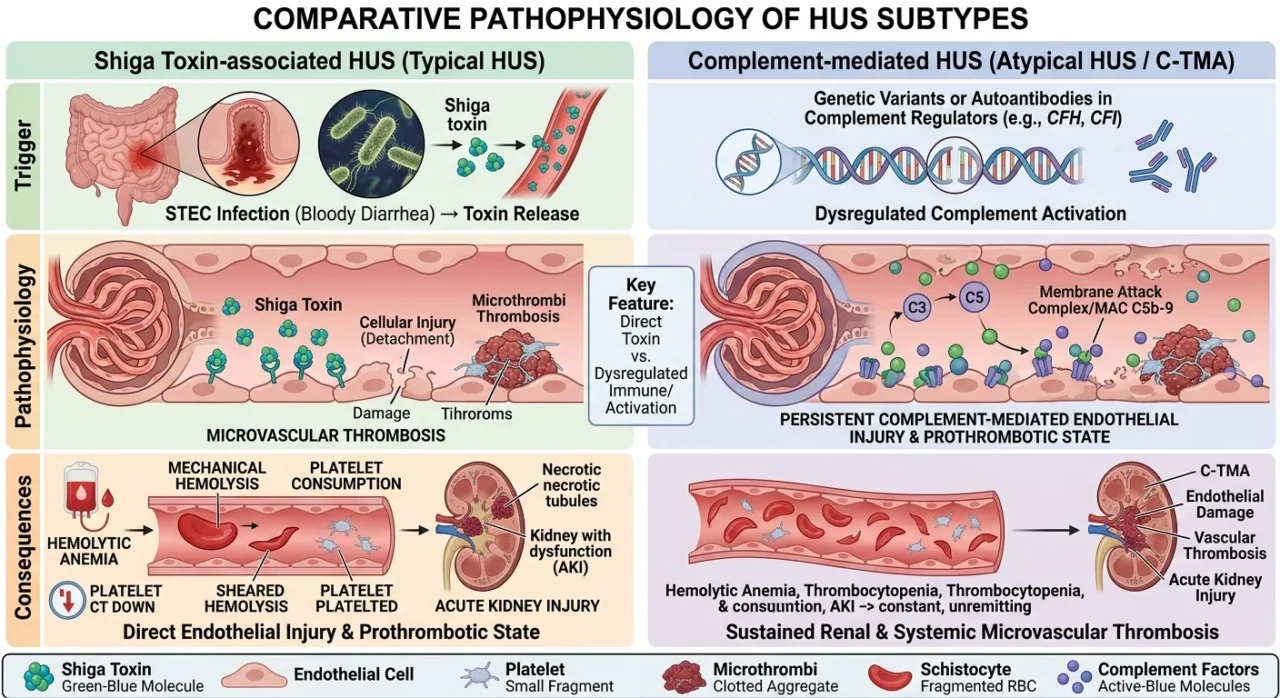
The resulting microvascular thrombosis causes mechanical hemolysis, platelet consumption, and acute kidney injury, typically without a preceding diarrheal illness and with a tendency toward recurrence and systemic involvement.
Secondary Thrombotic Microangiopathies develop in association with an underlying condition or external trigger, including malignancy, pregnancy, autoimmune disease, infection, transplantation, or drug exposure.
Their pathogenesis often involves endothelial injury and activation of prothrombotic pathways.
Secondary TMAs can be classified according to their underlying triggers:
Malignant hypertension-associated TMA – Severe hypertension causes endothelial injury particularly in the kidneys and brain. This endothelial injury promotes platelet activation and microvascular thrombosis, leading to thrombocytopenia and MAHA. Renal and neurological involvement are common.
Pregnancy-associated TMA – May occur in association with preeclampsia and HELLP syndrome, while pregnancy can also trigger or unmask iTTP and complement-mediated TMA.
Transplant-associated TMA (TA-TMA) – Occurs most commonly after hematopoietic stem-cell transplantation and, less frequently, solid-organ transplantation. Endothelial injury may result from conditioning regimens, immunosuppressive drugs, infections, graft-versus-host disease, and complement activation.
Drug-induced TMA – Certain medications can cause TMA through immune-mediated mechanisms or direct endothelial toxicity. Reported associations include calcineurin inhibitors, quinine, certain chemotherapeutic agents, antiplatelet drugs, and other medications.
Cancer-associated TMA – Can occur in patients with advanced malignancy, particularly with metastatic adenocarcinoma and bone marrow involvement. Tumor-related endothelial injury and microvascular obstruction may produce a TMA-like picture, sometimes accompanied by disseminated intravascular coagulation.
Autoimmune disease-associated TMA – Systemic autoimmune disorders, including systemic lupus erythematosus and antiphospholipid syndrome, may cause endothelial injury and complement activation leading to TMA.
Infection-associated TMA – Severe infections can produce endothelial injury, systemic inflammation, complement activation, and coagulation abnormalities that may result in a TMA phenotype.
Secondary TMAs has also been described in association with conditions such as pancreatitis, severe metabolic disorders, and other states associated with significant endothelial injury.
Here’s a concise introductory paragraph you can use to open the “Clinical trials” section of your article:
The therapeutic landscape of thrombotic microangiopathies has shifted from largely supportive care to mechanism‑driven, targeted interventions, with an expanding portfolio of clinical trials across both primary and secondary TMAs.
In primary TMAs, research has focused on refining immunosuppression and caplacizumab use in immune TTP, optimizing long‑term complement inhibition in atypical HUS, and exploring upstream complement inhibitors beyond C5.
In secondary TMAs – particularly transplant‑associated TMA, hypertensive emergency–associated TMA, and drug‑induced TMA – recent trials have tested complement‑targeted agents such as narsoplimab, ravulizumab, and eculizumab, aiming to reduce the historically high mortality and improve organ recovery.
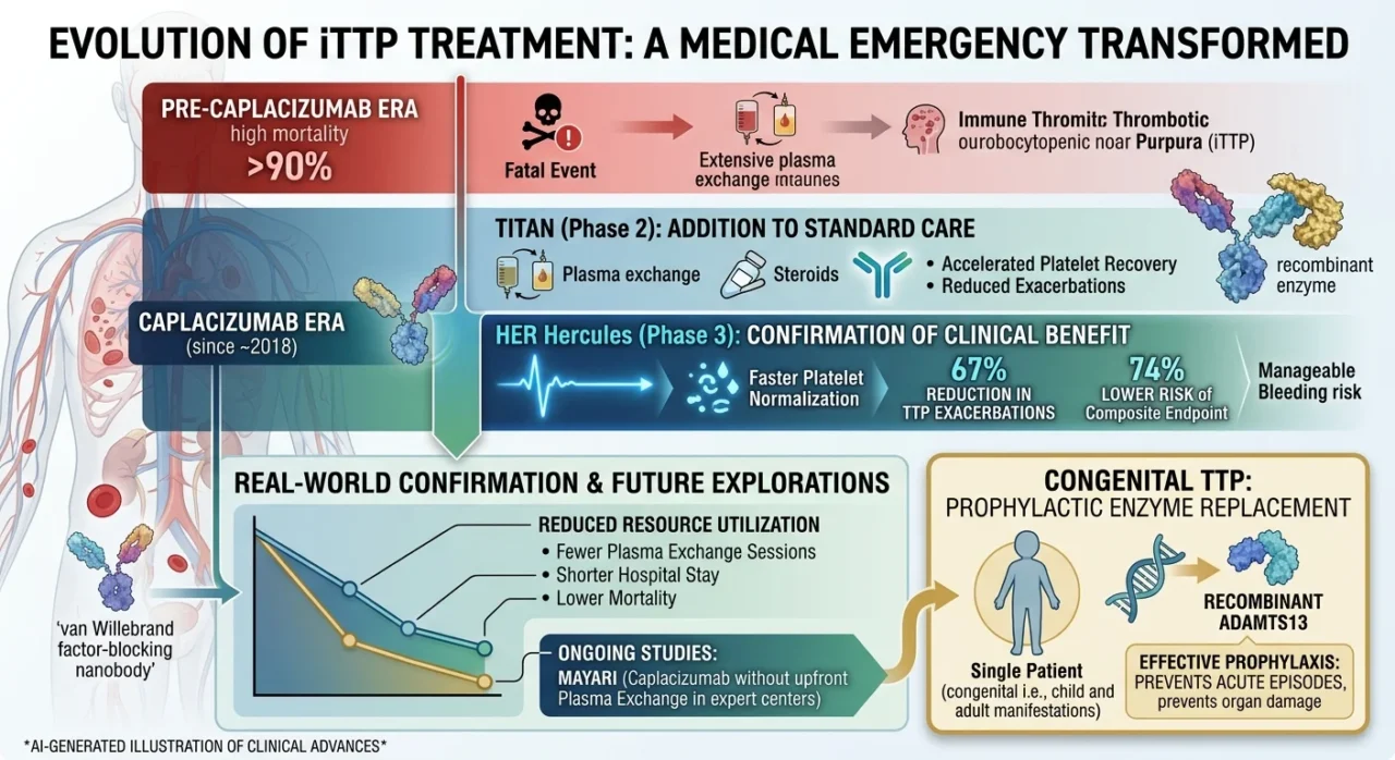
Clinical trials in Immune Thrombotic Thrombocytopenic Purpura (iTTP) have transformed a once highly fatal disease into a treatable emergency.
The TITAN Phase 2 trial first showed that adding caplacizumab – a von Willebrand factor–blocking nanobody – to plasma exchange and immunosuppression accelerated platelet recovery and reduced early exacerbations.
The pivotal HERCULES Phase 3 study confirmed these benefits, demonstrating faster platelet normalization, a 67% reduction in TTP exacerbations/relapses, and a 74% lower risk of death, recurrence, or major thromboembolism during treatment, with manageable bleeding as the main safety issue.
Real‑world cohorts and meta‑analyses through 2025–2026 have reinforced these findings, reporting fewer plasma exchange sessions, shorter hospital stays, and lower mortality with caplacizumab.
Ongoing studies, such as MAYARI, are exploring caplacizumab‑based regimens without upfront plasma exchange in expert centers, while in congenital TTP, recombinant ADAMTS13 has emerged as an effective prophylactic enzyme replacement that prevents acute episodes.
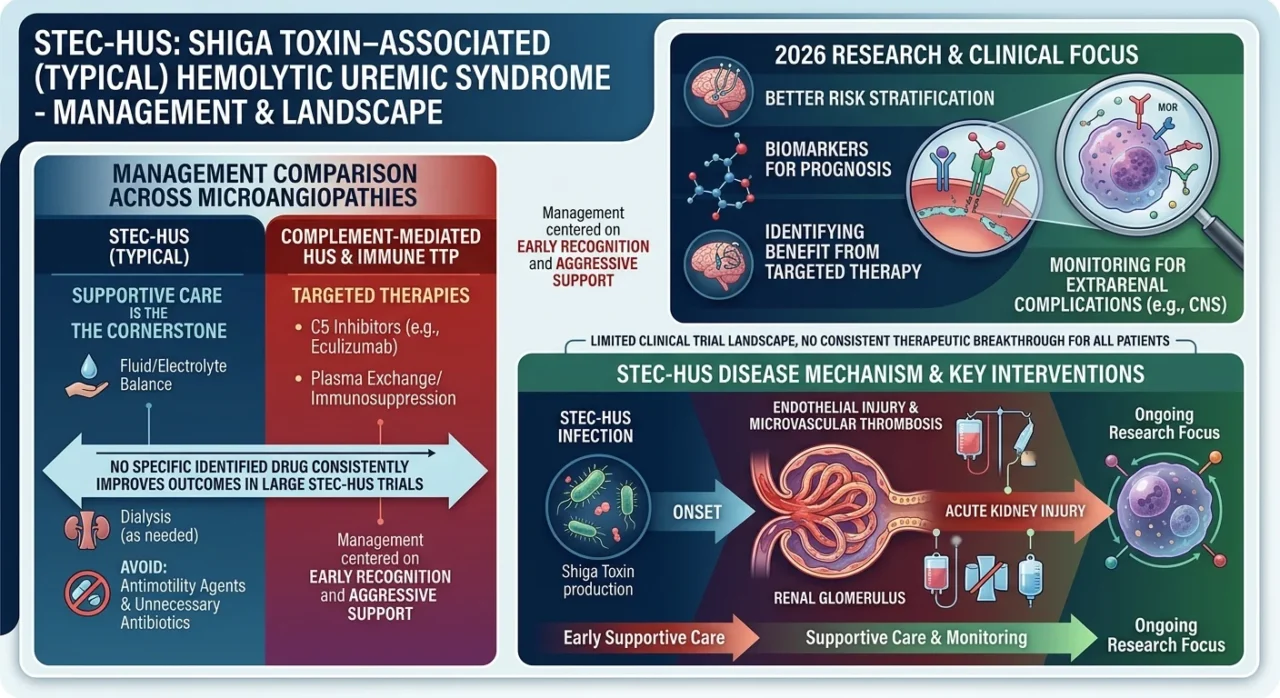
In Shiga Toxin–Associated (Typical) Hemolytic Uremic Syndrome, the clinical trial landscape remains limited compared with other thrombotic microangiopathies.
Unlike immune TTP or complement‑mediated HUS, no large randomized trials have identified a specific drug that consistently improves kidney or survival outcomes in classic STEC‑HUS.
Management is therefore centered on early recognition and aggressive supportive care – careful fluid and electrolyte management, dialysis when needed, and avoidance of antimotility agents and unnecessary antibiotics – while monitoring for severe extrarenal complications such as CNS involvement.
As of 2026, research efforts are focused on better risk stratification and biomarkers to identify the minority of children and adults who might benefit from targeted therapy, but supportive care remains the cornerstone of treatment for typical STEC‑HUS.
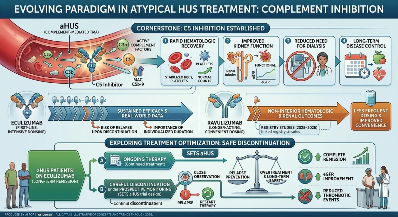
In Atypical Hemolytic Uremic Syndrome (complement‑mediated TMA), clinical trials over the past decade have established complement inhibition as the cornerstone of therapy. Pivotal studies of the C5 inhibitor eculizumab demonstrated rapid hematologic recovery, improvement in kidney function, and reduced need for dialysis in both adults and children with aHUS, leading to its approval and widespread adoption as first‑line treatment.
Long‑term extension and real‑world data have shown sustained disease control with ongoing therapy, while also highlighting risks of relapse after discontinuation and the importance of individualized treatment duration.frontiersin
More recently, the longer‑acting C5 inhibitor ravulizumab has been evaluated in aHUS, with trials showing non‑inferior hematologic and renal outcomes compared with eculizumab and the convenience of less frequent dosing.
Observational cohorts and registry studies through 2025–2026 reinforce these findings, reporting high rates of complete or partial remission, improved eGFR, and reduced thrombotic events in patients maintained on C5 inhibitors.
At the same time, prospective trials such as SETS aHUS are exploring whether carefully selected patients can safely discontinue eculizumab under close monitoring, aiming to balance relapse prevention against overtreatment and long‑term safety.
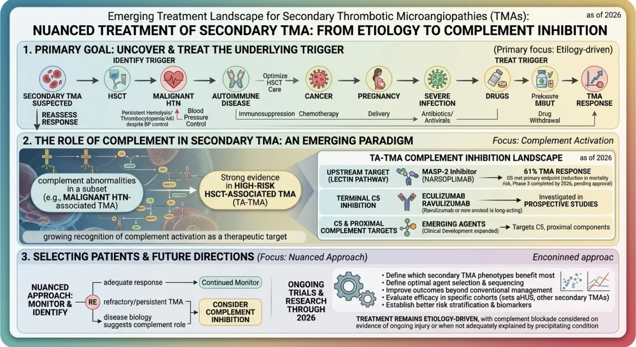
Secondary thrombotic microangiopathies (TMAs) arise in association with conditions such as hematopoietic stem-cell transplantation (HSCT), malignant hypertension, autoimmune disease, cancer, pregnancy, severe infection, and certain drugs. Unlike primary complement-mediated TMA, management is primarily directed at identifying and treating the underlying trigger.
However, the distinction is not always straightforward, as complement activation may contribute to endothelial injury in several forms of secondary TMA.
The clearest evidence for complement-directed therapy has emerged in HSCT-associated TMA (TA-TMA), particularly in patients with high-risk disease and multiorgan involvement.
Prospective studies have investigated terminal complement inhibition with eculizumab and ravulizumab, while other approaches have targeted upstream or alternative complement pathways.
In a pivotal single-arm study, the lectin-pathway inhibitor narsoplimab produced a TMA response in 61% of evaluable adults with TA-TMA, supporting the role of complement-directed strategies in selected patients.
More recently, clinical development has expanded to agents targeting C5 and proximal complement components, reflecting growing recognition of complement activation as a therapeutic target in TA-TMA.
In malignant hypertension-associated TMA, aggressive blood-pressure control remains the cornerstone of treatment. Importantly, however, malignant hypertension can also occur as a manifestation of underlying complement-mediated TMA.
Persistent microangiopathic hemolysis, thrombocytopenia, or kidney injury despite adequate blood-pressure control should therefore prompt reassessment for an alternative diagnosis, including complement-mediated TMA.
Observational studies have identified complement abnormalities in a subset of patients with malignant hypertension-associated TMA, suggesting that complement inhibition may be appropriate in carefully selected cases rather than routinely in all patients.
For other secondary TMAs—including those associated with systemic autoimmune disease, cancer, pregnancy, infection, or drugs—the evidence for complement inhibition remains substantially less established and is largely based on case reports, small series, and early-phase studies.
Consequently, treatment should remain etiology-driven, with complement blockade considered when there is convincing evidence of ongoing complement-mediated endothelial injury or when the clinical course is not adequately explained by the precipitating condition.
The emerging treatment landscape therefore supports a more nuanced approach to secondary TMA: control the underlying trigger, monitor the response closely, and identify patients whose disease biology suggests a role for complement inhibition.
Ongoing trials of complement-targeted therapies may help define which secondary TMA phenotypes benefit most, how treatment should be selected, and whether complement inhibition can improve outcomes beyond conventional management.
Where Is the Field Going?
The next phase of TMA research is likely to focus less on finding a single treatment for all patients and more on identifying which biological mechanisms are driving disease in each individual.
Complement inhibition is moving toward increasingly targeted approaches, with studies investigating C5, C3, factor B, and other components of the complement pathway.
At the same time, researchers are working to identify biomarkers that can detect disease earlier, predict severity, and help determine which patients are most likely to benefit from complement blockade.
This shift may be particularly important in secondary TMAs, where the presence of a trigger does not necessarily explain the entire disease process.
Pregnancy, severe hypertension, transplantation, and other conditions can themselves cause endothelial injury while also unmasking complement-mediated disease in susceptible patients.
The challenge for the coming years will therefore be to distinguish patients who require treatment of the underlying trigger alone from those who may benefit from additional mechanism-specific therapy.
Ultimately, the future of TMA management is likely to be defined by earlier diagnosis, better biomarkers, more precise patient selection, and individualized treatment duration.
Rather than treating TMA as a single clinical syndrome, the field is moving toward matching each patient’s underlying disease biology with the therapy most likely to prevent irreversible organ damage.
FAQ
1. What defines a thrombotic microangiopathy (TMA)?
TMA is a clinicopathologic syndrome characterized by microangiopathic hemolytic anemia, thrombocytopenia, and organ injury resulting from systemic microvascular thrombosis. Typical laboratory findings include schistocytes, elevated LDH and low haptoglobin.
2. Which laboratory test best distinguishes TTP from other TMAs?
ADAMTS13 activity is the key diagnostic test. Severe deficiency (<10%) supports TTP, while higher activity shifts the differential toward STEC-HUS, complement-mediated HUS, or secondary TMA.
3. When should caplacizumab be started in suspected immune TTP?
Caplacizumab should be started as soon as iTTP is suspected, together with plasma exchange and immunosuppression, without waiting for ADAMTS13 results. Early treatment accelerates platelet recovery and reduces exacerbations and major complications.
4. What is the first-line therapy for complement-mediated HUS?
C5 inhibition with eculizumab or ravulizumab is the first-line targeted therapy. Treatment should not be delayed while awaiting genetic testing.
5. How do eculizumab and ravulizumab differ in practice?
Both inhibit C5. Ravulizumab has a longer half-life, allowing less frequent administration and potentially facilitating outpatient treatment.
6. Is there a specific drug therapy for STEC-HUS?
No established specific pharmacological therapy is available for classic STEC-HUS. Management remains supportive, including fluid and electrolyte management, dialysis when indicated, and monitoring for neurological and other extrarenal complications.
7. How should pregnancy-associated TMA be approached?
Pregnancy-associated TMA requires differentiation between preeclampsia/HELLP, iTTP, and complement-mediated TMA. ADAMTS13 activity and clinical context are central to diagnosis and treatment selection.
8. What is the role of complement inhibition in HSCT-associated TMA?
Complement-directed therapies, including eculizumab and ravulizumab, have been investigated in high-risk HSCT-associated TMA. Narsoplimab, a MASP-2 inhibitor, has also been studied in this setting.
9. When should malignant hypertension-associated TMA prompt evaluation for complement-mediated TMA?
Persistent hemolysis, thrombocytopenia, or renal injury despite adequate blood-pressure control should prompt reconsideration of the diagnosis and evaluation for complement-mediated TMA.
10. What is the overarching principle for secondary TMAs?
Treat the underlying trigger. Complement inhibition should be considered selectively when there is evidence supporting complement-mediated endothelial injury, while management of most drug-induced, cancer-associated, and autoimmune TMAs remains primarily etiology-directed.
Written by Robert Tadevosyan.
Stay updated with Hemostasis Today.
-
Oct 19, 2026, 19:44Where You Live Should not Determine Whether You Receive Care – World Federation of Hemophilia
-
Oct 19, 2026, 19:42Belen Bianco: Prevention, Early Suspicion and an Interdisciplinary Approach Save Lives
-
Oct 19, 2026, 19:40Christina Chrysohoou: Catheter-Based LAAC vs OAC for Stroke Prevention in Atrial Fibrillation
-
Oct 19, 2026, 19:38Matthew Flick: A Two-Day Research Meeting on Fibrinogen, Hemostasis and Thrombosis
-
Oct 19, 2026, 19:37Hayat Saeed: Excited to Share Our Publication on the FGL1–Hepcidin Axis in Iron Homeostasis and Disease
-
Oct 19, 2026, 19:08IPAW 2026 Comes to a Close, but the Work Continues – PPTA
-
Oct 19, 2026, 17:44Paul Riley: Moderating a Webinar on Anticoagulation Monitoring
-
Oct 19, 2026, 16:36Christopher Pittman: Blood Clot Prevention Cannot End at Hospital Discharge
-
Oct 19, 2026, 16:13Javier M. Floren: When Immune Cells Move With Their Environment