

Anis Mahjoubi: MRI Findings of Krukenberg Tumors
Anis Mahjoubi, MR Clinical Application Specialist, shared a post on LinkedIn:
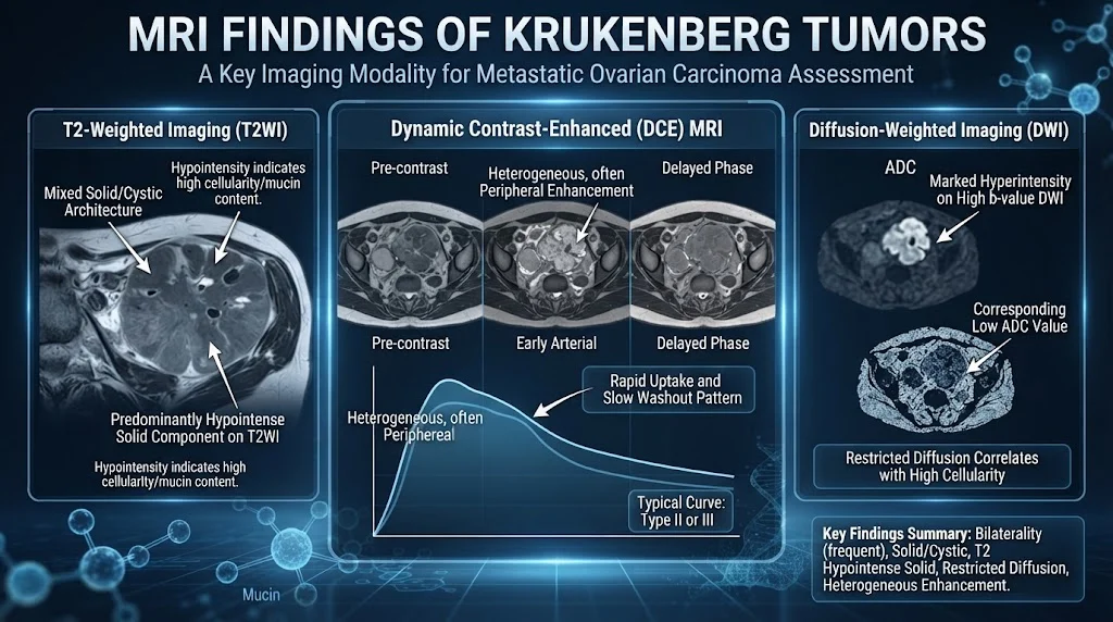
“MRI signs of Krukenberg tumors (ovarian metastases with signet ring cells, usually from gastric or colorectal primaries):
MRI features are often characteristic but not fully specific. Key findings include:
Morphologic features :
Bilateral in approximately 80 percent of cases (highly suggestive when present).
Well defined or sharp margins.
Oval or rounded configuration often preserved (even when large).
Predominantly solid, mixed solid cystic, or (less often) mostly cystic.
Smooth external contour; lobulated appearance common.
Ascites frequently associated.
Signal intensity characteristics :
Solid components (due to dense stromal or fibrous reaction):
T1 weighted: Hypointense to intermediate.
T2 weighted: Characteristically hypointense (low signal) , this is one of the most helpful clues, especially when bilateral, with sharp margins and oval shape. The hypointensity corresponds to dense collagenous stroma.
Variable overall signal if edema or mucin is present.
Cystic or mucinous components:
T2 weighted: Hyperintense (high signal from mucin or fluid).
T1 weighted: Variable (can be hyperintense if thick mucin or proteinaceous or hemorrhagic content).
Non enhancing.
Contrast enhancement and diffusion
Post or gadolinium (T1 C plus): Solid components and walls of intratumoral cysts show strong or vivid heterogeneous enhancement.
Dynamic curves often show rapid early uptake followed by washout (type 3 curve).
DWI or ADC: Restricted diffusion in the solid components (high signal on high b value DWI, low ADC values).
Additional helpful signs
Uniform cystic locules or ‘moth eaten’ cystic areas within solid tissue.
Hypointense solid peripheral or randomly distributed components on T2.
Look for the primary tumor (gastric wall thickening, colonic mass, etc.) and other metastases.
Differential considerations on MRI :
Primary ovarian carcinoma (especially mucinous).
Fibroma or fibrothecoma (very T2 hypointense but usually unilateral and less enhancing).
Other ovarian metastases or lymphoma.
Note: MRI alone cannot definitively distinguish Krukenberg tumors from primary ovarian malignancies. Clinical context (known primary, younger age, bilaterality) and histology remain essential. Always correlate with CT or PET for staging and search for the primary site.”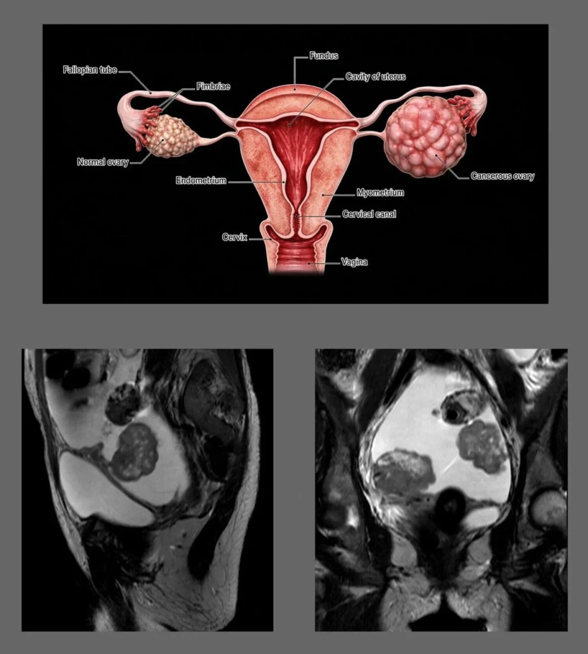
Stay updated with Hemostasis Today.
-
Sep 21, 2026, 15:08Fikreab Tesfaye Tefera: 10 Practical Take-Home Points for Diagnosing Iron Deficiency Anemia
-
Sep 21, 2026, 14:10Aswin K Mohan: Lessons from TRANSCON 2026: Building Resilient Blood Supply Systems Before Crisis Hits
-
Sep 21, 2026, 14:00Vincent Lu: Beyond PT and aPTT – How Hemostasis Evolves Across a Lifetime
-
Sep 21, 2026, 13:56Alex Knapp: Emerging Technologies and Advances in Hemophilia Care
-
Sep 21, 2026, 13:51Abdalhakim Shubietah: pLAAO Fails Noninferiority to OAC in Landmark Meta-Analysis of 7 RCTs
-
Sep 21, 2026, 13:21In His Last UNGA, António Guterres Delivers a Urgent Call for Global Unity
-
Sep 21, 2026, 13:14Fatih Mehmet Gul: Are We Moving Faster Than Our Ability to Ask ‘Should We?’
-
Sep 21, 2026, 13:08Sangita Reddy: How Does Our Own Healthcare Journey in India Look Through the Same Lens?
-
Sep 21, 2026, 12:53Conan McIlwrath: Strengthening Haemophilia Care Through International Collaboration