

AI in Hemostasis Laboratories: From Automated Clot Detection to Clinical Decision Support
The integration of artificial intelligence (AI) and machine learning (ML) into specialized clinical laboratory medicine is moving beyond proof-of-concept research toward increasingly practical applications. In hemostasis, where clinical decisions often depend on time-sensitive and technically complex laboratory measurements, AI has the potential to improve pre-analytical quality control, extract additional information from coagulation assays, and support interpretation of increasingly complex clinical datasets.
Recent reviews of AI in thrombosis and hemostasis have identified growing applications in laboratory diagnostics, risk prediction, anticoagulant management, and clinical decision support, while also highlighting persistent limitations in external validation, interoperability, interpretability, and prospective clinical evaluation.
Pre-Analytical and Operational Enhancements
The pre-analytical phase remains a major source of error in coagulation testing. Hemostasis samples can be affected by underfilling of citrate tubes, clots, hemolysis, icterus, lipemia, sample handling, and other collection-related variables.
Automated Clot and Sample-Quality Detection
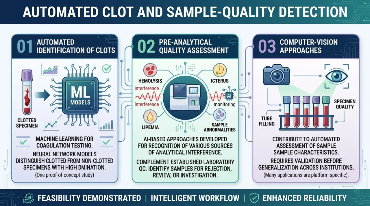
Machine learning has already been investigated for automated identification of clotted specimens in coagulation testing. In one proof-of-concept study, neural-network models trained on coagulation testing data distinguished clotted from non-clotted specimens with high discrimination, demonstrating the feasibility of incorporating ML into automated sample-quality assessment.
More broadly, AI-based approaches are being developed for pre-analytical quality assessment, including recognition of hemolysis, icterus, lipemia, sample abnormalities, and other sources of analytical interference. These systems could complement-not replace-established laboratory quality-control procedures by identifying samples that require rejection, review, or additional investigation.
Computer-vision approaches may also contribute to automated assessment of sample characteristics such as tube filling and specimen quality. However, many of these applications remain laboratory- or platform-specific and require validation before they can be generalized across institutions.
Beyond the Clotting Time: Waveform Analysis
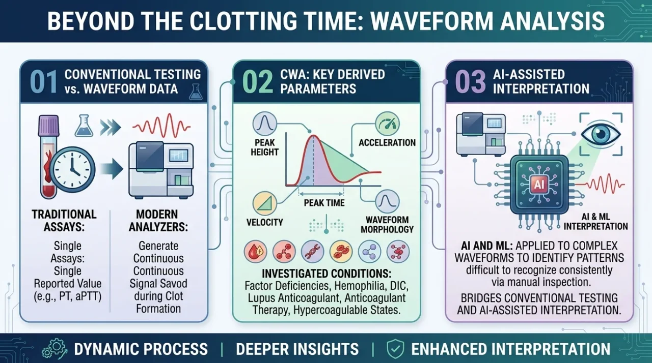
Routine coagulation assays traditionally reduce a dynamic biochemical process to a single reported value—for example, the PT or aPTT. Yet modern optical and mechanical analyzers generate a continuous signal during clot formation.
The resulting clot waveform contains information about the kinetics of fibrin formation that is not captured by clotting time alone. Clot waveform analysis (CWA) therefore represents an important bridge between conventional coagulation testing and AI-assisted interpretation.
Parameters derived from the waveform-including peak height, peak time, velocity, acceleration, and waveform morphology-have been investigated in conditions ranging from coagulation factor deficiencies and hemophilia to disseminated intravascular coagulation, lupus anticoagulant, anticoagulant therapy, and hypercoagulable states. Importantly, CWA itself is not synonymous with artificial intelligence.
Machine Learning and Waveform Pattern Recognition
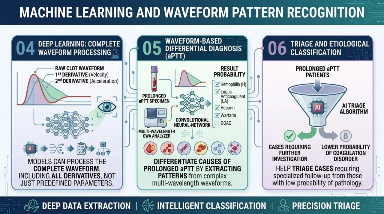
This is one of the most promising areas of AI-assisted hemostasis. Deep-learning models can process the complete coagulation waveform, including its first- and second-derivative curves, rather than relying on a small number of predefined parameters.
A recent study developed a convolutional neural-network model using multi-wavelength aPTT clot waveforms to distinguish several causes of a prolonged aPTT, including hemophilia, lupus anticoagulant, heparin, warfarin, and direct oral anticoagulant exposure.
Similarly, recent work has demonstrated that waveform-based algorithms can help triage patients with prolonged aPTT and distinguish cases requiring further etiological investigation from those at lower probability of an identifiable coagulation disorder.
These findings illustrate an important shift: AI does not necessarily need to replace the coagulation assay. Instead, it can extract additional information from data that the laboratory is already generating.
Platelet Function Testing: An Emerging Frontier
The same principle can potentially be applied to platelet function testing. Light transmission aggregometry and impedance aggregometry generate dynamic response curves rather than single numerical measurements. Machine-learning approaches could assist in recognizing patterns associated with drug effects, impaired platelet responses, or inherited platelet disorders.
However, compared with coagulation waveform analysis, AI applications in platelet aggregometry remain less mature. Differences in agonists, concentrations, instrumentation, pre-analytical conditions, and interpretation criteria create substantial challenges for model development and transferability.
Clinical Decision Support: From Laboratory Data to Patient Context
The potential value of AI extends beyond interpretation of individual laboratory tests. Hemostasis is inherently multidimensional: clinicians integrate laboratory measurements with medications, comorbidities, imaging, vital signs, previous results, and longitudinal clinical information.
Machine-learning models can therefore combine laboratory and electronic health-record data to identify complex patterns that may not be apparent from any single variable.
- Predicting Acute Traumatic Coagulopathy:
One example is acute traumatic coagulopathy (ATC), in which early recognition can influence hemorrhage management. A recent machine-learning study developed a Coagulation Risk Index using continuous physiological data collected during the first 15 minutes after hospital admission.
The model incorporated information from vital-sign and physiological waveform data and demonstrated strong performance for predicting ATC in bleeding trauma patients.
This example illustrates a broader concept: AI can potentially identify a coagulation phenotype before the complete conventional laboratory picture has developed. It is important, however, to distinguish such models from conventional laboratory-based DIC scores.
- Anticoagulation Management:
Anticoagulation is another area in which longitudinal data may be particularly valuable. For vitamin K antagonists such as warfarin, ML models have been developed to estimate maintenance dose requirements and anticipate INR behavior using combinations of demographic, clinical, laboratory, medication, and, in some models, pharmacogenetic variables.
The evidence is encouraging but not yet definitive. A recent systematic review found that most published ML studies of warfarin dosing were retrospective, while relatively few evaluated clinical safety outcomes such as major bleeding or thromboembolism.
This distinction is important: improved prediction of dose or INR does not automatically demonstrate improved patient outcomes. For direct oral anticoagulants, AI could theoretically integrate longitudinal renal function, hemoglobin trends, concomitant medications, and clinical events to identify patients at increased risk of bleeding or inappropriate exposure.
- VTE and Bleeding Risk Prediction:
AI-based risk models have also been developed for venous thromboembolism and bleeding prediction. By incorporating numerous variables and nonlinear interactions, ML models may identify patterns that conventional risk scores do not capture. Recent evidence suggests that some ML models demonstrate better discrimination than established clinical scores in selected populations.
The Challenges of Bringing AI Into the Hemostasis Laboratory
The technical potential of AI should not obscure the practical challenges of implementation.
- Interpretability and Human Oversight: Complex neural networks and ensemble models may produce accurate predictions without providing an easily understandable rationale. In hemostasis, where laboratory results can directly influence transfusion, anticoagulation, or urgent diagnostic decisions, clinicians need to understand the context and limitations of an AI-generated recommendation. AI should therefore function as decision support rather than an autonomous replacement for laboratory expertise.
- Standardization and Interoperability: A model trained on data from one analyzer, reagent system, laboratory population, or reference interval may not perform identically elsewhere. This is particularly relevant to clot waveform analysis. Reagent type can significantly influence waveform parameters, even when measurements are performed on the same analyzer platform. Such variation illustrates why multicenter datasets, standardized preprocessing, and external validation are essential before widespread implementation.
- External Validation and Model Drift: A model can perform extremely well on its development dataset and still fail when exposed to a different patient population or laboratory workflow. Changes in reagent lots, instruments, testing protocols, patient demographics, disease prevalence, or clinical practice can alter model performance. Continuous monitoring and periodic reassessment should therefore be considered part of the AI lifecycle rather than optional post-deployment activities.
- Regulation, Governance, and Accountability: The regulatory status of AI-enabled clinical software depends on its intended use and jurisdiction. Some clinical decision-support functions may fall outside medical-device regulation, while software that performs certain regulated functions may be subject to medical-device requirements. Current regulatory frameworks increasingly emphasize lifecycle management, intended use, validation, transparency, risk management, and human oversight for AI-enabled medical technologies.
- Bias and Representativeness: AI systems learn from the data used to develop them. If particular populations, institutions, disease states, or laboratory conditions are underrepresented, model performance may not be equitable across settings. This is particularly relevant for specialized hemostasis testing, where rare disorders and low-prevalence phenotypes can be difficult to represent adequately in training datasets. Transparent reporting of training populations, missing data, labeling procedures, and external performance is therefore essential.
AI is unlikely to make the hemostasis laboratory less important. Instead, its successful integration could make the laboratory more informative—moving from reporting isolated measurements toward delivering richer, context-aware interpretations of the patient’s hemostatic state.
The future of AI in hemostasis will therefore depend not simply on increasingly sophisticated algorithms, but on the quality of the data, the robustness of validation, the transparency of implementation, and the continued involvement of laboratory professionals in determining how these systems should be used.
FAQ
1. Can AI improve the quality of coagulation testing?
Yes. AI can help identify problematic samples, recognize clots and other pre-analytical issues, and support automated quality control.
2. Does AI provide information beyond a conventional PT or aPTT result?
Yes. AI-assisted waveform analysis can extract additional information from the dynamics of clot formation.
3. Can AI assist with platelet function testing?
Yes. Machine-learning approaches can analyze aggregometry patterns and may help identify drug effects or platelet-function abnormalities.
4. Does AI have a role in anticoagulation management?
Yes. Machine-learning models have been studied for warfarin dose prediction, INR management, and identification of anticoagulation-related risks.
5. Is AI ready for routine use in every hemostasis laboratory?
No. Differences in instruments, reagents, patient populations, and workflows mean that models require appropriate validation before implementation.
6. What changes when AI enters the hemostasis laboratory?
AI allows laboratories to analyze larger and more complex datasets and may reveal patterns that are difficult to detect with conventional rule-based approaches.
7. Is AI interpretation always reliable across different laboratories?
No. A model that performs well in one laboratory may require recalibration or further validation in another setting.
8. What remains difficult for AI?
Rare disorders, limited datasets, differences between laboratories, and changes in analytical methods can all challenge model performance.
9. Will AI replace laboratory specialists?
No. AI is primarily being developed as a tool to augment laboratory expertise rather than replace professional interpretation.
10. Is AI already capable of making independent hemostasis diagnoses?
No. Most current applications require human oversight, clinical validation, and appropriate regulatory and quality safeguards.
Written by Marieta Aleksanyan, MD
Stay updated with Hemostasis Today.
-
Oct 22, 2026, 18:04Basem Goueli: Blood Clots in the NFL
-
Oct 22, 2026, 17:57Syed Ahmed: Viral Triggers of Immune Thrombocytopenia and Thrombosis
-
Oct 22, 2026, 17:49Valerie Tutwiler: Bridging Engineering and Medicine to Advance Thrombosis Research
-
Oct 22, 2026, 17:41Jesus Ardila Novoa: How Patient Education Can Transform Hemophilia Care
-
Oct 22, 2026, 17:30Filipe Gonçalves: Advancing Bleeding Disorder Diagnosis Through Multidisciplinary Collaboration
-
Oct 22, 2026, 17:17Storage Pool Deficiency An Overview of a Rare Platelet Disorder – EHC
-
Oct 22, 2026, 17:10Matthew Hazell: Optimizing the Use of Rare Red Blood Cell Units
-
Oct 22, 2026, 17:03Smita Patil: New Insights Into Platelet Alpha-Granule Secretion
-
Oct 22, 2026, 16:48Ahmed Alotaibi: Rare Rh Phenotype and Severe Fetal Anaemia