


Thrombus Age Assessment Through Advanced Imaging and Biomarkers
Every thrombus carries a history, but it does not carry a timestamp.
When a patient presents with deep vein thrombosis or pulmonary embolism, clinicians often need to know not only whether a clot is present, but also when it likely formed.
That question influences decisions about thrombolysis, catheter-directed therapy, anticoagulation intensity, and the risk of post-thrombotic syndrome.
Yet despite major progress in thrombosis imaging and molecular biology, precisely dating a blood clot in a living patient remains one of the field’s unresolved challenges.
The traditional distinction between acute and chronic thrombosis is clinically useful but biologically incomplete.
A thrombus is not a fixed structure; it is a evolving biological material that changes its cellular composition, stiffness, fibrin architecture, inflammatory activity, and susceptibility to lysis over time. The clinical question is therefore shifting from a simple binary classification toward a more nuanced assessment of thrombus maturity, organization, and treatment responsiveness.
Why Thrombus Age Matters
Thrombus age can determine whether a clot is likely to lyse with pharmacological or catheter-directed therapy, whether endovascular removal is technically feasible, and how much long-term vein damage may follow.
In iliofemoral deep vein thrombosis, for example, early intervention is generally more effective when the thrombus is still relatively fresh and organized fibrosis has not yet developed.
The clinical stakes are particularly high in patients with extensive proximal DVT, massive pulmonary embolism, or thrombosis associated with extracorporeal circuits and vascular devices.
In these settings, distinguishing a recently formed, fibrin-rich and cell-rich thrombus from an old, fibrotic, partially recanalized lesion may change the entire treatment strategy.
At the same time, thrombus age cannot be inferred reliably from symptoms alone.
Patients may present late, symptoms may develop gradually, and some thrombi are clinically silent until they embolize or cause chronic venous obstruction. This gap between biological thrombus evolution and clinical presentation is precisely why biomarkers and advanced imaging have become such an active area of research.
The Biology of an Aging Clot
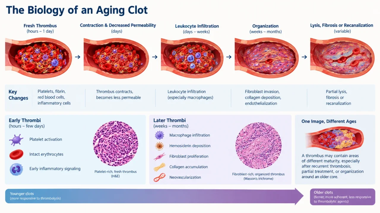
A newly formed venous thrombus is rich in platelets, fibrin, red blood cells, and inflammatory cells.
Over the following days and weeks, it contracts, becomes less permeable, undergoes leukocyte infiltration, and begins to organize through fibroblast invasion, collagen deposition, and endothelialization. Eventually, parts of the thrombus may lyse, while other regions become fibrotic or recanalized.
This process explains why older clots tend to be firmer, more adherent to the vessel wall, and less responsive to thrombolytic agents. It also explains why a single imaging feature can be misleading: a thrombus may contain areas of different maturity, especially after recurrent thrombosis, partial treatment, or organization around an older core.
Recent forensic and translational research has attempted to define clinically meaningful time windows using histological and molecular markers. Early thrombi show platelet activation, intact erythrocytes, and early inflammatory signaling, whereas later thrombi display macrophage infiltration, hemosiderin deposition, fibroblast proliferation, collagen accumulation, and neovascularization.
D-Dimer: Useful but Not a Clock
D-dimer remains the most widely used blood marker in suspected venous thromboembolism, but it cannot date a thrombus.
It is a degradation product of cross-linked fibrin and rises whenever fibrin formation and breakdown are active, including after surgery, trauma, infection, inflammation, pregnancy, and malignancy.
A high D-dimer level supports the presence of active coagulation and fibrinolysis, but it does not distinguish a clot formed yesterday from one formed several weeks ago.
Its principal clinical value remains exclusion of VTE in patients with low pretest probability and, in selected circumstances, assessment of recurrence risk or duration of anticoagulation.
The future may lie less in D-dimer alone and more in panels of biomarkers that reflect specific stages of thrombus evolution.
Investigators are studying neutrophil extracellular traps, extracellular DNA, polyphosphates, kallikrein–kinin activity, inflammatory cytokines, matrix metalloproteinases, and markers of fibrin turnover as potential indicators of thrombus biology.
Molecular Dating: Promising but Not Yet Clinical
The most recent advances have come from forensic medicine, where accurate post-mortem estimation of thrombus age can help establish the relationship between death and a thromboembolic event.
A 2025 review highlighted the potential of histological, immunohistochemical, and molecular markers to distinguish thrombi formed within days from those present for weeks or longer.
Candidate markers include neutrophils and neutrophil extracellular traps in early thrombi, macrophage-associated markers during organization, aquaporin expression in intermediate stages, and markers of collagen deposition and vascularization in older lesions.
Some studies suggest that clinically relevant intervals—such as within 3 days, 3 to 10 days, 10 to 21 days, and beyond 21 days—may eventually be distinguished through combined molecular profiling.
These findings are scientifically compelling, but they are not yet ready for routine clinical use. Most evidence comes from autopsy tissue, animal models, or ex vivo thrombi, and the molecular signature of a thrombus may vary according to its location, cause, host inflammatory state, and prior anticoagulant exposure.
Ultrasound: The First-Line Tool With Limits
Duplex ultrasound remains the cornerstone of DVT diagnosis because it is widely available, non-invasive, and highly accurate for detecting acute venous thrombosis. Acute thrombi are often associated with vein distension, compressibility, hypoechoic luminal material, and absent collateral vessels.
Chronic thrombosis more often produces an irregular thickened vein wall, echogenic intraluminal material, luminal narrowing, partial recanalization, fibrous cords, and collateral circulation.
However, conventional ultrasound is better at detecting thrombosis than at dating it. Echogenicity depends on machine settings, operator technique, thrombus depth, and the age distribution of the clot. More importantly, subacute thrombi may display overlapping features of both acute and chronic disease, particularly in patients with prior DVT or incomplete thrombus resolution.
For this reason, ultrasound remains indispensable for diagnosis but insufficient as a standalone method for precise thrombus-age assessment.
Its greatest future contribution may come from functional extensions that measure the mechanical properties of the clot rather than merely its appearance.
Elastography: Measuring Clot Stiffness
As a thrombus organizes, it becomes progressively stiffer. This simple biological principle underlies one of the most promising bedside approaches to thrombus-age assessment: ultrasound elastography.
Strain elastography and shear-wave elastography measure tissue deformation in response to mechanical force. In DVT, increasing thrombus stiffness generally correlates with greater organization and older thrombus age.
A 2023 systematic review found that nearly all included studies reported increasing stiffness with DVT duration, supporting the biological rationale for elastography as a maturity marker.
The appeal of elastography is practical. It can potentially be incorporated into a standard vascular ultrasound examination, offering a quantitative measure of clot consistency without exposing the patient to radiation or contrast. Shear-wave elastography, in particular, may help separate fresh from organized thrombi and could eventually guide selection for catheter-directed thrombolysis or endovascular thrombectomy.
Nevertheless, the method is not yet standardized. Studies differ in elastography technique, thrombus-age definitions, anatomical location, and patient selection.
Until large prospective trials establish validated cutoff values and demonstrate impact on clinical outcomes, elastography should be considered a promising adjunct rather than a definitive dating tool.
MRI: Imaging the Clot’s Composition
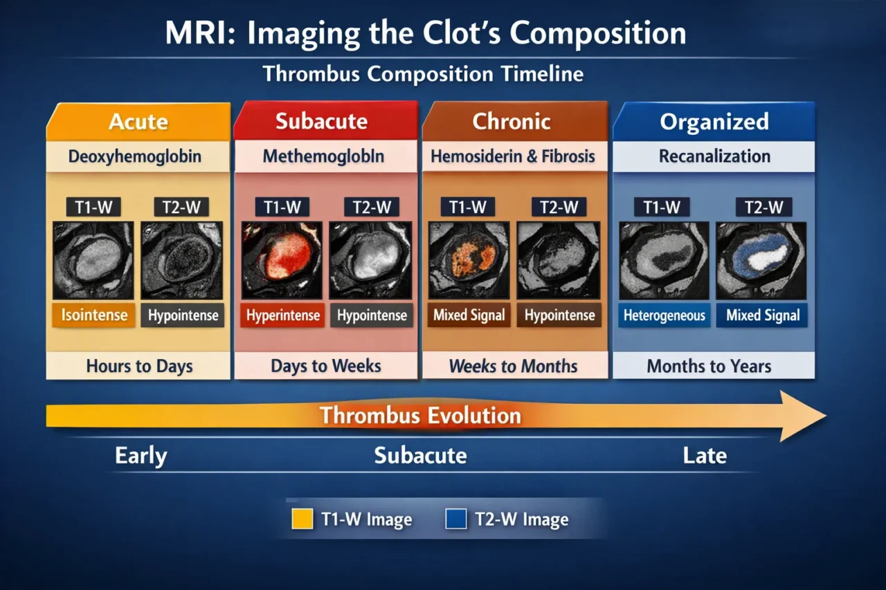
Magnetic resonance imaging offers a more detailed view of thrombus composition than conventional ultrasound. As red blood cells within a thrombus degrade, hemoglobin evolves from oxyhemoglobin to deoxyhemoglobin, intracellular methemoglobin, extracellular methemoglobin, and eventually hemosiderin.
These biochemical changes alter signal intensity on T1- and T2-weighted sequences, creating a potential biological timeline.
In broad terms, very early thrombi may be isointense on T1-weighted images and hypointense on T2-weighted images because of deoxyhemoglobin. Subacute thrombi often become hyperintense on T1-weighted sequences as methemoglobin accumulates. Chronic, organized thrombi show more heterogeneous signal patterns related to fibrosis, neovascularization, and recanalization.
Magnetic resonance venography may be especially valuable before endovascular therapy for iliofemoral DVT. A thrombus-age scoring system based on MRV features has been used to identify patients more likely to benefit from catheter-directed thrombolysis and to predict resistance to thrombolytic treatment.
Yet MRI is not a simple clot clock. Signal intensity varies with sequence, field strength, thrombus location, flow artifacts, and mixed maturity within the same lesion. In cerebral venous thrombosis, for example, early clots can be difficult to distinguish from normal flowing blood, underscoring the importance of interpreting MRI findings in clinical context.
Emerging Imaging: From Structure to Function
The next phase of thrombus imaging aims to move beyond anatomy and measure the biology of the clot.
Quantitative susceptibility mapping can estimate iron content and therefore red-cell burden, while advanced diffusion and magnetization-transfer techniques may provide information about water content, cellular density, and extracellular matrix organization.
In ex vivo studies of venous thrombi, quantitative susceptibility mapping and shear-wave elastography have been correlated with histological composition and susceptibility to recombinant tissue plasminogen activator.
These findings suggest that imaging may eventually predict not only thrombus age but also the likelihood of successful thrombolysis.
Nuclear imaging adds another promising dimension. FDG PET/CT can detect inflammatory activity within recent thrombi, with uptake generally declining as the clot organizes. Experimental tracers targeting fibrin, platelets, or fibrinolytic activity are also being developed to visualize specific molecular components of the thrombus.
These techniques remain investigational, but they represent a conceptual shift: instead of asking whether a vessel is blocked, future imaging may ask what the thrombus is made of, how inflammatory it is, and whether it is likely to respond to lysis.
A Multimodal Future
No single test is likely to solve the problem of thrombus dating.
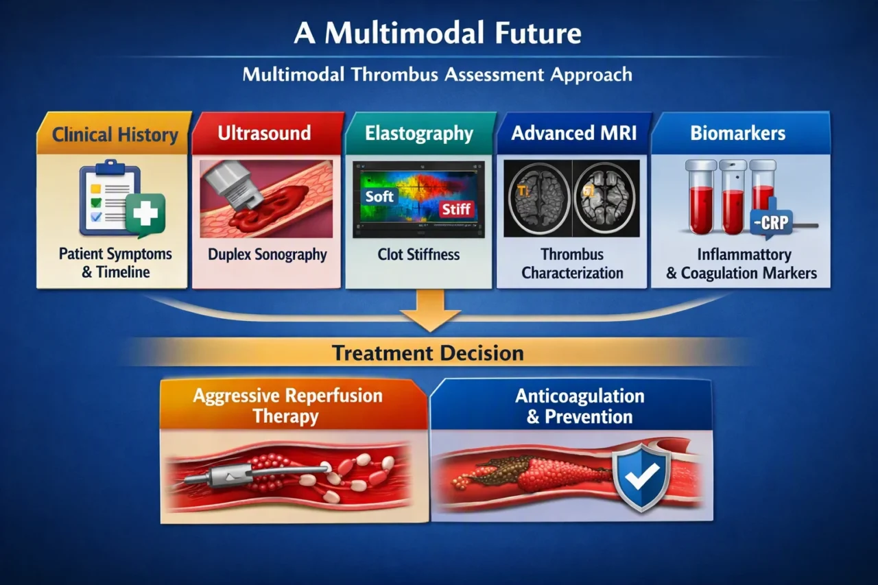
The most realistic future is a multimodal approach that combines clinical chronology, conventional ultrasound, quantitative elastography, advanced MRI, and selected biomarkers.
In this model, clinical history would remain the foundation, while imaging and molecular data would refine the estimate. A patient with a soft, hypoechoic, non-occlusive thrombus, high inflammatory activity and a short symptom interval may be an ideal candidate for aggressive reperfusion therapy.
In contrast, a stiff, organized, partially recanalized thrombus with low inflammatory activity may be better managed with anticoagulation and prevention of post-thrombotic sequelae.
The goal is not simply to label a clot as acute or chronic. It is to understand its biological state well enough to match treatment to the mechanism and stage of disease.
The Bottom Line
Can we date a blood clot? Not with minute-level precision, and not yet with a single validated clinical test. But the field is moving rapidly from subjective visual assessment toward quantitative measures of thrombus stiffness, composition, inflammatory activity, and lysis susceptibility.
D-dimer tells us that fibrin turnover is occurring, ultrasound tells us where the clot is, elastography tells us how stiff it has become, and MRI tells us something about its internal composition. Molecular biomarkers may eventually add the missing layer: a biological estimate of when the thrombus began and how it is evolving.
The future of thrombus-age assessment will likely be integrative rather than reductionist. Instead of replacing clinical judgment, these tools may give clinicians a more precise biological context for one of the most consequential decisions in thrombosis medicine: whether to watch, anticoagulate, lyse, or mechanically remove the clot.
Frequently Asked Questions
1. Can we determine the exact age of a blood clot?
No, but clinicians can often estimate whether it is acute, subacute, or chronic.
2. Why does thrombus age matter?
It influences thrombolysis response, endovascular treatment feasibility, embolization risk, and post-thrombotic syndrome.
3. Can D-dimer date a thrombus?
No. It reflects fibrin turnover but is elevated in many non-thrombotic conditions.
4. How does an acute thrombus appear on ultrasound?
As a distended, non-compressible vein with hypoechoic or anechoic material.
5. What suggests chronic thrombosis?
Echogenic material, vein-wall thickening, luminal narrowing, recanalization, fibrous cords, and collateral vessels.
6. Why is elastography promising?
It measures thrombus stiffness, which generally increases as the clot organizes.
7. Is elastography ready for routine use?
Not yet; standardized cutoffs and large prospective trials are still needed.
8. How does MRI help?
It reflects hemoglobin degradation, water content, fibrin organization, and cellular changes within the thrombus.
9. Can MRI give an exact age?
No. It helps stage the thrombus but cannot reliably determine its exact age.
10. What is the future approach?
A multimodal assessment combining clinical history, ultrasound, elastography, MRI, and molecular biomarkers.
Written by Anna Stepanyan, MD
Stay updated with Hemostasis Today.
-
Oct 10, 2026, 10:55Tzu-Fei Wang: How Should We Approach Anticoagulation for Cancer-Associated Gonadal and Renal Vein Thrombosis?
-
Oct 10, 2026, 10:50Robert Sidonio Joins ATHN Advanced Clinical Research Fellowship Program
-
Oct 10, 2026, 10:45Federica Fogacci: Co-Moderating at CA Meet 2026 in Padua – Elevating Lifestyle Interventions in Cardiology
-
Oct 10, 2026, 10:40Juan Cristóbal Morales Muñoz: Pharmacokinetic Profiling of Factor VIII Concentrates in Chilean Patients
-
Oct 10, 2026, 10:32Randall Schaefer: What Do Blood Product Wastage Rates Really Tell Us in Prehospital Care?
-
Oct 10, 2026, 10:26Simone Strasser: ROTEM-Guided Transfusion Cuts Blood Product Use in Patients with Cirrhosis
-
Oct 10, 2026, 10:18Jérôme S.: WHO Launches Global Roadmap to Combat Hypertensive Disorders of Pregnancy
-
Oct 10, 2026, 10:07Maha Othman: Look Forward to an Amazing ISTH 2027 in Vancouver!
-
Oct 10, 2026, 10:02Rucha Patil: National Convening Highlights Opportunities for Equitable Hemophilia Care Across India