


Serena Williams and Pulmonary Embolism: From Personal Crisis to Lessons in Venous Thromboembolism
Serena Williams is one of the most accomplished athletes in modern history – a 23-time Grand Slam singles champion and former world No. 1 whose career has been defined by exceptional physical performance and resilience.
Yet in 2011, at the age of 29, she developed a pulmonary embolism, confronting a potentially life-threatening complication of venous thromboembolism despite her young age and extraordinary level of fitness.
Six years later, following childbirth by cesarean delivery, she experienced pulmonary embolism again.
Her medical history provides a compelling illustration of a fundamental principle in thrombosis: physical fitness does not eliminate the risk of venous thromboembolism.
Rather, thrombotic risk is dynamic, shaped by the interaction of individual susceptibility with transient and acquired risk factors such as surgery, immobility, pregnancy, and the postpartum period.
The story of Serena Williams therefore extends beyond an individual medical event. It offers a clinically relevant perspective on pulmonary embolism, recurrent VTE and the importance of recognizing thrombosis across changing physiological and clinical circumstances.
The First Pulmonary Embolism
In February 2011, Williams developed a pulmonary embolism shortly after undergoing treatment for a foot injury. She had previously undergone foot procedures after cutting her foot on broken glass and had spent an extended period away from competitive tennis.
Williams subsequently described leg swelling and difficulty breathing before doctors identified blood clots in her lungs. She received anticoagulant treatment and required continued medical monitoring during her recovery.
This episode established a major clinical risk factor for the future: a previous VTE substantially changes the thrombotic risk profile of a patient. A history of VTE is particularly important when subsequent high-risk situations such as pregnancy, surgery, or prolonged immobility occur.
A Second PE After Pregnancy
In September 2017, Williams gave birth to her daughter, Alexis Olympia Ohanian Jr., by emergency cesarean section after fetal heart-rate abnormalities during labor.
The following day, she developed sudden shortness of breath. Because of her previous history, Williams immediately suspected another pulmonary embolism. Imaging subsequently demonstrated several small blood clots in her lungs, and intravenous heparin was started.
The clinical course was complicated by bleeding after anticoagulation. Her cesarean incision opened during coughing, and she subsequently required additional surgery for a large abdominal hematoma.
This illustrates a fundamental challenge in VTE management: anticoagulation is essential for treating a potentially life-threatening PE, but the same treatment can increase bleeding risk, particularly around recent surgery.
Why Pregnancy Increases VTE Risk
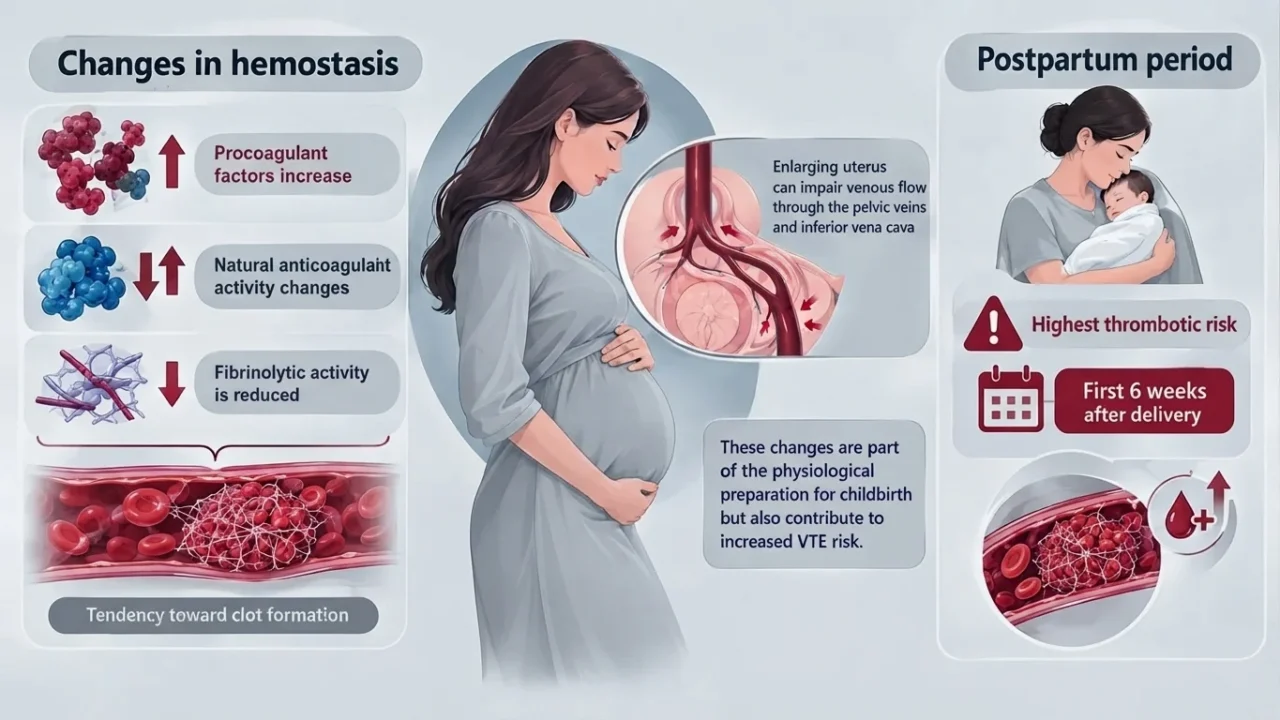
Pregnancy is accompanied by profound changes in hemostasis. Procoagulant factors increase, natural anticoagulant activity changes, and fibrinolytic activity is reduced, creating a physiological state that favors clot formation.
At the same time, the enlarging uterus can impair venous flow through the pelvic veins and inferior vena cava. These changes are part of the physiological preparation for childbirth but also contribute to increased VTE risk.
The risk does not immediately disappear after delivery. The postpartum period carries particularly high thrombotic risk, with the first six weeks after delivery representing the period of greatest concern.
Cesarean Delivery Adds Another Risk
Cesarean delivery is an additional recognized risk factor for pregnancy-associated VTE. Surgery can contribute through tissue injury, activation of coagulation, and reduced mobility during recovery.
The combination becomes particularly important when other risk factors are present. In Williams’ case, her previous pulmonary embolism represented an important pre-existing risk factor before pregnancy, while pregnancy and cesarean delivery added further thrombotic challenges.
Importantly, this does not mean that every patient with these factors will develop VTE. Risk assessment is individualized, and prophylaxis decisions depend on the complete clinical context and the balance between thrombosis and bleeding risk.
The Role of Previous VTE
A previous VTE is one of the most clinically important predictors of future thrombotic events. For patients with a history of VTE who become pregnant, anticoagulation planning therefore requires careful assessment before, during, and after pregnancy.
Current European guidance identifies low-molecular-weight heparin (LMWH) as the preferred drug class for prevention and treatment of pregnancy-associated VTE. Patients with confirmed acute VTE generally require therapeutic anticoagulation, while the intensity and duration of prophylaxis in other situations depend on individual risk.
Why Pulmonary Embolism Can Be Difficult to Recognize
PE can present with nonspecific symptoms, including sudden dyspnea, chest pain, tachycardia, dizziness, or hemoptysis. During pregnancy and the postpartum period, some symptoms may overlap with normal physiological changes or other complications.
This makes clinical awareness particularly important. Current guidelines emphasize prompt diagnostic assessment when PE is suspected rather than attributing unexplained respiratory symptoms to pregnancy or postoperative recovery.
Diagnosis Requires More Than a Single Test
The diagnostic approach to suspected PE depends on the patient’s clinical condition. In pregnancy, contemporary algorithms may incorporate clinical probability, D-dimer testing in selected patients, compression ultrasonography, and imaging such as CT pulmonary angiography when appropriate.
The 2025 European Society of Cardiology guidelines describe pregnancy-specific diagnostic strategies designed to reduce unnecessary radiation exposure while maintaining the ability to identify potentially life-threatening PE.
In stable patients, a stepwise approach can begin with clinical assessment and D-dimer testing, followed by compression ultrasonography when indicated. If PE cannot be excluded, chest imaging, including CT pulmonary angiography, may be required to establish the diagnosis.
Importantly, pregnancy can make the clinical assessment more challenging because symptoms such as shortness of breath, tachycardia, and leg discomfort may overlap with normal physiological changes. This makes structured diagnostic pathways particularly important when PE is suspected.
Anticoagulation and the Bleeding–Thrombosis Balance
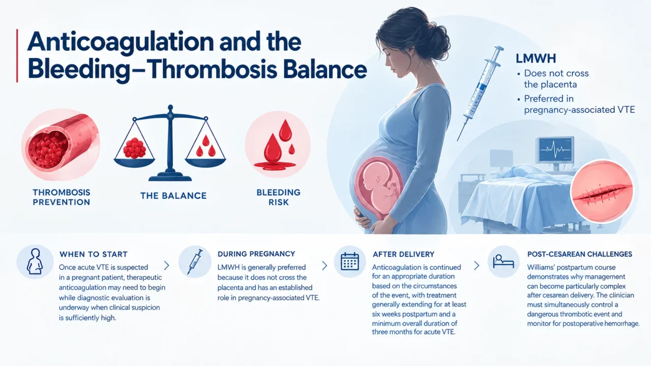
Once acute VTE is suspected in a pregnant patient, therapeutic anticoagulation may need to begin while diagnostic evaluation is underway when clinical suspicion is sufficiently high.
LMWH is generally preferred because it does not cross the placenta and has an established role in pregnancy-associated VTE.
After delivery, anticoagulation is continued for an appropriate duration based on the circumstances of the event, with treatment generally extending for at least six weeks postpartum and a minimum overall duration of three months for acute VTE.
Williams’ postpartum course demonstrates why management can become particularly complex after cesarean delivery. The clinician must simultaneously control a dangerous thrombotic event and monitor for postoperative hemorrhage.
The Broader Lesson From Williams’ Story
Williams’ experience is not evidence that pregnancy or cesarean delivery inevitably leads to PE. Rather, it illustrates how multiple recognized risk factors can accumulate within a short period.
Her history also demonstrates the importance of recognizing previous VTE when planning pregnancy and assessing thrombotic risk. A prior PE is not simply a historical detail; it can influence future anticoagulation and prevention strategies.
From an Athlete’s Story to a Hematologic Lesson
Pulmonary embolism can affect people across a wide range of ages and levels of physical fitness. Being a highly trained athlete does not eliminate the possibility of VTE when major acquired risk factors are present.
Williams’ case therefore extends beyond celebrity medicine. It provides a recognizable example of how several risk factors can interact over time, including surgery, reduced mobility during recovery, pregnancy, cesarean delivery, and a previous history of VTE. Her experience also illustrates the clinical challenge of balancing anticoagulation with bleeding risk, particularly in the postpartum and postoperative setting.
More broadly, her history highlights an important principle in thrombosis medicine: VTE risk is determined by the overall clinical context rather than by physical fitness alone. A person who is otherwise young and active may still develop thrombosis when transient or persistent risk factors substantially alter the balance between coagulation and anticoagulation.
Recognizing the Warning Signs
Sudden unexplained shortness of breath, chest pain, coughing with blood, unexplained tachycardia, syncope, or signs of DVT should prompt urgent medical evaluation. Symptoms may vary considerably, and PE does not always present with the full classic clinical picture.
In patients with risk factors such as recent surgery, pregnancy or the postpartum period, previous VTE, prolonged immobility, or other provoking conditions, new cardiopulmonary symptoms should be assessed promptly rather than attributed automatically to the underlying situation.
PE can be life-threatening, but early recognition and appropriate anticoagulant treatment can substantially change the clinical course. The key clinical message is not to diagnose PE from symptoms alone, but to recognize when the possibility is serious enough to require timely diagnostic evaluation.
FAQ
1. What is a pulmonary embolism?
A pulmonary embolism occurs when a blood clot, usually originating from the deep veins, travels to the pulmonary circulation and obstructs blood flow through the lungs.
2. When did Serena Williams first experience PE?
Williams experienced a pulmonary embolism in 2011 after treatment for a foot injury. She subsequently received anticoagulant treatment.
3. Did Serena Williams experience another PE after pregnancy?
Yes. After giving birth by emergency cesarean section in 2017, she developed acute shortness of breath and was subsequently diagnosed with several small pulmonary emboli.
4. Why was her previous PE important during pregnancy?
A previous VTE is an important risk factor for recurrence. Pregnancy and the postpartum period further increase thrombotic risk, making individualized anticoagulation planning important.
5. Why does pregnancy increase the risk of VTE?
Pregnancy produces a physiological hypercoagulable state, while mechanical compression of pelvic veins and changes in venous flow further contribute to thrombotic risk.
6. Is the postpartum period still a high-risk period for VTE?
Yes. VTE risk remains elevated after delivery and is particularly high during the first six weeks postpartum.
7. Does cesarean delivery increase VTE risk?
Yes. Cesarean delivery is a recognized risk factor for pregnancy-associated VTE, particularly when combined with other thrombotic risk factors.
8. What anticoagulant is generally preferred during pregnancy?
Low-molecular-weight heparin is generally preferred for prevention and treatment of pregnancy-associated VTE.
9. Can anticoagulation cause bleeding?
Yes. Anticoagulants reduce thrombosis but increase bleeding risk. This balance can become particularly challenging after surgery, as illustrated by Williams’ postpartum complications.
10. What is the main hematologic lesson from Serena Williams’ case?
Her history illustrates how previous VTE, pregnancy, cesarean delivery, postoperative immobility, and the physiological hypercoagulability of pregnancy can interact to increase thrombotic risk, while anticoagulation simultaneously creates an important bleeding consideration.
Written by Hermine Sayiyan, MD
Stay updated with Hemostasis Today.
-
Oct 5, 2026, 14:55Marco Moscarelli: 3D Reconstruction Enhances Volumetric Assessment of Post-TAVI Thrombosis
-
Oct 5, 2026, 13:24Abdul Muqtadir Abbasi: Diagnostic Pearls for Evaluating Isolated Thrombocytopenia in Residents
-
Oct 5, 2026, 13:16Katerina Pavenski: Rethinking What ‘Normal’ Means for Iron Deficiency
-
Oct 5, 2026, 13:09Rick Matthews: Balancing Established VTE Partnerships with Innovative Interventional Technologies
-
Oct 5, 2026, 13:01Nawsherwan Mohammad: Recognizing TTP Before the ADAMTS13 Result Arrives
-
Oct 5, 2026, 12:51Bruno Pougault: The Hidden Challenges Behind Anti Xa Testing
-
Oct 5, 2026, 12:41Syed Huzaifa Khan: Rethinking CTRP12 as a Biomarker for In Stent Restenosis
-
Oct 5, 2026, 12:29Mirvat Alasnag: Creating a Space for Women in Cardiovascular Medicine
-
Oct 5, 2026, 11:59Khaoula Mazouzi: The Potential of Haemoglobin Vesicles as an Oxygen Carrier