

Bernard-Soulier syndrome in the Genomic Era: From GP Ib-IX-V Dysfunction to Precision Diagnosis
Bernard-Soulier syndrome (BSS) is a rare inherited platelet disorder characterized by mucocutaneous bleeding, thrombocytopenia, and giant platelets. Its defining defect lies in the glycoprotein Ib-IX-V (GPIb-IX-V) complex, a receptor that enables platelets to capture von Willebrand factor (VWF) and initiate adhesion at sites of vascular injury.
The central defect in BSS is a failure of platelet adhesion at the very first step of primary hemostasis. Even when platelets circulate in the bloodstream, they cannot efficiently tether to the damaged vessel wall, compromising the formation of an effective platelet plug.
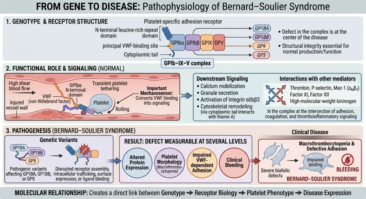
The GPIb-IX-V complex comprises four subunits—GPIbα, GPIbβ, GPIX, and GPV—encoded by GP1BA, GP1BB, GP9, and GP5, respectively.
These subunits work together to ensure proper receptor assembly, transport to the platelet surface, and function. GPIbα contains the principal VWF-binding site within its extracellular N-terminal domain, while its cytoplasmic tail interacts with filamin A, linking the receptor to the platelet cytoskeleton.
Variants affecting GP1BA, GP1BB, or GP9 can disrupt receptor assembly, surface expression, or ligand binding, undermining platelet adhesion.
Under normal conditions, this receptor operates in a mechanically demanding environment. At sites of vascular injury, exposed VWF captures circulating platelets under high-shear conditions, allowing them to tether transiently to the damaged vessel wall.
This interaction does more than anchor platelets: it enables them to sense mechanical forces generated by blood flow and initiate intracellular signaling that supports calcium mobilization, granule secretion, and activation of integrin αIIbβ3. These events help transform initial platelet capture into stable adhesion and aggregation.
GPIb-IX-V also connects platelet adhesion with coagulation and inflammation through interactions with thrombin, factor XI, factor XII, high-molecular-weight kininogen, P-selectin, and Mac-1 (αMβ2).
Its biological significance therefore extends beyond a single receptor–ligand interaction, placing it at the interface of primary hemostasis, coagulation, and thromboinflammatory responses.
In BSS, disruption of this receptor compromises the earliest stages of platelet plug formation. Abnormal receptor function also affects platelet production and contributes to the characteristic macrothrombocytopenia.
The resulting phenotype reflects the combined consequences of defective VWF-dependent adhesion and reduced platelet numbers, manifesting clinically as easy bruising, epistaxis, gingival bleeding, and excessive menstrual bleeding.
The significance of BSS lies in the direct link between molecular architecture and clinical hemostasis: a pathogenic variant in a single receptor component can alter protein assembly or function, impair platelet interaction with VWF, and produce a recognizable combination of giant platelets, thrombocytopenia, and bleeding.
This relationship makes BSS a compelling model for understanding how receptor biology translates into platelet dysfunction and inherited bleeding disease.
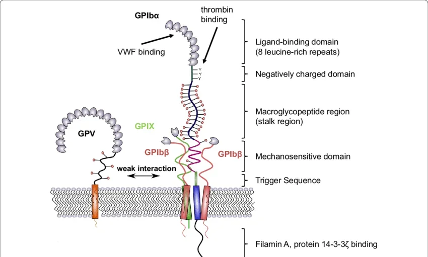
Structure of GPIb-IX. The largest subunit of the complex is GPIbα, which consists of a N-terminal binding domain (made up of 8 leucine-rich repeats), a negatively charged domain, a sialomucin region (macroglycopeptide domain), the mechanosensitive domain (MSD), and proximal to the membrane, the trigger sequence. Intracellularly, GPIbα interacts with protein 14-3-3ζ and filamin A which links the complex to the cytoskeleton. The transmembrane domain provides a close interaction with GPIbβ and GPIX to form a stable parallel four-helical bundle structure. This complex structure is further strengthened by the interplay of GPIbβ and GPIX extracellular domains with the MSD in GPIbα. GPV is weakly associated with GPIb-IX by polar interactions and can be replaced by other receptors
The Molecular Spectrum of BSS
The three genes most strongly associated with classical Bernard-Soulier syndrome are GP1BA, GP1BB, and GP9, encoding GPIbα, GPIbβ, and GPIX, respectively.
Because these proteins assemble into a single receptor, pathogenic variants in any of these genes can ultimately disrupt GPIb-IX-V function, but the molecular consequences depend on which subunit is affected and whether the variant interferes with receptor synthesis, assembly, trafficking, surface expression, or ligand binding.
Among these genes, GP1BA is particularly important because GPIbα contains the principal binding site for VWF and forms the extracellular and cytoplasmic interfaces through which the receptor participates in adhesion and signaling.
Biallelic pathogenic GP1BA variants can therefore produce severe impairment of GPIb-IX-V function and the classical BSS phenotype, whereas certain monoallelic variants produce macrothrombocytopenia with a milder or distinct clinical phenotype, illustrating that not every GP1BA defect results in classical BSS.
Variants in GP1BB and GP9 affect subunits required for the proper assembly and surface expression of the receptor.
Defects in either gene can markedly reduce the amount of functional GPIb-IX-V reaching the platelet membrane, producing the combination of giant platelets, thrombocytopenia, and impaired platelet adhesion characteristic of classical BSS.
The molecular defect is therefore closely linked to receptor abundance: when assembly or trafficking is sufficiently disrupted, the platelet surface becomes profoundly deficient in the receptor required for normal VWF-dependent adhesion.
The relationship becomes more complex when the effect of individual variants is considered.
Some pathogenic variants cause near-complete loss of receptor expression, whereas others permit residual expression or alter specific receptor functions without completely eliminating surface GPIb-IX-V.
Consequently, platelet count, platelet size, receptor expression, ristocetin-induced agglutination, and bleeding severity may not correlate perfectly, meaning that the clinical phenotype alone cannot always predict the underlying molecular defect.
The importance of this genotype-phenotype variability is illustrated by the broader spectrum of GPIb-IX-V disorders.
Certain monoallelic GP1BA variants are associated with inherited macrothrombocytopenia, while specific alterations affecting the VWF-binding properties of GPIbα can produce platelet-type von Willebrand disease, demonstrating that different alterations within the same receptor can produce clinically distinct disorders.
BSS should therefore be viewed within a wider molecular spectrum of GPIb-IX-V abnormalities rather than as a single genetically uniform disease.
Large-scale genetic studies further demonstrate the extent of this heterogeneity.
In an international analysis of 211 BSS families, Savoia and colleagues identified 112 different variants across GP1BA, GP1BB, and GP9, including recurrent variants and evidence of founder effects in particular populations.
Such diversity explains why two patients with apparently similar BSS phenotypes may carry different molecular defects, while patients with variants in the same gene may show different degrees of receptor deficiency and clinical severity.
This creates an important diagnostic principle: identifying a variant is only the beginning of molecular diagnosis.
A genetic result becomes clinically meaningful when it is interpreted alongside the inheritance pattern, platelet morphology, platelet function, GPIb-IX-V surface expression, and bleeding phenotype, particularly when a previously unreported variant or variant of uncertain significance is encountered.
The molecular spectrum of BSS therefore provides the bridge between platelet biology and genomic diagnosis: the gene determines the molecular defect, the molecular defect alters the receptor, and the altered receptor produces the measurable platelet and clinical phenotype.
Establishing this chain of evidence is what allows genomic testing to move beyond simply finding variants and toward a genuinely precision-based diagnosis of Bernard-Soulier syndrome.
Clinical Phenotype and Conventional Diagnosis
The clinical presentation of Bernard-Soulier syndrome reflects the central role of GPIb-IX-V in primary hemostasis, with bleeding typically dominated by mucocutaneous manifestations such as epistaxis, gingival bleeding, easy bruising, menorrhagia, and excessive bleeding after surgery or trauma, although severity varies substantially between affected individuals.
Severe cases may present early in life with recurrent spontaneous bleeding, whereas milder phenotypes can remain unrecognized until hemostatic stress exposes the underlying platelet defect.
The characteristic hematological combination of thrombocytopenia and giant platelets provides an important diagnostic clue, but platelet size should be interpreted together with the clinical history because automated hematology analyzers may underestimate the platelet count when very large platelets fall outside the instrument’s counting range.
Examination of the peripheral blood smear is therefore particularly valuable, as it can reveal the striking macrothrombocytopenia of BSS and simultaneously direct the differential diagnosis toward other inherited disorders characterized by large or giant platelets.
Once an inherited macrothrombocytopenia is suspected, platelet function testing provides the first functional link to the underlying receptor defect.
The characteristic abnormality in BSS is markedly impaired or absent ristocetin-induced platelet agglutination, reflecting defective interaction between VWF and platelet GPIbα, while responses to other agonists are generally preserved because the primary defect lies in platelet adhesion rather than the downstream aggregation machinery.
This pattern helps distinguish BSS from disorders in which platelet aggregation itself is primarily impaired, although pre-analytical variables, severe thrombocytopenia, and the technical limitations of platelet function testing can complicate interpretation.
The diagnostic picture becomes more specific when platelet-surface glycoproteins are assessed by flow cytometry.
Reduced or absent expression of GPIbα, GPIX, and GPV supports a defect within the GPIb-IX-V complex and can distinguish BSS from several other inherited platelet disorders, while assessment of additional markers such as GPIIb/IIIa can help define whether the abnormality is restricted to the GPIb-IX-V pathway.
Importantly, flow cytometry can also identify patients with residual receptor expression, which is relevant when interpreting variants associated with partial rather than complete loss of receptor function.
Taken together, clinical bleeding, macrothrombocytopenia, peripheral smear findings, ristocetin-induced agglutination, and GPIb-IX-V surface expression establish a strong phenotypic framework for BSS, but they do not necessarily identify the precise molecular lesion.
This limitation becomes particularly important in patients with overlapping phenotypes, because inherited macrothrombocytopenias caused by defects in MYH9, ACTN1, TUBB1, DIAPH1, and other genes can produce thrombocytopenia and enlarged platelets without a primary defect in GPIb-IX-V.
The distinction from platelet-type von Willebrand disease is particularly important because both disorders involve the VWF-GPIbα axis but arise from fundamentally different molecular mechanisms: BSS results from quantitative or functional deficiency of the platelet GPIb-IX-V receptor, whereas platelet-type VWD results from a gain-of-function defect in GPIbα that increases platelet binding to VWF.
Similarly, distinguishing BSS from immune thrombocytopenia may be difficult when the inherited disorder has not previously been recognized, particularly if macrothrombocytopenia is overlooked or the patient is evaluated primarily on the basis of a low platelet count.
These limitations explain why the diagnosis of BSS increasingly extends beyond the classical laboratory phenotype.
Phenotyping establishes that the platelet is abnormal; flow cytometry can indicate which receptor system is affected; genomic testing can determine why it is abnormal.
The transition from conventional testing to molecular diagnosis therefore does not represent a replacement of classical hematology, but rather the addition of a genetic layer capable of resolving cases in which phenotype and laboratory findings alone cannot define the underlying defect.
This diagnostic progression leads directly to the genomic era of BSS: once a GPIb-IX-V defect is suspected, the next question is no longer simply whether the patient has Bernard-Soulier syndrome, but which genetic alteration has disrupted the receptor and whether that variant explains the observed phenotype.
Conventional and Genomic Diagnosis
The diagnosis of BSS begins with recognition of a characteristic platelet phenotype but requires demonstration of a defect in the GPIb-IX-V axis for confident classification.
The typical combination is mucocutaneous bleeding, persistent macrothrombocytopenia, and giant platelets on peripheral blood examination.
Platelet counts are often underestimated by automated analyzers because very large BSS platelets may exceed the size-discrimination thresholds used for conventional platelet counting; therefore, examination of the blood film is not simply confirmatory but can determine whether an apparent thrombocytopenia represents true reduction in platelet number or analytical misclassification of giant platelets.
The functional hallmark of BSS is defective ristocetin-induced platelet agglutination.
Ristocetin promotes the interaction between VWF and the N-terminal A1-binding region of platelet GPIbα; therefore, absent or markedly reduced agglutination indicates disruption of the VWF-GPIbα interaction, provided that the assay is interpreted in the context of platelet count and VWF status.
In classical biallelic BSS, ristocetin-induced agglutination is typically absent or severely reduced, whereas responses to agonists that activate downstream platelet signaling through other receptors are comparatively preserved.
This distinguishes a primary adhesion/receptor defect from disorders such as Glanzmann thrombasthenia, in which the principal abnormality lies downstream at integrin αIIbβ3-mediated aggregation.
Ristocetin testing, however, does not identify BSS at the molecular level and is not entirely disease-specific.
The same VWF-GPIbα axis is involved in platelet-type von Willebrand disease (PT-VWD), in which gain-of-function variants of GPIbα increase the affinity of the receptor for VWF rather than abolishing receptor function.
Consequently, interpretation of ristocetin-induced agglutination must be integrated with VWF antigen, VWF activity, platelet morphology, and receptor-expression studies rather than treated as an isolated diagnostic endpoint.
Flow cytometry provides the critical phenotypic bridge between abnormal platelet function and the molecular lesion.
Antibodies directed against CD42b (GPIbα), CD42a (GPIX), and CD42d (GPV) can quantify the abundance of the GPIb-IX-V complex on the platelet surface.
In classical biallelic BSS, surface expression of these components may be profoundly reduced or essentially absent, reflecting defective assembly, intracellular trafficking, or stability of the receptor complex.
By contrast, patients with residual receptor expression may show partial reduction despite a clinically meaningful defect, making quantitative flow cytometry particularly useful for distinguishing complete receptor deficiency from hypomorphic phenotypes.
This distinction is especially important because the amount of surface GPIb-IX-V provides functional information that platelet aggregometry alone cannot supply.
In a well-characterized BSS cohort, affected patients had GPIbα expression below 10% of control values together with absent or severely impaired ristocetin-induced aggregation, whereas some heterozygous carriers showed intermediate receptor expression without the full clinical phenotype.
Such observations demonstrate that BSS is not determined simply by the presence of a variant: the extent to which that variant reduces receptor abundance or function contributes directly to the measurable platelet phenotype.
The conventional diagnostic framework nevertheless has an important limitation: it establishes a phenotype of GPIb-IX-V dysfunction, but not necessarily its genetic cause.
This becomes problematic in inherited macrothrombocytopenias, where MYH9-related disease, ACTN1-associated thrombocytopenia, TUBB1-related thrombocytopenia, DIAPH1-related disease, and other disorders can produce large or giant platelets with variable bleeding phenotypes.
Conversely, monoallelic defects involving GPIb-IX-V may produce a considerably milder phenotype than classical biallelic BSS and can be mistaken for immune thrombocytopenia, particularly when the peripheral smear findings are overlooked.
Genomic testing therefore becomes most informative when it is performed as an extension of phenotypic characterization rather than as an independent diagnostic shortcut.
In a patient with macrothrombocytopenia, impaired ristocetin-induced agglutination, and reduced CD42a/CD42b expression, sequencing of GP1BA, GP1BB, and GP9 directly interrogates the three principal genes responsible for classical BSS.
The molecular distribution is itself heterogeneous: analysis of 211 families identified 112 different variants, with mutations distributed among GP1BA, GP1BB, and GP9 and with most affected individuals carrying biallelic variants.
Importantly, most pathogenic variants in this series were predicted to prevent normal expression of the receptor complex at the platelet membrane rather than directly abolish VWF binding.
The choice of sequencing strategy depends on the diagnostic context.
Targeted sequencing of GP1BA, GP1BB, and GP9 remains highly efficient when the phenotype strongly indicates BSS because it provides deep coverage of the relevant loci without generating the large number of incidental variants produced by broader testing.
However, a phenotype-first strategy is not always sufficient: patients with unexplained inherited thrombocytopenia may have overlapping morphological features, and a broader inherited platelet-disorder panel can simultaneously interrogate genes responsible for platelet production, cytoskeletal organization, granule biology, receptor signaling, and coagulation.
High-throughput sequencing has consequently shifted molecular diagnosis from sequential testing of individual candidate genes toward parallel analysis of multiple disease-associated loci.
For particularly unresolved cases, whole-exome sequencing (WES) and whole-genome sequencing (WGS) extend this approach beyond established BSS genes.
Their principal value is not merely the ability to detect another GP1BA, GP1BB, or GP9 variant, but the possibility of identifying an alternative molecular diagnosis when the observed phenotype is not fully explained by defects in the canonical BSS genes.
This is relevant because inherited platelet disorders are genetically heterogeneous and because phenotypic convergence is common: macrothrombocytopenia, bleeding, and abnormal platelet function can arise from defects in genes with entirely different biological roles.
DNA-based diagnosis therefore allows the diagnostic process to move from a receptor-centered hypothesis toward an unbiased assessment of the underlying genetic architecture.
The principal difficulty then shifts from variant detection to variant interpretation.
Identification of a rare GP1BA, GP1BB, or GP9 variant does not establish pathogenicity by itself, particularly when the variant is novel or classified initially as a variant of uncertain significance.
The strongest molecular diagnosis is obtained when independent evidence converges: an appropriate inheritance pattern, a compatible platelet phenotype, reduced or abnormal GPIb-IX-V expression, impaired ristocetin-induced agglutination, segregation within the family, and a variant predicted or demonstrated to disrupt receptor expression, assembly, stability, or function.
A published case illustrates this principle directly: a candidate BSS diagnosis was strengthened when markedly reduced platelet GPIb/IX expression and absent ristocetin-induced agglutination were used as functional evidence to support reinterpretation of a genetic variant initially classified as uncertain.
Thus, conventional and genomic diagnosis should not be viewed as competing diagnostic eras.
Peripheral morphology identifies the macrothrombocytopenic phenotype; ristocetin testing localizes the functional defect to the VWF-GPIbα interaction; flow cytometry determines whether the GPIb-IX-V complex is quantitatively deficient; and sequencing establishes the molecular lesion responsible for that abnormal receptor phenotype.
Precision Diagnosis: Integrating Phenotype, Protein and Genotype
The molecular diagnosis of Bernard-Soulier syndrome becomes most robust when three independent levels of evidence converge: the clinical and platelet phenotype, the abundance and function of the GPIb-IX-V complex, and the underlying genotype.
This distinction is important because sequencing identifies DNA variation, whereas the disease itself is produced by a defect at the protein and cellular levels.
A pathogenic GP1BA, GP1BB, or GP9 variant should therefore be interpreted according to its demonstrated or predicted effect on receptor assembly, trafficking, surface expression, ligand interaction, or platelet function rather than treated as diagnostic in isolation.
This phenotype-protein-genotype framework is central to the molecular diagnosis of inherited platelet disorders.
The phenotypic layer establishes whether the patient’s platelet abnormality is compatible with disruption of GPIb-IX-V.
Classical BSS is characterized by macrothrombocytopenia, giant platelets, mucocutaneous bleeding, and severe impairment of ristocetin-induced agglutination.
In a systematically characterized series of 13 patients, GPIbα expression was consistently below 10% of control values and platelet aggregation in response to ristocetin was absent or severely reduced, providing a close relationship between receptor deficiency and functional phenotype.
Importantly, the same study identified homozygous variants in all three canonical BSS genes, demonstrating that markedly different molecular lesions can converge on a similar platelet phenotype.
The protein layer adds information that neither clinical assessment nor sequencing can provide independently.
Flow cytometry can quantify surface CD42b (GPIbα), CD42a (GPIX), and CD42d (GPV), allowing the investigator to determine whether the mutation produces a quantitative receptor defect.
This distinction is mechanistically important: a truncating or destabilizing variant in GP1BB or GP9 may prevent stable assembly of the complex and consequently reduce surface expression of multiple GPIb-IX-V components, whereas a structurally intact receptor carrying a functional alteration in GPIbα may retain substantial surface expression despite impaired VWF-dependent function.
Thus, two patients carrying pathogenic variants within the same general receptor pathway can have very different flow-cytometric profiles depending on whether the dominant molecular consequence is defective receptor biogenesis or defective receptor function.
The relationship between protein expression and genotype can be particularly informative when interpreting missense variants.
A rare amino-acid substitution is difficult to classify solely from its population frequency or computational prediction, but evidence that the corresponding patient has a selective reduction of GPIb-IX-V expression, abnormal receptor processing, or a demonstrable defect in VWF-dependent platelet function provides biological evidence linking the variant to disease.
Conversely, preservation of normal receptor abundance and function in an individual carrying a rare variant may argue against a major loss-of-function mechanism, although the interpretation depends on the specific domain and molecular mechanism involved.
Functional characterization is therefore most valuable when it tests a biologically plausible consequence of the variant rather than simply demonstrating that the patient has an abnormal platelet phenotype.
The genotypic layer then establishes the inherited molecular lesion and its segregation within the family.
Classical BSS is predominantly an autosomal-recessive disorder, and the large international analysis of 211 families demonstrated that approximately 85% of probands carried homozygous variants, with the remaining cases largely representing compound heterozygosity.
Across GP1BA, GP1BB, and GP9, 112 different variants were identified, illustrating that there is no single molecular signature of BSS.
Most variants were predicted to abolish or markedly reduce membrane expression of the receptor complex, whereas a smaller proportion affected the interaction between GPIbα and VWF directly.
This distinction becomes particularly important when a patient carries only one heterozygous GP1BA variant.
A single variant in an established BSS gene cannot automatically be dismissed as an incidental carrier state, because specific monoallelic GP1BA variants have been associated with clinically significant macrothrombocytopenia and altered receptor behavior.
In one reported family, a heterozygous GP1BA p.Cys33Tyr variant segregated with macrothrombocytopenia and a mild bleeding phenotype; structural modeling, flow cytometry, immunoblotting, and electron microscopy provided additional evidence for an abnormal GPIbα protein.
Such cases demonstrate why genotype interpretation must account for the molecular mechanism and not rely exclusively on the classical assumption that BSS requires two clearly pathogenic variants.
The converse situation is equally important: a convincing genotype should explain the observed protein phenotype.
If two pathogenic variants are identified in GP1BA but platelet-surface GPIb-IX-V expression is normal and ristocetin-induced agglutination is preserved, the molecular result requires re-evaluation rather than automatic assignment of a BSS diagnosis.
Possible explanations include an incorrect variant classification, an alternative genetic diagnosis, incomplete phenotypic characterization, or a variant whose pathogenic mechanism is not adequately captured by routine expression studies.
This is one reason why genetic diagnosis should be considered an iterative process in which genomic findings are tested against independently measured biological phenotypes.
The same principle applies to variants of uncertain significance (VUS).
Rarity alone does not establish pathogenicity, and absence from population databases is insufficient evidence that a GP1BA, GP1BB, or GP9 variant causes BSS.
Stronger evidence is obtained when multiple observations converge: the variant is compatible with the expected inheritance pattern; it affects a functionally important region or produces a predicted loss-of-function effect; it segregates with disease; the patient has the characteristic macrothrombocytopenic phenotype; GPIb-IX-V expression is abnormal; and platelet function demonstrates the expected defect.
Large-scale inherited platelet-disorder studies emphasize precisely this problem: rare variants must be interpreted in the context of population frequency, phenotype, segregation, and functional evidence rather than classified solely on the basis of rarity.
A useful example of this integrated approach is provided by a recent case involving GP1BB and a 22q11.2 deletion.
The patient had macrothrombocytopenia, flow cytometry demonstrated reduced platelet GPIbα, and gene-panel sequencing identified a novel GP1BB variant; the combination of cytogenetic, protein-expression, phenotypic, and sequence-level evidence established the diagnosis.
The case illustrates an important point for precision diagnosis: the genomic abnormality was clinically meaningful because it was concordant with the expected biological consequence at the platelet-receptor level.
Challenges, Differential Diagnosis and Unanswered Questions
Despite the increasing availability of genomic testing, Bernard-Soulier syndrome remains diagnostically challenging because its defining phenotype—thrombocytopenia with enlarged platelets and mucocutaneous bleeding—is not genetically specific.
The diagnostic problem is therefore not simply to recognize macrothrombocytopenia, but to determine whether the abnormality originates from defective GPIb-IX-V biology or from another pathway regulating platelet production, cytoskeletal organization, or platelet function.
This distinction is particularly important because several inherited thrombocytopenias can closely resemble BSS on the peripheral smear while requiring entirely different genetic and clinical evaluation.
MYH9-related disease is one of the most important differential diagnoses.
MYH9 encodes non-muscle myosin heavy chain IIA, and pathogenic variants can produce macrothrombocytopenia with giant platelets and variable bleeding, closely overlapping the hematological phenotype of BSS.
However, MYH9-related disease may be accompanied by extrahematological manifestations—including sensorineural hearing loss, renal disease, cataracts, and characteristic leukocyte inclusions—which can provide diagnostic clues not explained by a primary GPIb-IX-V defect.
The distinction illustrates why platelet size alone has limited discriminatory value: the same morphological phenotype can result from disruption of completely different intracellular mechanisms.
Platelet-type von Willebrand disease (PT-VWD) represents a different and particularly important diagnostic problem because it involves the same molecular interface that is defective in BSS.
PT-VWD results from gain-of-function variants in GP1BA that increase the interaction between platelet GPIbα and VWF.
The resulting enhanced VWF binding produces increased ristocetin-induced platelet agglutination, preferential loss of high-molecular-weight VWF multimers, and often thrombocytopenia with enlarged platelets.
Thus, BSS and PT-VWD can both involve GP1BA and both produce thrombocytopenia, but they represent opposite functional disturbances of the VWF-GPIbα axis: loss of receptor function in BSS versus excessive receptor-ligand interaction in PT-VWD.
This distinction is clinically consequential because conventional VWF testing may not reliably separate the two disorders.
Specialized analysis of platelet-dependent VWF binding, VWF multimer distribution, ristocetin-induced agglutination, and ultimately molecular analysis of GP1BA may be required.
A diagnosis based solely on a low platelet count and abnormal ristocetin response can therefore be misleading unless the direction and mechanism of the abnormal GPIbα-VWF interaction are established.
Platelets fail to aggregate normally in response to most physiological agonists because fibrinogen-mediated platelet-platelet bridging is defective, whereas ristocetin-induced agglutination is generally preserved.
In BSS, the defect occurs upstream at the GPIb-IX-V/VWF adhesion axis, producing the characteristic loss of ristocetin-induced agglutination with relative preservation of aggregation through other pathways.
The two disorders therefore illustrate how agonist-specific platelet testing can localize the defective receptor system before molecular testing is performed.
The differential diagnosis becomes substantially broader when the principal abnormality is macrothrombocytopenia without a clearly defined platelet-function defect.
ACTN1-associated thrombocytopenia, TUBB1-related thrombocytopenia, DIAPH1-related disease, FLNA-associated thrombocytopenia, and other inherited platelet disorders can produce enlarged platelets through abnormalities in megakaryocyte cytoskeletal architecture or platelet formation rather than defective GPIb-IX-V expression.
In these cases, the absence of a profound GPIb-IX-V defect should prompt broader genetic analysis rather than forcing the phenotype into a BSS diagnosis.
The distinction from immune thrombocytopenia (ITP) is another practical challenge.
An unrecognized inherited macrothrombocytopenia may be interpreted as acquired thrombocytopenia, particularly when the family history is negative or unavailable and the patient’s bleeding phenotype is relatively mild.
Repeatedly low platelet counts, disproportionate platelet size, lifelong thrombocytopenia, affected relatives, or poor concordance between platelet count and bleeding severity should raise suspicion for an inherited disorder.
Misclassification is not merely semantic: unnecessary immunosuppression or other ITP-directed treatment can follow when the underlying abnormality is genetic.
A further limitation is that genetic diagnosis itself does not eliminate diagnostic uncertainty.
Sequencing may identify a rare missense variant in GP1BA, GP1BB, or GP9 whose pathogenicity cannot be established from sequence data alone.
This is particularly problematic for variants that are absent from population databases but do not clearly produce a loss-of-function mechanism.
In such cases, computational predictions may suggest structural damage, but they cannot substitute for evidence demonstrating altered receptor expression, abnormal assembly, impaired intracellular trafficking, defective VWF binding, or segregation with disease.
The unresolved category of the VUS therefore remains relevant even in the genomic era.
Structural variation represents another potential blind spot. Standard short-read sequencing is highly effective for single-nucleotide variants and many small insertions or deletions, but larger deletions, duplications, copy-number changes, and complex genomic rearrangements may require additional analytical approaches.
The reported BSS case involving a 22q11.2 deletion together with a novel GP1BB variant demonstrates this problem directly: the molecular diagnosis depended on recognizing a chromosomal deletion in addition to identifying the sequence-level variant affecting the remaining allele.
For unresolved cases, therefore, a negative targeted gene panel should not necessarily be interpreted as evidence against BSS or an inherited platelet disorder if the assay has limited sensitivity for copy-number or structural variants.
Another unanswered question concerns the extent to which genotype predicts clinical severity. The molecular defect clearly influences receptor abundance and function, but bleeding severity is not determined by receptor expression alone.
Patients with substantial reductions in GPIb-IX-V can show different bleeding phenotypes, and factors such as platelet count, residual receptor function, additional genetic modifiers, age, hormonal status, and hemostatic challenges may contribute to clinical variability.
Consequently, a molecularly defined diagnosis should not automatically be translated into an assumed bleeding risk without considering the patient’s longitudinal phenotype.
The relationship between receptor quantity and clinical phenotype also remains incompletely resolved.
Flow cytometry can demonstrate profound reduction of CD42b, CD42a, or CD42d, but the precise threshold at which residual receptor expression becomes clinically sufficient for adequate primary hemostasis is not established across all variants.
Similarly, two variants producing comparable reductions in surface GPIb-IX-V may affect different structural domains and therefore have different effects on mechanotransduction, ligand binding, or intracellular signaling.
This creates a limitation for genotype-based prediction: receptor abundance is measurable, but receptor behavior is not always captured by abundance alone.
A related challenge is the interpretation of monoallelic GP1BA variants. Classical BSS is generally recessive, yet specific heterozygous GP1BA variants can produce inherited macrothrombocytopenia or other phenotypes without the complete clinical picture of classical BSS.
Determining whether such variants represent mild BSS, a distinct GPIbα-associated thrombocytopenia, or another mechanistically defined disorder requires integration of inheritance, receptor expression, functional phenotype, and variant-specific evidence rather than applying a simple dominant/recessive classification.
Finally, an important unresolved issue is how far genomic diagnosis should extend beyond established disease genes.
Increasing numbers of patients with unexplained inherited thrombocytopenia undergo broad sequencing, creating an expanding population in whom rare variants are detected without a definitive functional phenotype.
The future challenge is therefore not simply to sequence more patients, but to connect genomic findings with quantitative platelet phenotyping, receptor proteomics, functional assays, family segregation, and eventually experimentally validated genotype-phenotype relationships.
In BSS, the most informative diagnostic model may ultimately be one in which the sequence variant is interpreted together with a molecular phenotype of the platelet rather than as an isolated genomic observation.
The differential diagnosis of BSS therefore exposes a broader limitation of precision medicine: greater genomic resolution does not automatically produce greater diagnostic certainty.
The clinically meaningful diagnosis emerges when the genetic alteration, receptor abnormality, platelet phenotype, inheritance pattern, and clinical presentation are mutually consistent.
Where these layers disagree, the discordance itself becomes diagnostic information and should trigger reassessment of the variant, the phenotype, or the possibility of an alternative inherited platelet disorder.
Future Perspectives: Toward Molecularly Resolved BSS
“The future of Bernard-Soulier syndrome diagnosis will depend less on simply detecting variants and more on defining their functional consequences within the GPIb-IX-V complex.
Sequencing can identify GP1BA, GP1BB, or GP9 variants, but a sequence change alone cannot establish whether it disrupts receptor folding, assembly, trafficking, surface expression, VWF binding, or intracellular signaling.
Molecularly resolved BSS therefore requires genomic findings to be integrated with quantitative receptor and platelet-function data.
Broader genomic approaches will be important for patients whose phenotype remains unexplained after conventional gene-panel testing.
Whole-genome sequencing, copy-number analysis, and long-read sequencing may identify structural, intronic, or complex variants that standard short-read sequencing misses.
The reported combination of a 22q11.2 deletion and a GP1BB variant illustrates why structural variation can be essential to establishing the complete molecular diagnosis.
Functional genomics will be particularly important for variants of uncertain significance, especially rare missense variants.
Depending on the predicted mechanism, relevant evidence may include quantitative CD42a/CD42b/CD42d expression, receptor assembly and trafficking studies, VWF-binding assays, and experimental characterization of mutant GPIb-IX-V.
Such testing can connect a sequence variant to a measurable molecular defect and provide evidence for pathogenicity that computational prediction alone cannot establish.
Future diagnosis will also require better genotype-phenotype databases linking individual variants with receptor expression, platelet function, inheritance, and bleeding severity.
This could allow variants to be classified according to their actual biological consequences rather than simply their sequence characteristics.
High-throughput functional screening may further expand this approach by systematically defining the effects of rare GPIb-IX-V substitutions that are unlikely to accumulate sufficient clinical evidence through conventional case reporting.
Ultimately, a molecularly resolved diagnosis of BSS should specify not only the causal variant but also how that variant disrupts GPIb-IX-V and how the molecular defect explains the patient’s platelet phenotype.
The field is therefore moving from gene identification toward mechanistic diagnosis, linking DNA sequence to receptor biology and ultimately to clinical bleeding.
BSS provides a particularly clear model of this transition from phenotype-based classification to precision hematology.
Frequently Asked Questions
1. What is Bernard-Soulier syndrome?
Bernard-Soulier syndrome (BSS) is a rare inherited platelet disorder caused primarily by defects in the platelet GPIb-IX-V complex. It is characterized by mucocutaneous bleeding, macrothrombocytopenia, giant platelets, and impaired platelet adhesion to von Willebrand factor (VWF).
2. Which genes cause Bernard-Soulier syndrome?
Classical BSS is primarily associated with GP1BA, GP1BB, and GP9, encoding GPIbα, GPIbβ, and GPIX, respectively. GP5, which encodes GPV, forms part of the receptor complex but is not considered a principal established cause of classical BSS.
3. Why are platelets unusually large in Bernard-Soulier syndrome?
The GPIb-IX-V complex contributes not only to platelet adhesion but also to normal megakaryocyte and platelet biology. Severe disruption of the complex is associated with abnormal platelet production and the characteristic macrothrombocytopenia of BSS.
4. What is the characteristic platelet-function abnormality in BSS?
The defining functional abnormality is markedly reduced or absent ristocetin-induced platelet agglutination, reflecting defective interaction between VWF and platelet GPIbα. Responses to other agonists may be relatively preserved because the primary defect involves platelet adhesion rather than integrin αIIbβ3-mediated aggregation.
5. How is BSS distinguished from platelet-type von Willebrand disease?
Both disorders involve the VWF-GPIbα axis, but their molecular mechanisms are fundamentally different. BSS is usually caused by loss or severe reduction of GPIb-IX-V function, whereas platelet-type VWD results from gain-of-function GP1BA variants that increase platelet binding to VWF.
6. Can flow cytometry help diagnose BSS?
Yes. Flow cytometry can quantify platelet-surface GPIbα (CD42b), GPIX (CD42a), and GPV (CD42d). Markedly reduced expression supports a quantitative defect of the GPIb-IX-V complex and can provide functional evidence when interpreting a candidate genetic variant.
7. Is finding a GP1BA, GP1BB, or GP9 variant sufficient to diagnose BSS?
Not necessarily. A rare variant does not automatically establish pathogenicity. The strongest diagnosis results from concordance between the genotype, inheritance pattern, receptor expression, platelet function, morphology, and clinical phenotype. This is particularly important when the detected variant is a missense variant or a variant of uncertain significance.
8. When should genetic testing be used in suspected BSS?
Genetic testing is particularly valuable when conventional testing demonstrates a GPIb-IX-V defect but does not establish its molecular cause, when the phenotype overlaps with other inherited macrothrombocytopenias, or when a variant needs confirmation and interpretation. Targeted testing of GP1BA, GP1BB, and GP9 may be sufficient in strongly suggestive cases, whereas broader panels, WES, or WGS may be appropriate for unresolved cases.
9. Can BSS be caused by structural genomic abnormalities?
Yes. Although many cases result from sequence-level variants, structural abnormalities can also contribute. A reported patient with BSS had a 22q11.2 deletion involving GP1BB together with a novel GP1BB variant, demonstrating that copy-number abnormalities may be important when conventional sequencing does not fully explain the phenotype.
10. What does “molecularly resolved” BSS mean?
A molecularly resolved diagnosis goes beyond identifying a variant. It establishes a coherent chain from genetic alteration – receptor defect – platelet phenotype – clinical bleeding. The goal of precision diagnosis is therefore not simply to determine which gene is altered, but to define how the specific molecular defect disrupts GPIb-IX-V biology and produces the patient’s disease.
Written by Robert Tadevosyan
Stay updated with Hemostasis Today.
-
Oct 14, 2026, 14:17Abdul Muqtadir Abbasi: PNH Treatment – Simplified for Medical Students
-
Oct 14, 2026, 14:07Barbara Adams Krolak: Highlighting Women’s Unique Risks for Blood Clots
-
Oct 14, 2026, 14:01Mohammad Alashqar: A Year in Thrombosis – What Changed?
-
Oct 14, 2026, 13:43Felicity Appiah: Understanding the Bethesda System for Cervical Cytology
-
Oct 14, 2026, 11:58Cesare Medri: A New Learning Achievement in Haemoglobin Disorder Care
-
Oct 14, 2026, 07:36Daniel Pereira Monteiro: ASCAT 2026 A Day of Science, Connection and Community
-
Oct 14, 2026, 07:20Rachael Jackman: Understanding Models of Transfusion Medicine at AABB 2026
-
Oct 14, 2026, 07:09Dianne E. van der Wal: New Academic Chapter in Platelet Science and Biomedical Education
-
Oct 14, 2026, 06:55Mahdieh Dagina Pedersen: A Meaningful Milestone in Hemophilia A Research and Oral Drug Development