


Sally Mandour: Ringed Sideroblasts in Peripheral Blood
Sally Mandour, Associate Professor, Head of Hematology Unit at Clinical Pathology Department of National Liver Institute, Monofeya University, shared a post on LinkedIn:
”Ringed sideroblasts in peripheral blood!
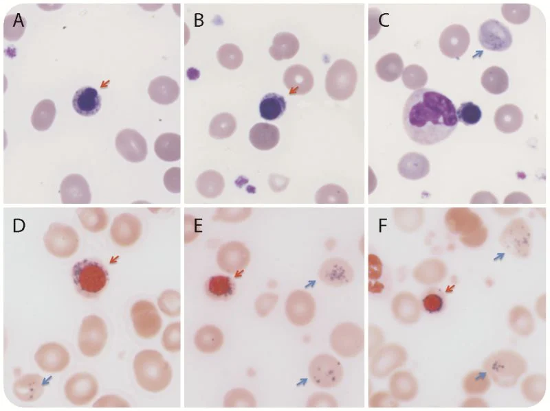
Ring sideroblasts are erythroblasts with iron-loaded mitochondria visualized only by Prussian blue staining (Perls’ reaction) as a perinuclear ring of blue granules it’s interesting to know that, in very rare conditions you can spot them transiently even in peripheral blood !
Giemsa staining of peripheral blood smears (panels A-C, original magnification ×1000) demonstrated numerous nucleated red blood cells (54/100 white blood cells) with 27 percent ring sideroblasts (red arrow). Numerous inclusions (Pappenheimer bodies, blue arrow) were also present in the erythrocytes.
Prussian blue staining confirmed the inclusions were iron positive (panels D-F, original magnification times 1000).
This is a case of hemolytic anaemia showed normoblastaemia and RBCs inclusions which were confirmed as ringed sideroblasts and iron granules (papenhiemer bodies) on Perls’ staining.”
Other posts featuring Sally Mandour on Hemostatis Today.
-
Nov 13, 2026, 14:58Lorena Arranz: The Power of Patient Engagement in Advancing MPN Research
-
Nov 13, 2026, 14:46Shehab Mohamed: SGLT2 Inhibitors in Anemia Management
-
Nov 13, 2026, 14:25Dino Mehic: Targeting TFPI in Unexplained Bleeding Disorders
-
Nov 13, 2026, 14:19Bahar Artım Esen: Rituximab in Antiphospholipid Syndrome
-
Nov 13, 2026, 14:13Tagreed Alkaltham: What If One Blood Donation Could Produce Units Of Blood?
-
Nov 13, 2026, 13:27Shirley D’Sa: Connecting European and Mayo Clinic Experts in MGCS Research and Care
-
Nov 13, 2026, 13:20Josie Sirna: Representing the Thalassemia Community at Canada’s Rare Disease Conference in Halifax
-
Nov 13, 2026, 13:14Annalisa Ruggeri: 18 Years in Haematology – Celebrating a Journey at San Raffaele
-
Nov 13, 2026, 13:08Did You Know That 20–30% of Female Hemophilia Carriers Experience Bleeding Symptoms? – Haemophilia Foundation Australia