

Daniel Ben-Mordechay: Portal Vein Thrombosis Every POCUS User Should Recognize
Daniel Ben-Mordechay, Chief Executive Officer at Orimed GTD, shared a post on LinkedIn:
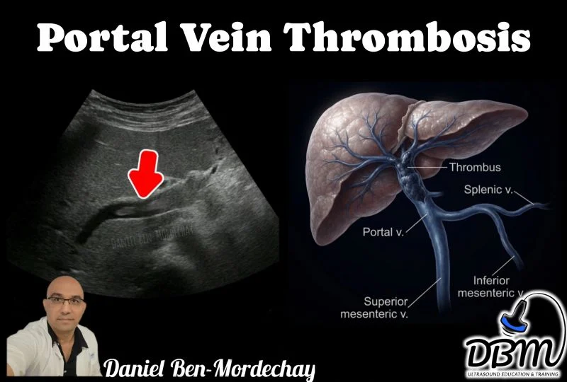
”Portal Vein Thrombosis: A Diagnosis Every POCUS User Should Recognize
Portal vein thrombosis (PVT) is a potentially serious vascular condition that can compromise hepatic blood flow and lead to portal hypertension, intestinal ischemia, or progressive liver dysfunction if not recognized and managed promptly.
Ultrasound is often the first-line imaging modality and plays a pivotal role in the initial evaluation.
On B-mode ultrasound, the thrombus typically appears as echogenic material within the lumen of the portal vein. In acute cases, the thrombus may be hypoechoic, making it more difficult to detect. The vein may appear dilated, and the thrombus can partially or completely occlude the vessel.
Color Doppler is essential in the assessment of suspected PVT.
A thrombosed segment demonstrates absent or markedly reduced intraluminal flow, while Spectral Doppler confirms the loss or significant alteration of normal hepatopetal portal venous flow.
Common risk factors include:
- Liver cirrhosis.
- Hepatocellular carcinoma and other malignancies.
- Hypercoagulable disorders.
- Pancreatitis.
- Intra-abdominal infection or inflammation.
- Recent abdominal surgery or trauma.
POCUS can rapidly raise suspicion for portal vein thrombosis, particularly in patients with unexplained abdominal pain, signs of portal hypertension, or known risk factors. However, it is important to remember that POCUS is a screening tool. Suspected PVT should always be confirmed with a comprehensive Doppler examination and, when clinically indicated, contrast-enhanced CT or MRI.
Understanding the normal sonographic appearance of the portal vein is the foundation of diagnosis. Equally important is recognizing abnormal intraluminal echoes, absent Doppler flow, and associated secondary findings. Early detection can significantly influence patient management and improve clinical outcomes.
Have you encountered portal vein thrombosis during your ultrasound examinations? Share your experience and teaching tips in the comments.
Other posts featuring Daniel Ben-Mordechay on Hemostasis Today.
-
Oct 17, 2026, 17:27John Strouse: Building Standards for Whole-Person Sickle Cell Care Through Collaboration
-
Oct 17, 2026, 17:20Jean-Jacques Kiladjian: Exploring the Future of Polycythemia Vera Treatment at MPN Horizons 2026
-
Oct 17, 2026, 17:10Habib Bennaceur: Strengthening Collaboration in Haemophilia, Sickle Cell Disease and Thalassaemia in India
-
Oct 17, 2026, 16:58Danny Hsu: Building Connections at the AUSNBled Collaborative Dinner in Auckland
-
Oct 17, 2026, 16:48Kalyan Roy: Enhancing Patient Experience Through Learning and Collaboration
-
Oct 17, 2026, 16:43Wilfried Dinh: Antiplatelet Therapy in 2026: Duration is Becoming a Dynamic Decision
-
Oct 17, 2026, 16:37Ian Setchfield: Dr. Evans Presents on Safe Anticoagulation Prescribing at East Kent Hospitals
-
Oct 17, 2026, 16:33Lewis Thomas: Connecting Through Shared Experiences at ASCAT 2026
-
Oct 17, 2026, 16:29Nilesh Jethwa: Yasmin Adverse Event Reports Exceed 23,000 Over Two-Year Period