Jan Sloves Highlights New Protocol Making Transvaginal Ultrasound a Game-Changer for PeVD
Jan Sloves, President and Consultant at Vascular Imaging Professionals LLC, reposted Francine Freitas Fernandes’s post on LinkedIn:
“We should all recognize the importance of precise, patient-centered imaging in the diagnosis and management of Pelvic Venous Disorders (PeVDs) and Chronic Pelvic Pain. The New JVSVL article lays out a Step-by-Step Protocol for Transvaginal Ultrasound (TVUS), offering a reproducible, practical method to assess pelvic veins and venous reflux, key contributors to pain & recurrent varicosities in women.
Title: Transvaginal ultrasound approach for diagnosing pelvic venous disorders
Authors: Francine Freitas Fernandes, Joana Storino, Fanilda Souto Barros, Manuella Barreto Silva, Nathalia Cardoso Oliveira, Isabela Rodrigues Tavares

Read full article here.
What Stands Out in this Work:
- TVUS enables real-time assessment of periuterine, perivaginal, gonadal, and iliac veins, with high sensitivity for reflux and post-thrombotic changes
- The protocol emphasizes provocative maneuvers (Valsalva, cough, pelvic floor contraction) to “bring out” hidden reflux that conventional imaging may miss
- This approach adds diagnostic value beyond standard transabdominal ultrasound, CT, or MR, especially for women with complex anatomy or recurrent lower limb varicosities
- TVUS’s operator-dependent challenges are offset by careful anatomical landmarking and dynamic Doppler techniques described in detail
- Crucially, PeVDs remain underdiagnosed, often presenting as pelvic pain, perineal heaviness, or even recurrent leg varicosities. Integrating TVUS into routine workups can help us close this gap and deliver targeted care
Bottom line: TVUS is a game-changer for vascular and interdisciplinary teams managing pelvic vein pathology in women. I encourage colleagues to review this protocol and consider its practical application in daily practice, particularly for complex venous patients who need more than a standard scan.
Improved imaging means improved outcomes!”
Quoting Francine Freitas Fernandes’s post:
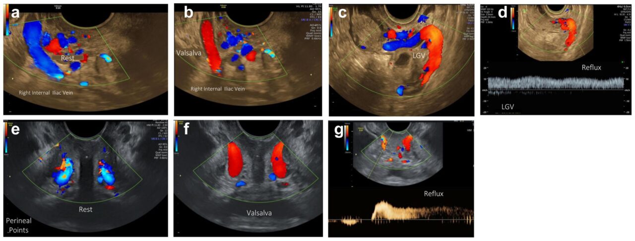
“Excited to share our ultrasound images with the world! Journal of Vascular Surgery Venous and Lymphatic Disorders
TRANSVAGINAL ULTRASOUND is the apple of our eye when it comes to PELVIC VENOUS DISORDERS, an extremely valuable diagnostic tool!
It’s definitely worth incorporating into everyday clinical practice.”
Quoting Journal of Vascular Surgery Venous and Lymphatic Disorders’s post
“Do you treat pelvic venous disorders?! Then check out this article out of Brazil where authors present a step-by-step protocol for transvaginal US for assessing pelvic veins!

Stay updated with Hemostasis Today.
-
Feb 24, 2026, 16:01Is It Safe to Briefly Stop Anticoagulation After VTE? – RPTH Journal
-
Feb 24, 2026, 15:58Anastasia Conti: Honored to Receive the Roche Foundation Grant for Independent Research in Onco-Hematology
-
Feb 24, 2026, 15:55Courtney Lawrence: Targeted Donor Screening Reduces Transfusion-Transmitted Babesia Cases
-
Feb 24, 2026, 15:30Aswin K Mohan: Practical Tips for Preparing Before, During, and After Blood Donation
-
Feb 24, 2026, 15:24Stéphanie Forté: The Hidden Burden of Stroke in Adults Living with Sickle Cell Disease
-
Feb 24, 2026, 15:13Tagreed Alkaltham: Equity in Emergency Care Through a Blood Bank Lens
-
Feb 24, 2026, 14:57Joseph Raffaele: Can Improving Mitochondrial Quality in Immune Cells Alter Immune Aging Markers?
-
Feb 24, 2026, 14:56Jan Sloves: Turning Routine Duplex into a Multiparametric Evaluation of Limb Edema
-
Feb 24, 2026, 14:31Augustina Isioma Ikusemoro: What Happens After You Donate Blood?

