

Two Emerging Mechanisms Linking Sickle Cell Disease to Thrombosis
Sickle cell disease (SCD) is not merely a microvascular occlusive disorder; it is a systemic thromboinflammatory disease in which chronic hemolysis, endothelial injury, leukocyte activation, platelet reactivity, and coagulation activation converge to create a persistent prothrombotic state.
Although vaso-occlusion remains central to SCD pathophysiology, patients also have an elevated risk of venous thromboembolism (VTE), including deep vein thrombosis and pulmonary embolism.
A systematic review and meta-analysis found increased risks of venous thrombosis and pulmonary embolism in adults with SCD, while cohort data estimate a first VTE incidence of 5.2 events per 1,000 person-years and a cumulative incidence of 11.3% by age 40 years.
Two recent studies broaden this mechanistic framework.
Wang and colleagues provide experimental evidence that cell-free sickle hemoglobin (HbS) can directly promote venous thrombosis, while Borba-Junior and colleagues identify the podoplanin (PDPN)/C-type lectin-like receptor-2 (CLEC-2) axis as a potential driver of platelet–monocyte thromboinflammation in patients with SCD.
Together, these findings reframe SCD-associated thrombosis as a multicellular process involving erythrocyte-derived danger signals, inflammatory leukocytes, platelets, endothelium, fibrin and the vasculature.
The Thromboinflammatory Landscape
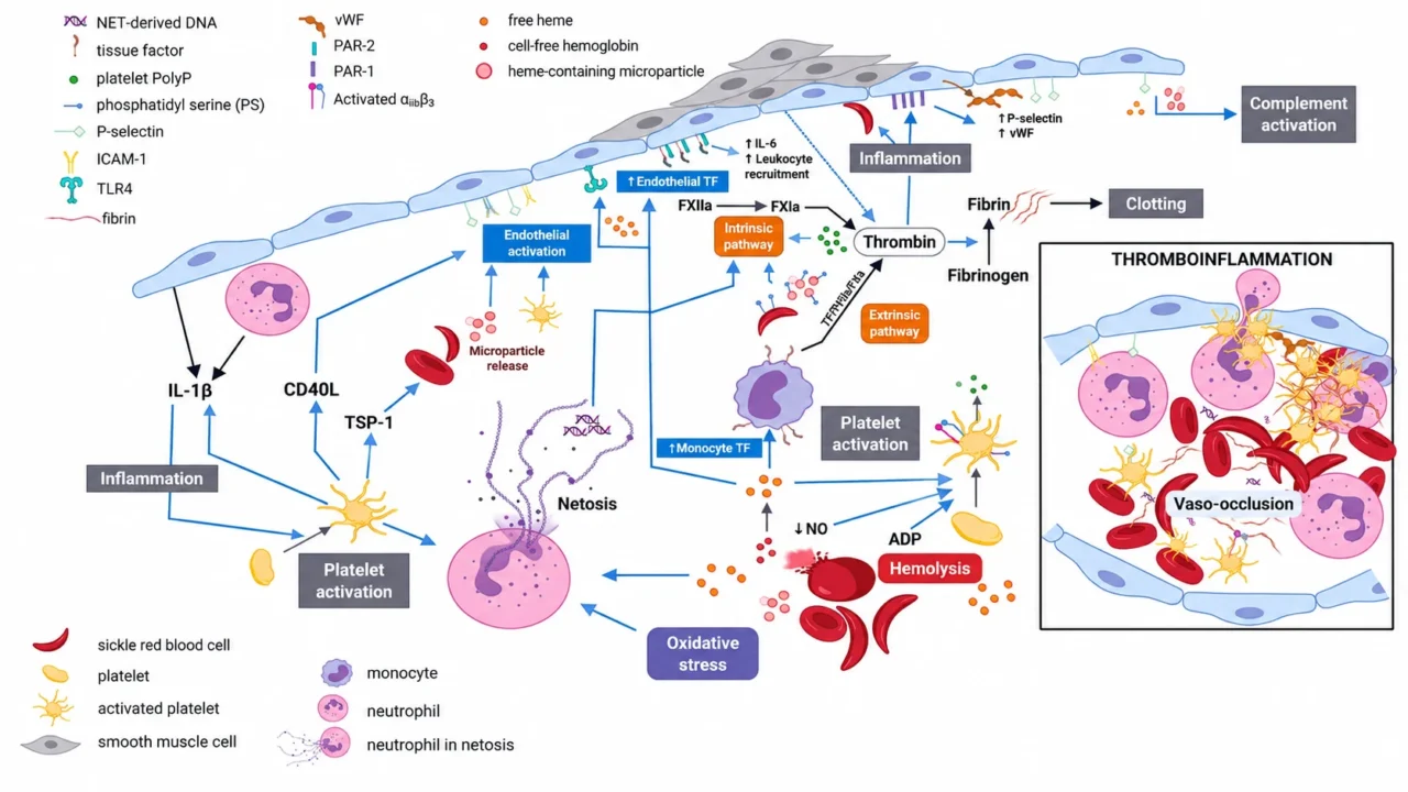
The prothrombotic phenotype of SCD reflects all three elements of Virchow’s triad: hypercoagulability, endothelial dysfunction, and altered blood flow.
Hemolysis releases cell-free hemoglobin and heme, which consume nitric oxide, generate oxidative stress, activate endothelial cells, and promote inflammatory signaling.
Sickled erythrocytes also expose procoagulant phosphatidylserine, adhere to activated endothelium, and can impair fibrinolysis by making fibrin more resistant to tissue plasminogen activator-mediated degradation.
In parallel, leukocytes and platelets amplify one another’s activation. Monocytes can express tissue factor and support thrombin generation, while activated neutrophils release neutrophil extracellular traps (NETs), which provide a scaffold for platelet adhesion, coagulation activation, and thrombus growth.
Endothelial activation further increases adhesion-molecule expression and tissue-factor release, linking inflammation directly to hemostasis.
Thus, thrombosis in SCD is best understood as thromboinflammation: the coordinated activation of innate immunity and coagulation rather than a consequence of red-cell obstruction alone.
Cell-Free HbS As A Direct Procoagulant
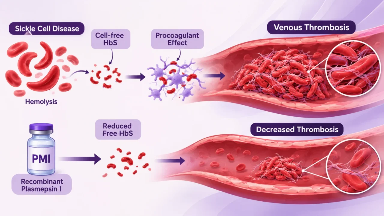
Intravascular hemolysis has traditionally been viewed mainly as a source of nitric oxide depletion and endothelial dysfunction. Wang, Silaghi, and Eitzman extend this concept by showing that cell-free HbS may also act as a direct procoagulant mediator of venous thrombosis. Their study, published in Journal of Thrombosis and Haemostasis in 2026, combined SCD mouse models with in vitro coagulation experiments.
The investigators found that addition of HbS shortened clotting time relative to normal hemoglobin, supporting a direct effect on coagulation kinetics. In SCD mice, venous thrombi exhibited a distinctive architecture: rigid, elongated erythrocytes interconnected by fibrin-like structures, with frequent erythrocyte membrane disruption.
These observations suggest that free HbS may not only initiate coagulation but also alter the physical stability and composition of the developing thrombus.
Most notably, treatment with recombinant plasmepsin I (PMI), a hemoglobin-degrading enzyme, reduced circulating free hemoglobin and decreased thrombus burden in the experimental model.
This “hemoglobinase” strategy therefore provides proof of concept that lowering cell-free HbS may reduce venous thrombogenesis.
The finding is mechanistically compelling because it connects a hallmark of SCD—chronic hemolysis—to a potentially targetable upstream driver of thrombosis.
Why HbS Matters Beyond Hemolysis
Cell-free hemoglobin and heme are increasingly recognized as danger-associated molecular patterns. Free heme can promote oxidative stress, inflammasome activation, neutrophil activation, NET formation, and endothelial adhesion, thereby creating a microenvironment favorable to thrombus formation.
Wang and colleagues add an important layer to this model: HbS may have a direct procoagulant effect, rather than functioning solely through indirect vascular and inflammatory pathways.
This distinction has therapeutic significance.
Current approaches that reduce hemolysis or improve nitric oxide biology may mitigate endothelial dysfunction, but directly degrading circulating HbS could potentially interrupt a more proximal thrombogenic signal.
However, PMI remains an experimental intervention; its safety, immunogenicity, dosing, effects on anemia and tissue oxygen delivery, and relevance to human VTE remain undefined.
The PDPN/CLEC-2 Pathway
The second emerging mechanism concerns the interface between inflammation and platelet biology. Podoplanin (PDPN) is a transmembrane mucin-like glycoprotein expressed by several cell types, including monocytes, whereas CLEC-2 is a platelet-expressed C-type lectin-like receptor. PDPN–CLEC-2 engagement can promote platelet activation, and in inflammatory settings it may help recruit platelets into complexes with leukocytes.
Borba-Junior and colleagues evaluated this pathway in 50 patients with SCD—35 with HbSS genotype and 15 with HbSC genotype—alongside 25 healthy controls. They observed higher circulating levels and cellular expression of PDPN and CLEC-2, particularly in patients with the SS genotype. Soluble PDPN and CLEC-2 were higher in HbSS than HbSC patients, and the product of soluble CLEC-2 and D-dimer was also increased in HbSS disease.
The study also found increased leukocyte–platelet aggregates expressing CLEC-2 in patients with SCD. CLEC-2 was localized to platelets, while PDPN was more evident in monocyte–platelet aggregates, consistent with a model in which PDPN-positive monocytes engage CLEC-2-positive platelets. This interaction may stabilize monocyte–platelet aggregates, amplify platelet activation, and provide a cellular bridge between inflammation and coagulation.
Evidence From Human SCD
A key strength of the PDPN/CLEC-2 study is that it was conducted in human blood samples rather than relying exclusively on experimental models. The number of CLEC-2-positive monocytes within platelet aggregates correlated with D-dimer, von Willebrand factor (VWF), and PDPN-positive monocytes—markers consistent with coagulation activation, endothelial activation, and thromboinflammation.
In vitro, blocking PDPN with a specific antibody reduced monocyte–platelet aggregate formation and platelet activation, measured by P-selectin expression.
This does not establish that PDPN blockade prevents thrombosis in patients, but it identifies a biologically plausible and experimentally testable pathway. The stronger PDPN/CLEC-2 signal in HbSS than HbSC disease also raises the possibility that genotype, hemolytic intensity, inflammatory burden, or vascular injury may modulate the relevance of this axis.
Two Complementary Mechanisms
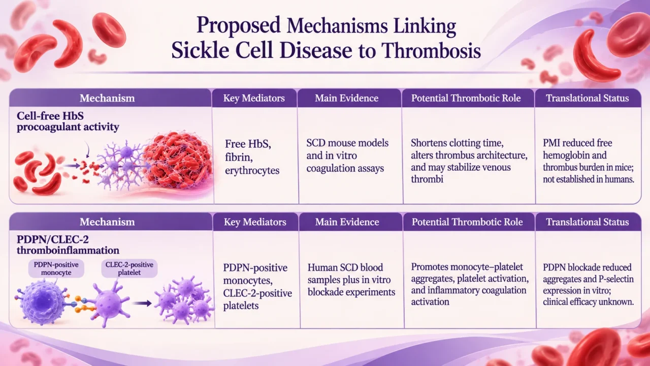
These mechanisms are complementary rather than competing. Cell-free HbS may represent an erythrocyte-derived, humoral prothrombotic signal, whereas PDPN/CLEC-2 represents a cellular pathway linking innate immune activation to platelet-driven thrombus formation. Both operate within a broader SCD milieu characterized by endothelial activation, tissue-factor expression, NET formation, platelet activation, and impaired fibrinolysis.
Therapeutic Implications
The most important implication of these studies is that SCD-associated thrombosis may be amenable to pathway-specific intervention rather than management solely through conventional anticoagulation.
If validated, hemoglobinase therapy could reduce the prothrombotic burden created by intravascular hemolysis, while PDPN/CLEC-2 targeting could disrupt a specific platelet-leukocyte inflammatory circuit.
However, neither approach is currently ready for clinical use. PMI and PDPN blockade require rigorous preclinical and clinical evaluation for efficacy, bleeding risk, off-target immune effects, dosing, and applicability across SCD genotypes and clinical contexts. In particular, interventions that alter hemostasis must be assessed not only for thrombosis prevention but also for bleeding, anemia, infection risk, and effects on vaso-occlusive crises.
Biomarker Potential
These findings also suggest candidate biomarkers for risk stratification. Circulating free hemoglobin, soluble PDPN, soluble CLEC-2, monocyte–platelet aggregates, P-selectin, D-dimer, and VWF could potentially define a “thromboinflammatory phenotype” in SCD. The observed correlations between CLEC-2-positive monocytes in aggregates and D-dimer or VWF support this concept, although correlations do not prove causality or predictive value.
Future studies should determine whether these markers identify patients at heightened risk of VTE, pulmonary hypertension, stroke, or recurrent vaso-occlusive complications. Prospective, longitudinal cohorts with standardized biomarker sampling, clinical event adjudication, and adequate representation of HbSS, HbSC, and other SCD genotypes will be essential.
A Broader Model Of SCD Thrombosis
The central message is that SCD-associated thrombosis arises from an integrated biological network. Sickled erythrocytes and hemolysis generate cell-free HbS and heme; these products promote oxidative stress, endothelial dysfunction, and inflammatory signaling.
Leukocytes and platelets then form aggregates through pathways such as PDPN/CLEC-2, while tissue factor, NETs, VWF, and fibrin help convert vascular inflammation into thrombus formation.
By identifying cell-free HbS as a direct procoagulant mediator and PDPN/CLEC-2 as a thromboinflammatory cellular axis, these studies move the field beyond the traditional view of sickled cells simply blocking vessels.
They provide a mechanistic rationale for therapies that target hemolysis-derived danger signals and platele-leukocyte crosstalk-two promising, but still investigational, directions for reducing thrombotic complications in SCD.
FAQs
1. Why Is Sickle Cell Disease A Prothrombotic Condition?
SCD creates a sustained thromboinflammatory state through chronic hemolysis, endothelial activation, platelet reactivity, leukocyte activation, tissue-factor expression, and impaired fibrinolysis. These processes act together with sickled erythrocytes to promote both vaso-occlusion and venous thrombosis.
2. How Does Cell-Free HbS Promote Thrombosis?
Cell-free sickle hemoglobin can directly shorten clotting time, alter thrombus architecture, and stabilize fibrin-rich venous thrombi. It also amplifies hemolysis-driven inflammation and endothelial dysfunction, making it both a procoagulant and a vascular danger signal.
3. What Makes HbS Different From Normal Hemoglobin In Thrombosis?
HbS is released more readily during intravascular hemolysis and appears to exert procoagulant effects beyond those of normal hemoglobin. In experimental models, HbS shortened clotting time and produced thrombi with rigid, elongated erythrocytes linked by fibrin-like structures.
4. What Is Plasmepsin I And Why Is It Important?
Plasmepsin I (PMI) is a hemoglobin-degrading enzyme investigated as an experimental “hemoglobinase” therapy. By reducing circulating free HbS, PMI lowered thrombus burden in SCD mouse models, providing proof of concept for targeting hemolysis-derived prothrombotic signals.
5. What Is The PDPN/CLEC-2 Pathway?
PDPN is a podoplanin protein expressed on monocytes, while CLEC-2 is a receptor expressed on platelets. Their interaction promotes monocyte–platelet aggregate formation, platelet activation, and inflammatory coagulation activation in SCD.
6. Why Are Monocyte–Platelet Aggregates Important In SCD?
Monocyte–platelet aggregates link innate immune activation to platelet-driven thrombus formation. In SCD, CLEC-2-positive aggregates correlate with D-dimer and von Willebrand factor, supporting their role as cellular markers and potential mediators of thromboinflammation.
7. Which Biomarkers May Identify Higher Thrombotic Risk In SCD?
Circulating free hemoglobin, soluble PDPN, soluble CLEC-2, monocyte–platelet aggregates, P-selectin, D-dimer, and VWF may define a high-risk thromboinflammatory phenotype. Their clinical predictive value still requires prospective validation in large, longitudinal SCD cohorts.
8. Are Cell-Free HbS And PDPN/CLEC-2 Independent Mechanisms?
They are complementary components of one integrated thrombotic network. Cell-free HbS represents an erythrocyte-derived humoral procoagulant signal, whereas PDPN/CLEC-2 mediates platelet–monocyte cellular crosstalk; both converge on coagulation activation and thrombus formation.
9. Can These Pathways Become Therapeutic Targets?
They are promising therapeutic targets, but remain investigational. PMI reduced thrombus burden in mice, and PDPN blockade reduced monocyte–platelet aggregates and platelet activation in vitro; neither has demonstrated clinical efficacy or safety in patients with SCD.
10. What Must Be Proven Before These Findings Change Clinical Practice?
Researchers must demonstrate that targeting cell-free HbS or PDPN/CLEC-2 prevents VTE in humans without causing bleeding, anemia, immune toxicity, or interference with vaso-occlusive physiology. Large prospective studies must also validate whether the associated biomarkers predict thrombotic events and guide treatment.
Written by Elen Avetisyan, MD.
Stay updated with Hemostasis Today.
-
Nov 21, 2026, 13:06Christine Faressa: Precision PRP and Platelet Biology in Musculoskeletal Care
-
Nov 21, 2026, 13:04Eyad Jamileh: Atrial Fibrillation – Bridging the Global Gaps in Diagnosis and Stroke Prevention
-
Nov 21, 2026, 12:49Thibault Veyron: Optimizing Bivalirudin Treatment in HIT
-
Nov 21, 2026, 12:44Paulus Kirchhof: Left Atrial Appendage Occlusion vs DOACs
-
Nov 21, 2026, 12:19Jiyeon Kim: Human Platelet Lysate – Advancing Cell Therapy and Space Research
-
Nov 21, 2026, 12:13Chaza Alaji: DVT in Early Pregnancy – Why Anticoagulation Decisions Matter
-
Nov 21, 2026, 12:06Salih Ehsan: Why Iron Therapy May Not Work
-
Nov 21, 2026, 11:59Edward Lee Carter: DOACs vs Warfarin for LV Thrombus – Are We Getting Closer to an Answer?
-
Nov 21, 2026, 11:50Abdul Mannan: When G6PD Testing Misleads During Acute Hemolysis