

William Wallace/LinkedIn
Oct 23, 2025, 09:59
William Wallace: How Collagen Builds the Body’s Structural Framework
William Wallace, Director of Product Development and Scientific Affairs at ProHealth Longevity, shared a post on LinkedIn:
“How collagen builds the body’s structural framework
Collagen makes up nearly two-thirds of all the protein in the human body, acting as the biological scaffolding that gives skin, bone, cartilage, and connective tissue their strength and resilience. It’s not a single protein but a family of more than 30 types, each tailored to a specific tissue.
- From molecule to fiber
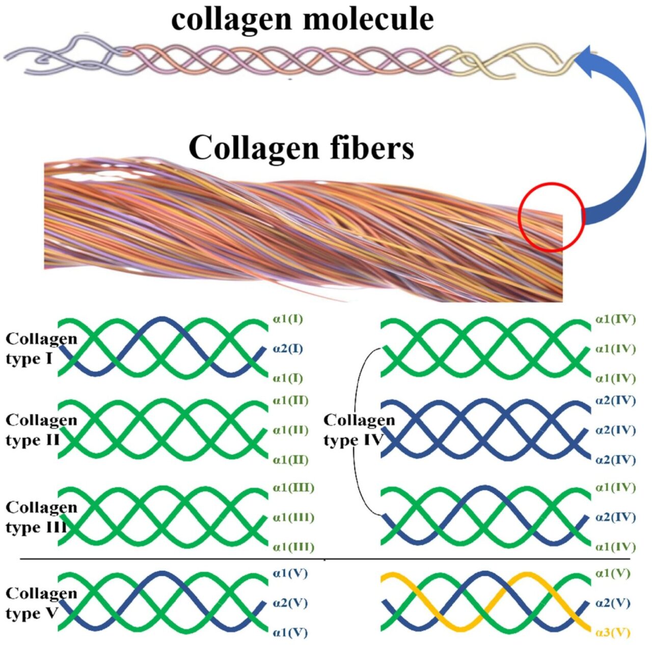
Every collagen molecule is a triple helix formed by three polypeptide chains (α-chains). Thousands of these helices bundle into fibrils, which then weave into collagen fibers—the cables that hold tissues together.
* Example: In tendons and ligaments, tightly packed type I collagen fibers resist stretching while still allowing flexibility under load. - Collagen type I — the dominant form
Type I collagen accounts for ≈90 % of the collagen in skin, ≈80 % of that in bone, and the majority in tendons and ligaments. It’s built from two α1(I) chains and one α2(I) chain, creating the strongest fibrillar form.
* Example: Its dense crosslinking provides tensile strength to the dermis and structural rigidity to bones. - Collagen type II — cartilage and joints
Type II collagen consists of three identical α1(II) chains and forms a fine mesh that cushions and stabilizes joints.
* Example: It’s the main structural protein of articular cartilage, giving flexibility while resisting compression. - Collagen type III and V — elasticity and regulation
Type III (three α1(III) chains) supports pliability in skin, blood vessels, and organs, often pairing with type I. Type V helps control fiber diameter and collagen organization, especially in the cornea, placenta, and skin.
* Example: Type III is abundant in arteries, allowing them to expand and recoil with blood flow. - Collagen type IV — the structural mesh
Type IV forms a non-fibrillar network in basement membranes, anchoring cells and filtering molecules in tissues like the kidney glomerulus and alveoli.
* Example: Its flexible lattice supports epithelial and endothelial layers without sacrificing permeability. - Nutrient support and stability
Vitamin C, copper, zinc, and iron are critical for hydroxylation and crosslinking reactions that stabilize collagen helices.
* Example: In vitamin C deficiency (scurvy), defective crosslinking leads to fragile skin, bleeding gums, and impaired wound healing.
From molecular helices to visible fibers, collagen’s hierarchy explains its dominance in the human body—it’s the primary structural protein that unites strength, flexibility, and repair across every organ system.”
Stay updated with Hemostasis Today.
-
Oct 9, 2026, 12:48Santiago Cepeda Chafla: CereBleed Simplifies Brain Hemorrhage Assessment
-
Oct 9, 2026, 12:34Abdul Muqtadir Abbasi: Pancytopenia Is a Finding, Not a Diagnosis
-
Oct 9, 2026, 12:24Mercy Lung’aho: When Diet Quality and Nutritional Biomarkers Tell Different Stories
-
Oct 9, 2026, 12:22Eugene Fan: Bridging Therapeutic Breakthroughs and Equitable Thalassaemia Care
-
Oct 9, 2026, 12:14Brittany Busse: A New Focus on Iron Deficiency Before Anemia
-
Oct 9, 2026, 12:13Akshat Jain: A New Textbook Bringing Global Perspectives on Sickle Cell Disease
-
Oct 9, 2026, 12:03Heather Case: Bridging Awareness and Action in Women’s Hemophilia Care
-
Oct 9, 2026, 11:58Brandon Luu: Can Iron Deficiency Cause Fatigue Without Anemia?
-
Oct 9, 2026, 11:51Sumaiya Dickens: Marginal Zone B Cells in Transfusion Medicine and Hemophilia A Research