


Seema Dawood/LinkedIn
May 31, 2026, 11:51
Seema Dawood: Principles of Blood Smear Examination and Interpretation
Seema Dawood, Medical Laboratory Technologist at The Aga Khan University Hospital, shared a post on LinkedIn:
“Principles of Blood Smear Examination and Interpretation
During blood smear examination and interpretation, correlation with the scattergram generated by an Automated Hematology analyzer is essential to enhance the accuracy, completeness, and consistency of Hematologic diagnosis.
- Scattergram analysis helps localize and characterize abnormal cell populations, allowing focused and targeted examination of the Peripheral blood smear.
- Supports confirmation and interpretation of analyzer flags, such as Immature Granulocytes (IG), Blast flags, Platelet clumping, and Abnormal cellular distribution patterns.
- Correlation with scattergram reduces the risk of misinterpretation, which may occur when blood smear evaluation is performed in isolation, particularly in cases with subtle or equivocal morphologic features.
Accurate Hematologic interpretation requires integration of information from both the Peripheral blood smear and the analyzer Scattergram.
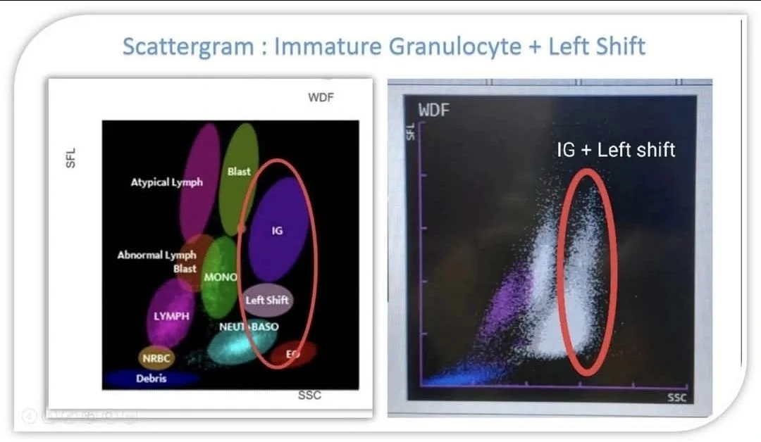
This picture: Blood Smear Confirmation (IG and Left Shift)
Blood Smear Correlation:
- As demonstrated in the image showing an IG flag with left shift, peripheral blood smear examination reveals a left shift characterized by an increased number of band forms and the presence of Immature Granulocytic cells, including Promyelocytes ,myelocytes and Metamyelocytes
- Interpretation: These findings are consistent with the presence of Immature Granulocytes (IG) associated with a left shift, which is commonly observed in Acute Infections, Inflammatory conditions, or bone marrow stimulation.”
Other posts featuring Seema Dawood on Hemostasis Today.
-
Oct 20, 2026, 16:51Suzan Nakasendwa: A Growing Stroke Challenge in Uganda
-
Oct 20, 2026, 16:38Steve Moon: How Does Smoking Cessation Duration Affect Ischemic Stroke Risk
-
Oct 20, 2026, 16:25Cedrice Mentor: Reducing Platelet Wastage Through Cold Storage
-
Oct 20, 2026, 16:09Thirunavukkarasu Angappan: Can a Purified Fibrinogen Concentrate Replace Cryoprecipitate
-
Oct 20, 2026, 16:01Kalyan Roy: Platelet Conservation Through Evidence-Based Transfusion Practice
-
Oct 20, 2026, 15:28Stacy E. Croteau: 33 Countries, One Mission – Better Bleeding Disorder Care
-
Oct 20, 2026, 15:19Teresa Barnes: What Does a Pulmonary Embolism Mean for Jalen Hurts?
-
Oct 20, 2026, 15:04Laura S.: Could Speaking Up Prevent Maternal Deaths from Thrombosis?
-
Oct 20, 2026, 14:44Pete Stibbs: VTE in Professional Athletes – When Fitness Is Not Enough