


Precious Cosmas: Sonographic Features and Practical Approach to Deep Vein Thrombosis Assessment
Precious Cosmas, Radiographer, Sonographer at University of Nigeria, shared a post on LinkedIn:
”DVT can be present in a leg without announcing itself loudly.
And sometimes, the difference between ‘I see a vein’ and ‘I understand what this vein is telling me’ comes down to knowing what to look for on ultrasound.
Let’s simplify Deep Vein Thrombosis (DVT).
So, what exactly is DVT?
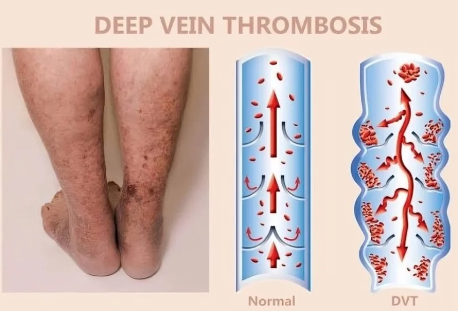
DVT is a blood clot that forms inside a deep vein, most commonly in the lower limb.
Think of a vein as a road carrying blood back to the heart.
A thrombus is like a roadblock, it can partially or completely interfere with normal blood flow.
What does DVT look like on ultrasound?
When assessing a suspected DVT, there are several important features we look for:
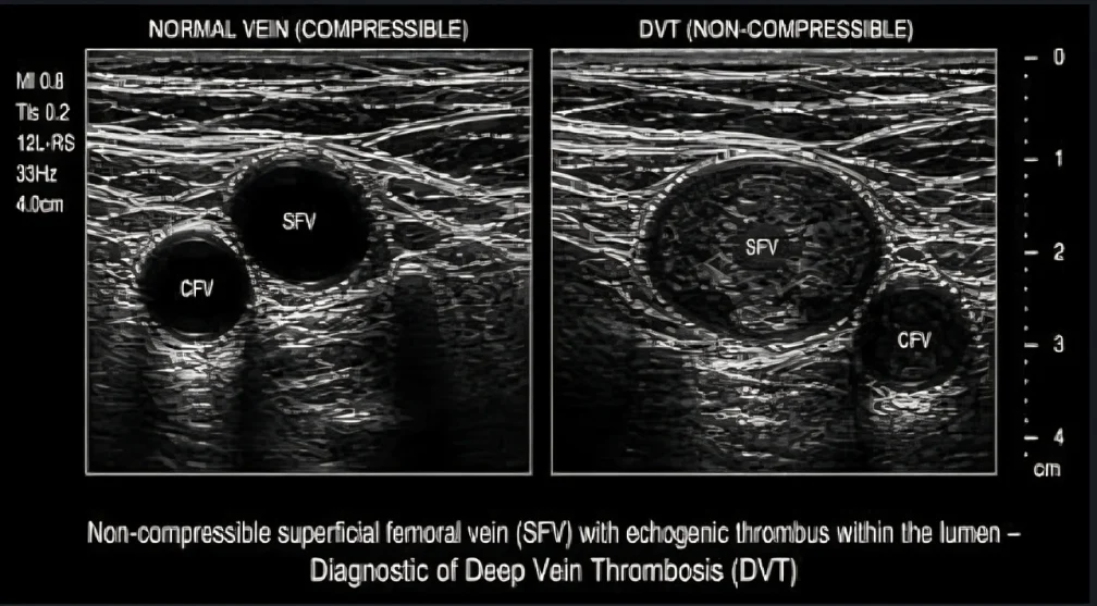
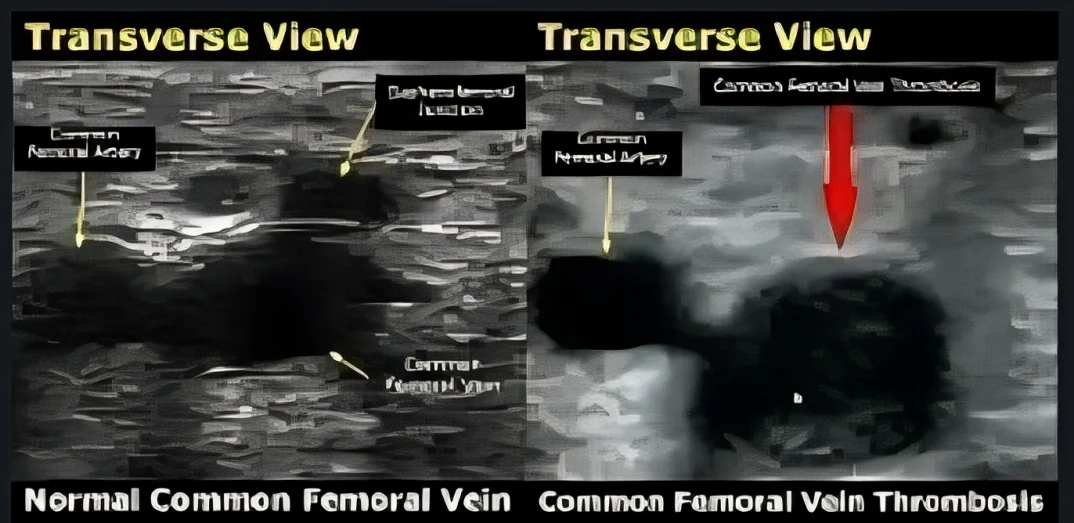
- Loss of compressibility. This is one of the most important findings. A normal vein should collapse when gentle pressure is applied with the transducer. If the vein doesn’t compress, think thrombus.
- Thrombus within the lumen. The clot may be visualized inside the vein as intraluminal material.
- Increased vein diameter. An acutely thrombosed vein appears larger than expected.
- Abnormal or absent Doppler flow. Color Doppler can demonstrate reduced or absent flow in the affected segment.
- Abnormal venous waveform. Loss of normal respiratory variation or other waveform abnormalities may provide additional information about venous obstruction.
The key takeaway:
Don’t just look at the vein. Test it.
Compression, grayscale assessment and Doppler evaluation work together to help us determine whether a vein is behaving normally.
For sonographers, mastering DVT assessment isn’t just about memorizing signs.
It’s about understanding normal venous anatomy and recognizing when the ultrasound findings deviate from normal.
Sonographers, what’s the first thing you check when evaluating a suspected DVT?
And if you’re learning vascular ultrasound, save this post, you’ll come back to it.”
Stay updated with Hemostasis Today.
-
Oct 13, 2026, 11:24Brandon Daniel D’Costa: Building Stronger Stakeholder Relationships Through Empathy and Open Communication
-
Oct 13, 2026, 11:08Renel Tique: 5th International Conference on Hemophilia and Rare Bleeding Disorders
-
Oct 13, 2026, 02:01Nour Al-Mozain: Is It Really Anti-D? – Unraveling Auto-Anti-LW in the Transfusion Laboratory
-
Oct 13, 2026, 01:51Berenice Villegas: The Role of Morphologic Evaluation in Body Fluid Differential Analysis
-
Oct 13, 2026, 01:44Debadrita Ray: Evaluating vWF and VITRO Score for Detecting CSPH in cACLD Patients
-
Oct 13, 2026, 01:34Nagesh Ramesh: Donating Blood for International Plasma Awareness Week
-
Oct 12, 2026, 21:51Sam Botchey: Orthobiologics Are Advancing but the Evidence Must Keep Pace
-
Oct 12, 2026, 21:50Serum Vitamin D, Platelet Function, and Bleeding in Primary ITP – Platelets Journal
-
Oct 12, 2026, 21:30Nika Samadzadeh Tabrizi: Looking Forward to the PACeS Trial on Anticoagulation for POAF