

Women and Hemostasis Across Hormonal Transition
How hormonal changes influence bleeding, coagulation and thrombotic risk throughout life
From the onset of menstruation to pregnancy and menopause, hormonal transitions influence the balance between bleeding and thrombosis.
Estrogen and progesterone regulate reproductive physiology, but their effects extend beyond the reproductive system, affecting coagulation proteins, natural anticoagulant pathways, fibrinolysis and the endometrium.
These changes support normal menstruation and help prepare the body for childbirth. However, they may also reveal an underlying bleeding disorder or contribute to venous thromboembolism (VTE).
For clinicians, the challenge is to distinguish physiological adaptation from clinically significant hemostatic dysfunction.
Heavy menstrual bleeding may be the first sign of an inherited bleeding disorder, while pregnancy and the postpartum period introduce important thrombotic risks. Later in life, menopausal hormone therapy requires careful consideration of its benefits and potential effects on coagulation.
Understanding women’s hemostasis therefore requires attention to both sides of the hemostatic balance: the risk of bleeding and the risk of thrombosis.
How Hormones Influence Hemostasis
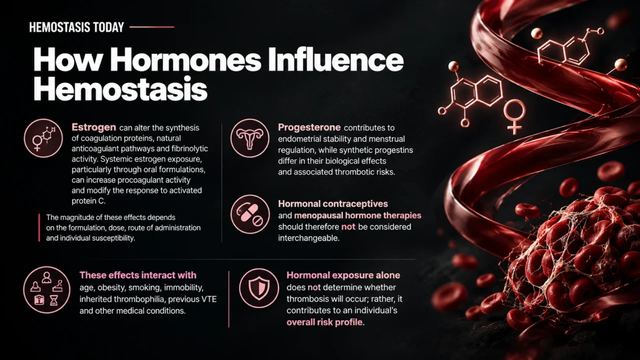
Estrogen can alter the synthesis of coagulation proteins, natural anticoagulant pathways and fibrinolytic activity. Systemic estrogen exposure, particularly through oral formulations, can increase procoagulant activity and modify the response to activated protein C.
The magnitude of these effects depends on the formulation, dose, route of administration and individual susceptibility.
Progesterone contributes to endometrial stability and menstrual regulation, while synthetic progestins differ in their biological effects and associated thrombotic risks.
Hormonal contraceptives and menopausal hormone therapies should therefore not be considered interchangeable.
These effects interact with age, obesity, smoking, immobility, inherited thrombophilia, previous VTE and other medical conditions.
Hormonal exposure alone does not determine whether thrombosis will occur; rather, it contributes to an individual’s overall risk profile.
Menstruation and Heavy Menstrual Bleeding
Menstruation is a tightly regulated hemostatic process. When pregnancy does not occur, progesterone levels decline, triggering endometrial breakdown and shedding.
Local vasoconstriction, platelet activity, coagulation, fibrinolysis and tissue repair work together to limit blood loss while allowing the endometrium to regenerate.
Heavy menstrual bleeding (HMB) is defined by its impact on physical, social, emotional, or material quality of life.
Although gynecological conditions such as fibroids, adenomyosis, and ovulatory dysfunction are important causes, an underlying hemostatic disorder may also be responsible.
When Menstrual Bleeding Signals a Hemostatic Disorder
Von Willebrand disease (VWD), platelet function disorders, and less common coagulation factor deficiencies may first become apparent at menarche.
HMB is particularly important when accompanied by recurrent epistaxis, easy bruising, prolonged bleeding after dental procedures, excessive surgical bleeding, postpartum hemorrhage, or a family history of abnormal bleeding.
The NICE guideline recommends considering coagulation disorder testing, including assessment for VWD, when HMB has been present since menstruation began and the personal or family history suggests an inherited bleeding disorder. A complete blood count is also recommended for women presenting with HMB.
Depending on the clinical presentation, assessment may include a detailed bleeding history, complete blood count, ferritin, and targeted coagulation studies.
VWD evaluation typically includes von Willebrand factor (VWF) antigen, VWF activity, and factor VIII activity. When initial results are normal but clinical suspicion remains high, further assessment for platelet function disorders, other factor deficiencies, fibrinolytic disorders, and connective tissue or hypermobility disorders may be appropriate.
The timing of testing matters. VWF and factor VIII levels can increase during acute bleeding, inflammation, physiological stress, and pregnancy, potentially masking mild VWD. A normal result obtained under these conditions does not always exclude the diagnosis.
Recognizing the hemostatic contribution to HMB is important because recurrent blood loss can lead to iron deficiency, anemia, fatigue, and reduced quality of life. Treating menstrual symptoms without identifying an underlying bleeding disorder may also leave women vulnerable during surgery, pregnancy, and childbirth.
An Emerging Non-Hormonal Approach
The LUNA study (NCT07326722) is evaluating transcutaneous auricular nerve stimulation in people aged 14–45 years with HMB, including those with mild type 1 VWD or low VWF levels. The study will assess menstrual blood loss and symptoms over three treatment cycles.
This device-based approach represents an area of ongoing investigation into non-hormonal options for managing menstrual bleeding.
Hormonal Contraception and Thrombotic Risk
Hormonal contraception can influence both menstrual bleeding and thrombosis. Combined hormonal contraceptives, which contain estrogen and a progestin, can regulate menstrual cycles and reduce HMB. However, estrogen-containing formulations are associated with an increased risk of VTE.
The magnitude of this risk varies according to estrogen dose, progestin type, and individual factors, including previous VTE and inherited thrombophilia. Contraceptive selection should therefore consider both the therapeutic benefits for menstrual symptoms and the individual’s thrombotic risk.
Progestin-only methods, particularly levonorgestrel-releasing intrauterine systems, are valuable alternatives for many women because they can reduce menstrual blood loss without systemic estrogen exposure.
Nevertheless, progestin-only formulations do not all have identical risk profiles, and the appropriate option depends on the patient’s clinical circumstances.
Estetrol-Based Contraception and Hemostatic Effects
Estetrol/drospirenone (E4/DRSP) has attracted attention as a combined oral contraceptive with potentially different effects on hemostasis. A 2026 systematic review and meta-analysis of randomized comparisons reported higher protein S activity and lower fibrinogen, plasminogen, and sex hormone-binding globulin levels with E4/DRSP than with ethinyl estradiol/drospirenone.
These findings suggest a potentially more favorable profile across selected laboratory markers. However, changes in surrogate hemostatic markers do not establish a reduction in clinical VTE events. Larger, long-term comparative studies are needed before concluding that this formulation carries a lower risk of thrombosis.
The key clinical distinction is between a favorable laboratory profile and proven clinical safety.
Pregnancy and the Postpartum Period
Pregnancy produces one of the most pronounced physiological shifts toward hypercoagulability. These adaptations help prepare the body for placental separation and delivery, when substantial blood loss may occur.
Several changes contribute to this state:
- Increased fibrinogen and coagulation factors, including factors VII, VIII, and X.
- Increased VWF and factor VIII levels.
- Reduced free protein S and acquired resistance to activated protein C.
- Reduced fibrinolytic activity, partly through changes in plasminogen activator inhibitors.
- Venous stasis resulting from compression of pelvic veins by the enlarging uterus.
Together, these changes support hemostasis during childbirth but also increase the risk of deep vein thrombosis and pulmonary embolism. Pregnancy-associated VTE remains an important cause of maternal morbidity and mortality.
Individualized Thromboprophylaxis During Pregnancy
The 2025 European Society of Cardiology guidelines on cardiovascular disease in pregnancy emphasize documented VTE risk assessment and individualized thromboprophylaxis.
For suspected VTE during pregnancy, the guidelines recommend initiating therapeutic low-molecular-weight heparin (LMWH) immediately, even before imaging, until VTE is excluded or confirmed.
LMWH remains the preferred anticoagulant for most VTE prevention and treatment indications during pregnancy because of its favorable maternal safety profile compared with unfractionated heparin.
Thromboprophylaxis decisions should consider previous VTE, the type of thrombophilia, cesarean delivery, obesity, immobility, infection, and other clinical factors. Thrombophilia alone does not mean that every pregnant woman requires anticoagulation; the expected benefit must be balanced against bleeding risk.
Why the Postpartum Period Matters
Delivery does not immediately restore the pre-pregnancy hemostatic state. Persistent coagulation changes, vascular injury, inflammation and reduced mobility can continue to promote thrombosis after childbirth.
VTE risk is particularly elevated during the first six weeks postpartum.
At the same time, childbirth presents a substantial bleeding challenge.
Postpartum hemorrhage may result from uterine atony, retained placental tissue, genital tract trauma, abnormal placentation, or coagulation abnormalities.
Women with inherited bleeding disorders require coordinated obstetric and hematological planning.
In VWD, VWF and factor VIII levels often rise during pregnancy, although the response varies by subtype and individual. These levels can fall after delivery, leaving some women vulnerable to delayed postpartum bleeding.
Tranexamic Acid and Emerging Evidence
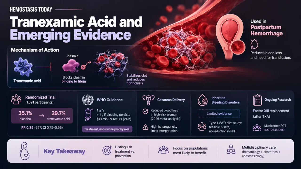
Tranexamic acid is an antifibrinolytic agent used in the management of postpartum hemorrhage.
A randomized, placebo-controlled trial involving 1,691 participants found that prophylactic tranexamic acid reduced postpartum hemorrhage from 35.1% to 29.7% (relative risk 0.85; 95.2% confidence interval 0.75–0.96).
However, this finding should be distinguished from current World Health Organization guidance, which supports intravenous tranexamic acid as treatment for postpartum hemorrhage rather than routine universal prophylaxis.
The recommended treatment dose is 1 g intravenously, with a second 1 g dose if bleeding persists after 30 minutes or recurs within 24 hours.
A 2026 meta-analysis also reported reduced blood loss with prophylactic tranexamic acid during cesarean delivery in high-risk pregnant women, although substantial heterogeneity across studies limits the interpretation of the findings.
Evidence specific to inherited bleeding disorders remains limited.
In a small pilot study involving women with type 1 VWD, tranexamic acid combined with recombinant VWF was feasible and safe but did not reduce postpartum hemorrhage compared with recombinant VWF alone.
A multicenter randomized trial is also investigating whether early factor XIII replacement, added to standard care after tranexamic acid, can reduce measured blood loss within 24 hours in women with postpartum hemorrhage.
These findings highlight the need to distinguish treatment of established hemorrhage from preventive strategies and to evaluate interventions in the populations most likely to benefit.
For women with known or suspected bleeding disorders, collaboration among hematology, obstetrics and anesthesiology is essential, particularly during pre-pregnancy assessment, delivery planning, and postpartum surveillance.
Perimenopause, Menopause and Hemostatic Changes
The menopausal transition is characterized by fluctuating ovarian hormone production, irregular ovulation and eventually a sustained decline in estrogen. These changes can alter menstrual regularity and bleeding patterns.
During perimenopause, unpredictable or heavy uterine bleeding may arise from hormonal fluctuations.
However, hormonal changes should not automatically be considered the only explanation.
Fibroids, endometrial abnormalities, thyroid disease, anticoagulant therapy and bleeding disorders may also contribute. Persistent or concerning bleeding requires appropriate gynecological assessment.
After menopause, menstrual bleeding ceases. Any postmenopausal bleeding warrants clinical evaluation rather than being attributed to hormonal changes alone.
Menopausal Hormone Therapy and VTE
Menopausal hormone therapy can improve symptoms associated with estrogen deficiency, but its effects on coagulation depend partly on the route of administration.
Oral estrogen undergoes first-pass hepatic metabolism and has a greater effect on several coagulation proteins than transdermal estradiol.
Observational evidence suggests that transdermal estradiol is associated with a more favorable VTE risk profile than oral estrogen, particularly in women with additional thrombotic risk factors.
The 2025 Menopausal Hormone Therapy Guidelines emphasize that VTE risk is influenced by age, time since menopause, and treatment route. Oral estrogen is contraindicated in women with a history of VTE or high-risk profiles under the guidance described in the source, while transdermal estrogen may be a safer alternative for selected high-risk women.
Transdermal administration should not be considered risk-free. Decisions about menopausal hormone therapy must account for previous VTE, cardiovascular risk, age, time since menopause, and other contraindications. Women with a uterus generally also require appropriate endometrial protection when using systemic estrogen.
The objective is not to avoid hormone therapy in every woman, but to select treatment through individualized assessment of symptoms, expected benefits, and potential risks.
Interpreting Hemostatic Tests Across Hormonal Transitions
Hormonal and physiological changes can influence laboratory results, making clinical context essential when investigating bleeding or thrombosis.
During acute bleeding, inflammation, or pregnancy, VWF and factor VIII levels may increase, potentially masking mild VWD.
Pregnancy also increases fibrinogen and several coagulation factors while reducing protein S levels. Oral estrogen exposure can alter hepatic synthesis of coagulation proteins and affect natural anticoagulant pathways.
The postpartum period presents a different challenge: coagulation activation and thrombotic risk may persist after delivery, even as pregnancy-associated physiological changes begin to resolve.
Routine coagulation tests must also be interpreted carefully.
Prothrombin time and activated partial thromboplastin time are useful in selected investigations, but normal results do not exclude VWD or platelet function disorders. Similarly, routine coagulation tests cannot reliably predict an individual’s VTE risk.
Testing should therefore answer a specific clinical question. When initial results are normal but the bleeding history remains strongly suggestive of an inherited disorder, repeat testing or specialized investigations may be necessary.
Toward Individualized Women’s Hemostatic Care
Effective management across hormonal transitions requires a coordinated approach to bleeding and thrombosis. Clinical assessment should consider menstrual and other bleeding symptoms, previous thrombotic events, family history, hormonal exposure, relevant comorbidities and appropriately selected laboratory investigations.
Recognizing menstrual health as part of routine hematological assessment is particularly important. Asking about HMB can uncover an inherited bleeding disorder years before a woman undergoes surgery or experiences pregnancy-related complications.
Likewise, assessing thrombotic risk during pregnancy and before prescribing estrogen-containing therapies can support safer clinical decisions.
The emerging direction of women’s hemostatic care is increasingly individualized: identifying bleeding disorders earlier, selecting hormonal treatments with their hemostatic effects in mind, and directing thromboprophylaxis toward women most likely to benefit.
Understanding these transitions is essential to recognizing when physiological changes become clinically significant-and to ensuring that both bleeding and thrombosis receive appropriate attention.
FAQ
1. How does estrogen affect coagulation?
Estrogen can alter several procoagulant proteins, natural anticoagulant pathways, and fibrinolytic activity. Its effects depend on the formulation, dose, route of administration, and individual risk factors.
2. Why can heavy menstrual bleeding indicate a bleeding disorder?
HMB may be an early manifestation of VWD, platelet function disorders or other coagulation abnormalities, particularly when it begins at menarche or occurs alongside other bleeding symptoms.
3. Does pregnancy increase the risk of venous thromboembolism?
Pregnancy induces a physiological hypercoagulable state, while venous stasis and vascular changes further increase the risk of VTE. The postpartum period is particularly important because thrombotic risk remains elevated after delivery.
4. Do all hormonal contraceptives carry the same thrombotic risk?
Thrombotic risk varies by contraceptive type and formulation. Estrogen-containing contraceptives are associated with an increased risk of VTE, whereas progestin-only methods generally have a lower risk. Individual medical history and pre-existing thrombotic risk factors should guide contraceptive selection.
5. Can pregnancy mask von Willebrand disease?
Yes. VWF and factor VIII levels often increase during pregnancy, potentially obscuring mild VWD. Repeat testing outside pregnancy may be necessary to establish the diagnosis.
6. Is transdermal estrogen associated with a lower VTE risk than oral estrogen?
Observational evidence suggests that transdermal estradiol has a more favorable thrombotic profile than oral estrogen. However, treatment decisions must still account for individual risk factors and contraindications.
7. Should all pregnant women with thrombophilia receive LMWH?
No. Thromboprophylaxis should be individualized according to previous VTE, the type of thrombophilia, additional risk factors, and bleeding risk.
8. Why is the postpartum period particularly important?
Coagulation changes persist after delivery, and vascular injury, inflammation, and venous stasis can contribute to continued thrombotic risk. Bleeding complications may also occur, particularly in women with inherited bleeding disorders.
9. Can hormonal therapy help women with heavy menstrual bleeding?
Yes. Depending on the cause and individual risk profile, combined hormonal contraception, progestin-only treatments, or a levonorgestrel-releasing intrauterine system may reduce menstrual blood loss.
10. When should bleeding be investigated rather than attributed to hormonal changes?
HMB beginning at menarche, recurrent unexplained iron deficiency, excessive bleeding after procedures, persistent abnormal uterine bleeding, and any postmenopausal bleeding warrant appropriate clinical assessment.
Written by Elen Avetisyan, MD.
-
Oct 21, 2026, 16:19Vanessa Carvalho: New Research on Platelet-Derived Extracellular Vesicles in Preeclampsia
-
Oct 21, 2026, 15:35Danny Hsu: Pharma’s 2025 Blockbusters Through a Hematologist’s Lens
-
Oct 21, 2026, 15:12Yue Zhang: Flow Cytometry for Translational Immunology and Cancer Research
-
Oct 21, 2026, 14:58Radoš Stefanović: How Red Blood Cells Adapt After Exercise
-
Oct 21, 2026, 14:08Vera Ghali: The Role of Hepcidin Assays in Diagnosing Iron Disorders
-
Oct 21, 2026, 13:52Negar Asdaghi: Could Cilostazol Improve Hematoma Clearance After ICH
-
Oct 21, 2026, 10:33Karen Schreiber: Beyond Clinical Outcomes – The Voices of Women with APS
-
Oct 21, 2026, 10:11Tau Braun: Clots Through the Copper-Zinc Model
-
Oct 21, 2026, 09:55Cristina Figueroa Villalba: Celebrating Excellence in Transfusion Medicine