

Hina Wajid: Venae Comitantes and DVT Ultrasound Assessment
Hina Wajid, Founder at Scan Point, shared a post on LinkedIn:
“Can One Vein Have DVT While the Other Is Normal?
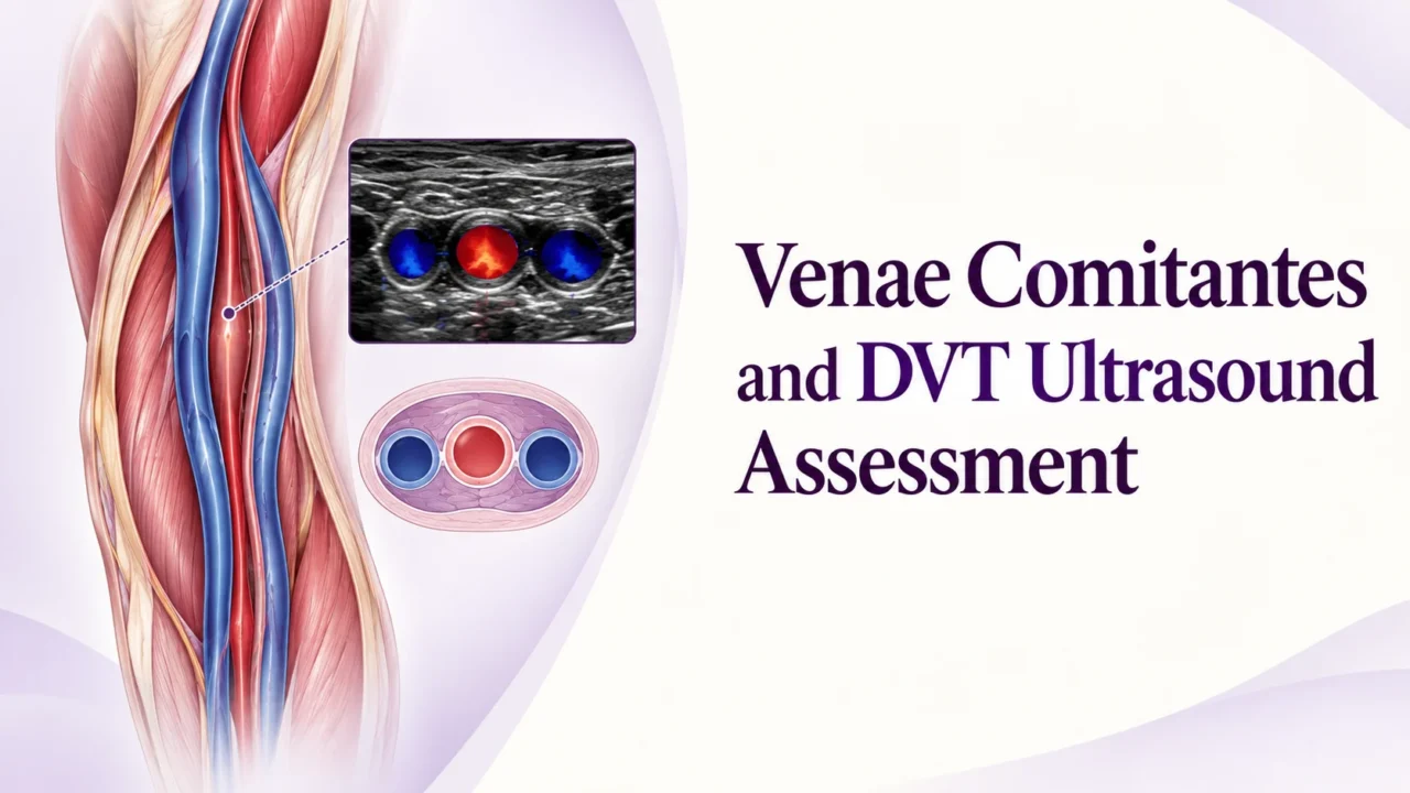
One Artery Can Have TWO Accompanying Veins
These paired veins are called Venae Comitantes!
They are commonly found alongside deep arteries in the upper and lower limbs.
Where Do We Find Them?
Upper Limb
Radial artery plus Radial veins
Ulnar artery plus Ulnar veins
Brachial artery plus Brachial veins
Lower Limb
Anterior tibial artery plus Anterior tibial veins
Posterior tibial artery plus Posterior tibial veins
Fibular artery plus Fibular veins
Ultrasound Appearance
In transverse view, you may see:
Vein Artery Vein (V A V)
Artery: Pulsatile
Veins: Normally compressible
Color Doppler: Helps identify blood flow
Important RVT Point!
During DVT scanning, remember that one accompanying vein may contain thrombus while the other remains patent.
Always examine both veins when present!
Memory Line: One VIP Artery plus Two Venous Bodyguards!
Join our ultrasound group for more updates:
Proceed to the video attached to the post.
Stay updated with Hemostasis Today.
-
Oct 24, 2026, 07:27The Importance of Emotional and Social Support in VWD – EHC
-
Oct 24, 2026, 07:18Keith Lamb: Prone Positioning early during ECMO improves outcomes
-
Oct 24, 2026, 05:59Nour Al-Mozain: Therapeutic Plasma Exchange in Sickle Cell Disease
-
Oct 24, 2026, 05:55Flora Peyvandi: A New Way of Thinking About Microthrombosis and Organ Dysfunction
-
Oct 24, 2026, 05:54Dwana Calhoun: How Emerging Tobacco Products Affect Hemostasis
-
Oct 24, 2026, 05:53Fanny Delettre: Sharing Clinical Research for the Continuous Improvement of Transfusion Practices
-
Oct 24, 2026, 05:51Raising Awareness of Acquired Haemophilia A Through Jenny’s Story – Haemophilia Foundation Australia
-
Oct 24, 2026, 05:50Narender Kumar: Honoured to Receive the THANZ International Travel Grant 2026 at Blood 2026
-
Oct 24, 2026, 05:46James Posada: Demonstrating the Benefits of Nuclease Therapy in Ischemic Stroke