


Ney Carter Borges: Vulnerable Plaque – Imaging and Clinical Significance
Ney Carter Borges, Member Cardiologist of Global Physician Association at Cleveland Clinic Florida, shared a post on LinkedIn:
“Vulnerable Plaque: Imaging and Clinical Significance
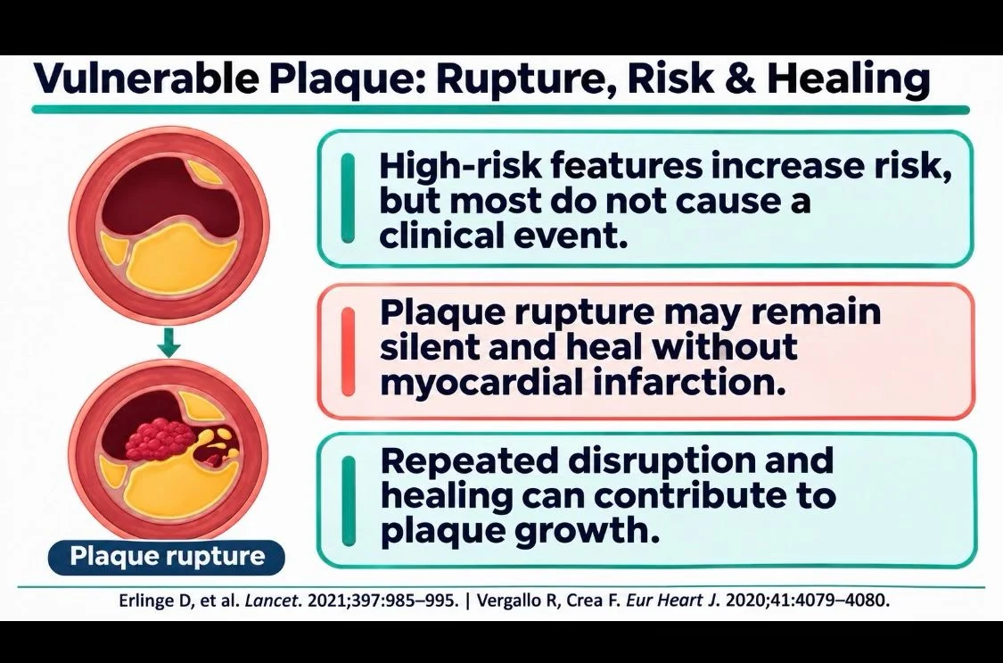
Vulnerable plaque describes atherosclerotic lesions with features that may favor disruption and thrombosis, regardless of severe luminal narrowing.
Large lipid-rich cores, thin fibrous caps, and inflammation are classic findings; rupture, erosion, or calcified nodules may trigger events.
Many plaque disruptions remain silent, heal, and contribute to lesion growth, so vulnerability does not mean an imminent infarction.
Imaging is complementary.
CCTA noninvasively depicts plaque burden, low attenuation, and remodeling; OCT uses light to assess the cap and surface; IVUS uses ultrasound to measure plaque burden and vessel remodeling; NIRS quantifies lipid content, often with IVUS; ¹⁸F-NaF PET highlights active microcalcification.
Except CCTA and PET, these require an intracoronary catheter; none precisely predicts which plaque will cause an event.
SCOT-HEART, PROSPECT II, and CLIMA linked imaging features to events, but individual prediction remains limited.
PRE¹⁸FFIR showed secondary prognostic associations, while its primary composite outcome was not significant.
Management centers on intensive lipid lowering and global risk-factor control.
PREVENT supports preventive PCI in selected patients with FFR-negative lesions and multiple high-risk features; it does not justify routine screening or stenting of all vulnerable plaques.
Its two-year composite was 0.4% versus 3.4%.
Imaging should guide interpretation, not replace assessment of atherosclerotic risk.”
Other posts featuring Ney Carter Borges on Hemostasis Today.
-
Nov 14, 2026, 10:00Benjamin Tur: Putting Iron Deficiency Anemia on the Global Agenda at the Paris Peace Forum’s Child Summit
-
Nov 14, 2026, 09:48Jack Shuang Hou: FDA Approves Bayer’s Sunharbo – First Factor XIa Inhibitor for Secondary Stroke Prevention
-
Nov 14, 2026, 09:42Samwel Mikaye: What Every Patient Should Know About DVT Symptoms and Treatment
-
Nov 14, 2026, 09:37Chronic Preoperative Corticosteroid Use and Postoperative Venous Thromboembolism Risk – Thrombosis and Haemostasis Journal
-
Nov 14, 2026, 09:25Carmelo Raffo: How Much Antithrombotic Therapy is Truly Necessary after LAAC?
-
Nov 14, 2026, 09:19Mary Cushman: Excellent to Have Jordan Shavit Visit Us for a Virtual Lab Meeting Today
-
Nov 14, 2026, 09:15Chancellor Donald: Heading to ASH 2026 in New Orleans? Let’s Connect on Faculty Opportunities at Tulane
-
Nov 14, 2026, 09:11Sajja Venkatesh: FDA Approved Asundexian, the First Factor XIa Inhibitor
-
Nov 14, 2026, 09:05Selina Evans: How Can Hospitals Make VTE Risk Assessment and Prevention Truly Routine?