


Ahmed Alotaibi: Which Macrophage Receptor ‘eats’ anti-E and Anti-K Coated RBCs?
Ahmed Alotaibi, Senior Specialist (Blood bank and blood transfusion) at Afif General Hospital – Riyadh Third Health Cluster (Ministry of Health), shared a post on LinkedIn:
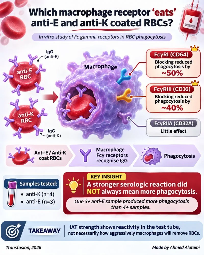
“Which macrophage receptor “eats” anti-E and anti-K coated RBCs?
When a clinically significant IgG alloantibody coats an RBC, we often say:
‘The cell will be cleared by macrophages.’
But how does the macrophage actually recognise it?
A new 2026 Transfusion study investigated this using anti-E, anti-C and anti-K coated RBCs.
Macrophages carry several Fcγ receptors that can recognise the Fc portion of IgG:
- FcγRI – CD64
- FcγRIIA – CD32A
- FcγRIII – CD16
The researchers blocked each receptor and measured how much RBC phagocytosis remained.
With standard anti-E and anti-C:
- Blocking FcγRI/CD64 reduced phagocytosis by about 50%.
- Blocking FcγRIII/CD16 reduced it by about 40%.
- Blocking FcγRIIA/CD32A had little effect.
Patient-derived antibodies showed a similar pattern.
They tested samples from:
- 4 patients with anti-K
- 3 patients with anti-E
For anti-K, blocking FcγRIII produced the greatest overall reduction in phagocytosis.
But one finding was especially interesting.
Among the anti-E samples:
A 3+ IAT reaction produced substantially more RBC phagocytosis than samples reacting 4+.
That is an important reminder:
A stronger serologic reaction does not necessarily mean greater RBC destruction.
Reaction strength tells us what happens in the test tube.
Actual RBC clearance also depends on factors such as the antibody itself, how it coats the RBC and how macrophage Fc receptors interact with it.
So the pathway is roughly:
Anti-E / anti-K binds RBC – IgG Fc recognised by CD64 and CD16 – macrophage phagocytosis
This was an in-vitro macrophage study, so it does not directly prove how much haemolysis would occur in a patient.
But it helps explain what happens after a clinically significant alloantibody finds its target.
The antibody marks the RBC.
The macrophage still has to decide whether to eat it.”
Stay updated with Hemostasis Today.
-
Nov 3, 2026, 16:53Jan Sloves: Key Factors for Achieving Durable Patency in Advanced Venous Stenting
-
Nov 3, 2026, 16:45Nina Weber: Review on Kidney Injury After Stem Cell Transplantation
-
Nov 3, 2026, 16:36Chris Baugh: EMplify Podcast Explores Antithrombotic Associated Bleeding in the Emergency Department
-
Nov 3, 2026, 16:32Carlo Zaninetti: Highlights from the XXIX Congress of the Italian Society on Haemostasis and Thrombosis
-
Nov 3, 2026, 15:10Lady Joan Branson’s Story – The Hidden Danger of Hospitalization
-
Nov 3, 2026, 14:38Emna Hammami: Exploring the Haemophilia Treatment Landscape at the EHC New Technologies Conference
-
Nov 3, 2026, 14:20EHC Marks the Final Edition of Its New Technologies Conference in Bucharest
-
Nov 3, 2026, 14:15Rhiannon Morris: Awarded CASS Foundation Travel Grant to Present Research at ASH 2026
-
Nov 3, 2026, 14:14Edward Barnard: New Study Identifies an Association Between O Negative Blood Group and Trauma Mortality