

Complement – Coagulation Crosstalk in Thrombosis
When we talk about thrombosis, the conversation usually starts with platelets, coagulation factors, thrombin and fibrin.
These are, after all, the systems we have traditionally associated with clot formation.
But the picture is becoming more complicated.
The immune system, and particularly the Complement Cascade, appears to play a much more important role in thrombosis than previously appreciated.
The relationship is not simply a matter of complement being activated alongside coagulation.
The two systems can directly influence one another.
Complement activation can stimulate endothelial cells and platelets toward a more prothrombotic state, while several coagulation-related proteases can affect complement activity in return.
At the same time, neutrophils and neutrophil extracellular traps (NETs) provide another connection between inflammation and clot formation.
Together, these interactions create a biological link between immune activation and thrombosis – a process increasingly described as immunothrombosis.
This helps put thrombosis in a broader context.
In diseases such as Complement-Mediated Thrombotic Microangiopathies, autoimmune disorders, cancer, severe infections, and cardiovascular disease, inflammation and thrombosis often develop side by side.
In some cases, they may actually reinforce each other through this interconnected network of complement, coagulation, platelets, endothelial cells, and immune cells.
The growing understanding of this relationship is also changing the therapeutic conversation.
Instead of looking at coagulation and complement as completely separate targets, researchers are asking whether the interaction between them can itself be targeted.
The challenge is finding a way to interrupt the pathological thrombo-inflammatory response while preserving the complement activity needed for host defense and the coagulation mechanisms required for normal hemostasis.
Complement Cascade
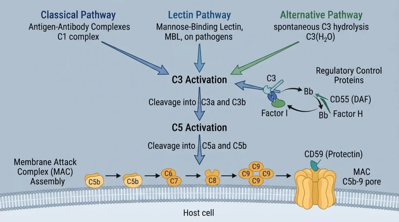
Complement can be activated through three main pathways:
- The classical pathway
- Lectin
- Alternative pathway
They begin in different ways, but ultimately converge on the activation of C3, a central component of the system.
Once C3 is activated, the cascade can rapidly amplify, generating fragments such as C3a and C3b and eventually leading to activation of C5.
This produces C5a, a potent inflammatory mediator, and C5b, which initiates assembly of the membrane attack complex (C5b–9).
Under normal conditions, this powerful system is tightly controlled by complement control proteins (CCP) such as CD55, CD59 and factor H.
This balance is important because complement needs to respond quickly to danger without causing unnecessary damage to healthy tissue.
Coagulation
The coagulation system is often introduced as a sequence of reactions designed to stop bleeding.
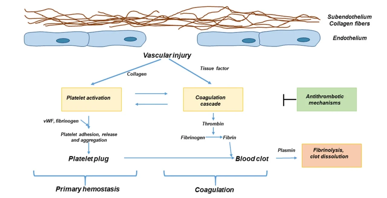
Following vascular injury, exposure of tissue factor and activation of coagulation factors initiate a series of enzymatic reactions that ultimately generate thrombin.
Thrombin then converts fibrinogen into fibrin, while also activating platelets and other coagulation components, allowing the developing thrombus to grow and become stabilized.
This process is tightly regulated.
Natural anticoagulant mechanisms, including antithrombin, the protein C–protein S system, and tissue factor pathway inhibitor, limit coagulation to the site and extent of vascular injury.
At the same time, the fibrinolytic system, particularly plasmin, helps remove fibrin once it is no longer needed. Under normal conditions, these mechanisms maintain a balance between clot formation and clot breakdown.
The picture changes when the endothelium becomes activated or injured.
Endothelial cells can lose their normal anticoagulant properties and acquire a more procoagulant phenotype, with increased expression of tissue factor and adhesion molecules and reduced activity of protective anticoagulant pathways.
Platelets are recruited and activated, while thrombin generation increases.
This creates an environment in which coagulation is no longer simply responding to bleeding but is being driven by inflammation and cellular activation.
Thrombin itself is an important part of this transition.
Noris, M., & Galbusera, M. (2022). The complement alternative pathway and hemostasis. Immunological Reviews, 313(1), 139–161.
Beyond converting fibrinogen to fibrin, it activates platelets through protease-activated receptors (PARs) and can stimulate endothelial and inflammatory responses.
Factor Xa and other coagulation proteases also have signaling functions that extend beyond their traditional roles in thrombin generation.
In this way, coagulation can actively influence the behavior of vascular and immune cells.
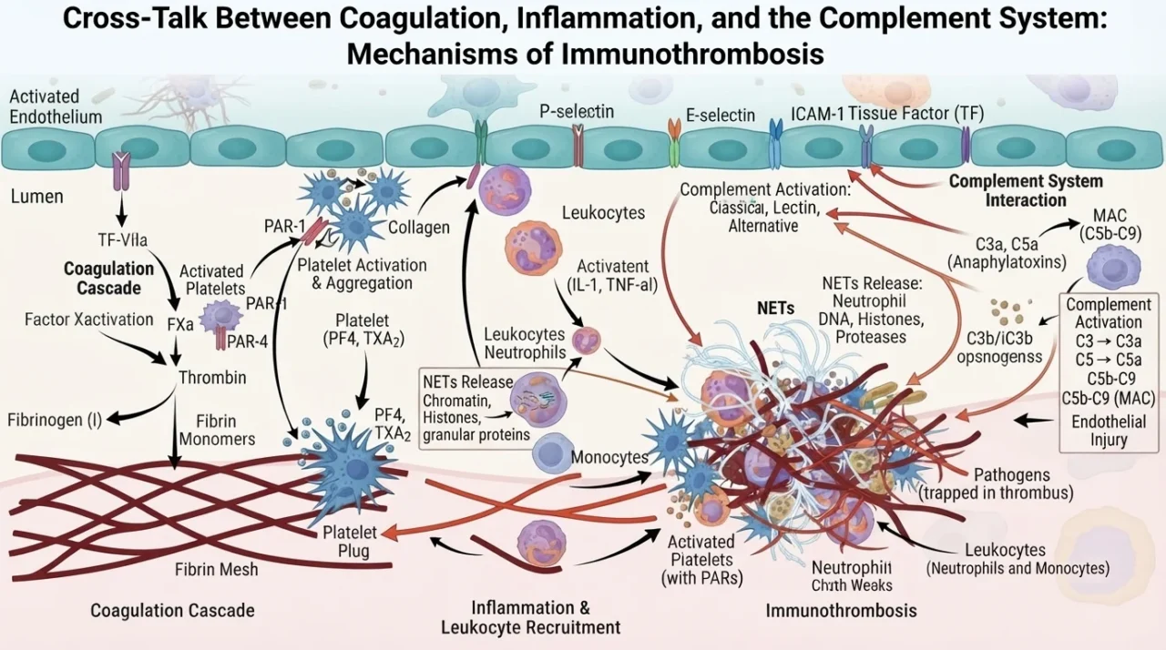
The coagulation system also interacts with inflammatory pathways through the formation of fibrin networks, platelet–leukocyte aggregates, and neutrophil extracellular traps (NETs).
NETs can provide a scaffold for platelet adhesion and coagulation, while platelets and coagulation factors can, in turn, influence immune-cell activation.
These interactions help explain the concept of immunothrombosis, in which coagulation participates in the body’s response to inflammation but can become harmful when excessive or sustained.
This broader view of coagulation is important for understanding its relationship with complement.
Complement activation can alter endothelial and platelet behavior and promote a procoagulant state, while coagulation proteases such as thrombin and plasmin can interact with components of the complement system.
The result is not two independent cascades, but a network in which complement, coagulation, platelets, endothelium, and innate immune cells can continuously influence one another.
The complement and coagulation systems are more closely connected than their traditional classification might suggest.
Both are organized as proteolytic cascades involving serine proteases and tightly regulated inhibitor proteins, and evidence suggests that some of their components evolved from shared ancestral genes.
Its defense system combines coagulation-like and complement-like activities, allowing it to respond to both vascular injury and invading microorganisms through an integrated mechanism.
Once complement and coagulation are viewed together, two questions naturally arise:
1. How does coagulation affect the complement cascade?
Several enzymes involved in coagulation and fibrinolysis can cleave complement components directly, providing routes of complement activation that do not depend on the conventional C3 convertases.
Thrombin was initially shown to activate both C3 and C5, raising the possibility that coagulation could directly trigger complement.
However, its activity toward C5 appears to differ from that of the classical C5 convertase.
Rather than efficiently cleaving C5 at the conventional R751 site, thrombin preferentially cleaves it at R947, producing C5aT and C5bT.
C5bT can subsequently participate in the formation of a C5bT–9 complex with lytic activity.
Although these findings demonstrate a molecular connection between thrombin and complement, experimental data suggest that thrombin may not be the main driver of complement activation in thrombosis.
Other proteases appear to be more efficient.
Factor XIa, factor Xa, factor IXa, and particularly plasmin have been shown to cleave C3 and C5 into biologically active fragments.
Plasmin is especially interesting because its ability to generate C3a and C5a can substantially exceed that of thrombin and factor Xa in experimental systems. Its cleavage of C5 can also generate a C5b-6 complex capable of assembling with C7, C8, and C9 into a functional membrane attack complex.
This creates an important connection between fibrinolysis and complement activation.
Plasmin is generated to break down fibrin, yet the same enzyme can simultaneously generate complement-derived inflammatory signals.
Supporting this concept, experimental models of arterial thrombosis have shown increased C3a and C5a following administration of a plasminogen activator.
Similar increases in complement activation products have been observed in patients with acute myocardial infarction following recombinant tissue plasminogen activator treatment.
Together, these findings suggest that some complement activation attributed to coagulation may actually occur further downstream, through plasmin generated during coagulation and fibrinolysis.
2. How complement influences coagulation?
The interaction also runs in the opposite direction.
Complement can promote a prothrombotic environment while simultaneously making an established clot more difficult to remove.
One important mechanism involves C3 and fibrin.
C3 can become incorporated into fibrin clots and interact with fibrinogen, producing a more consolidated clot that is less susceptible to fibrinolysis.
Experimental work has shown that C3 can bind the β-chain of fibrinogen, prolonging clot-lysis time, whereas disrupting the fibrinogen–C3 interaction can enhance fibrinolysis.
This provides a direct molecular link between complement activation and the persistence of a thrombus.
The relationship becomes even more interesting with C5a.
Rather than simply promoting inflammation, C5a can shift the local fibrinolytic balance toward clot preservation.
In human mast cells and basophils, C5a induces expression of plasminogen activator inhibitor-1 (PAI-1) while reducing the effective activity of tissue-type plasminogen activator (t-PA).
The result is a shift from a profibrinolytic to an antifibrinolytic phenotype, potentially making fibrin more resistant to degradation.
Complement can also promote the formation of new thrombus. C5a increases tissue factor expression on neutrophils, monocytes, and endothelial cells.
Because tissue factor is a major initiator of the coagulation cascade, this provides another route through which complement activation can increase thrombin generation and favor clot formation.
This does not mean that every coagulation event will trigger clinically meaningful complement activation, or that complement activation inevitably produces thrombosis.
The strength of each interaction appears to depend on the biological context.
Nevertheless, these findings show that complement and coagulation can converge at several critical points – thrombin generation, fibrin formation, tissue factor expression, and fibrinolysis – creating a network in which inflammation and thrombosis can reinforce one another.
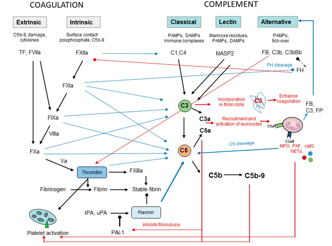
Noris, M., & Galbusera, M. (2022). The complement alternative pathway and hemostasis. Immunological Reviews, 313(1), 139–161.
The interaction between complement and coagulation becomes particularly relevant in diseases where inflammation, endothelial dysfunction, and thrombosis occur together.
Cardiovascular Disease
Cardiovascular disease provides some of the clearest evidence linking complement activation with thrombotic and atherosclerotic processes.
Several complement components have been associated with disease activity and cardiovascular outcomes, suggesting that they may have value as biomarkers as well as potential contributors to disease progression.
For example, C3a levels have been reported to be higher in patients with unstable angina than in those with stable angina, consistent with greater complement activation during more active coronary disease.
Elevated levels of the soluble membrane attack complex (sMAC) have also been independently associated with mortality and subsequent cardiovascular events after ST-elevation myocardial infarction (STEMI).
Similarly, higher circulating C5a has been linked to adverse cardiovascular outcomes in patients with genetically increased C5a levels.
The relationship extends to platelets.
Increased expression of C3aR and C5aR1, together with platelet activation markers such as P-selectin, has been associated with coronary artery disease. Experimental studies have further suggested that platelet C3aR signaling can influence outcomes after myocardial infarction.
Complement may also contribute to atherosclerosis itself. Higher circulating C3 and C4 levels have been associated with cardiovascular risk factors such as blood pressure, BMI, and lipid levels. Importantly, elevated C3 has been independently associated with future myocardial infarction and has been proposed to contribute to the progression of atherosclerotic lesions.
Stroke provides another example.
Increased plasma C3 and C3a have been observed in patients with stroke, while experimental complement depletion reduced infarct size after hypoxic–ischemic brain injury.
Systemic Inflammatory Response Syndrome and Sepsis
The complement–coagulation connection becomes particularly important in systemic inflammatory response syndrome (SIRS) and sepsis, where widespread inflammation can rapidly progress to endothelial injury, coagulation abnormalities, and multiorgan dysfunction.
During severe systemic inflammation, large amounts of C5a are generated.
Experimental studies have shown that C5a can increase tissue factor expression on monocytes, promoting a more thrombogenic state.
In animal models of sepsis, blocking C5a also reduced abnormalities in both coagulation and fibrinolysis, suggesting that excessive complement activation contributes to the disruption of hemostatic balance.
Importantly, this interaction is bidirectional. Coagulation activation can further stimulate complement, while complement-driven inflammation promotes additional coagulation and impaired fibrinolysis.
This creates a self-amplifying cycle that may contribute to progressive thrombosis and, in severe cases, disseminated intravascular coagulation (DIC).
Atypical Hemolytic Uremic Syndrome
In many patients, genetic or acquired abnormalities affecting complement regulation lead to uncontrolled activation on the vascular endothelium.
This makes aHUS a particularly clear example of how complement activation can translate into microvascular thrombosis.
Excessive complement activity damages endothelial cells, promotes platelet activation, and creates a prothrombotic environment in the small vessels.
Complement abnormalities are found in roughly half of patients, although their presence often represents a predisposition rather than a sufficient cause of disease; additional environmental or physiological triggers may be required to initiate clinical TMA.
Experimental evidence has further highlighted the importance of the terminal complement pathway.
In animal models, C5 deficiency protected against the development of TMA, providing an important rationale for targeting C5 therapeutically.
This was subsequently supported clinically by studies of eculizumab, which reduced microvascular thrombosis and was associated with substantial improvement in renal function in patients with aHUS.
At the same time, aHUS is a reminder that not every TMA is driven by complement.
Some patients have alternative mechanisms, such as DGKε deficiency, in which endothelial injury can occur independently of complement activation and therefore may not respond to C5 inhibition.
Paroxysmal Nocturnal Hemoglobinuria
Paroxysmal nocturnal hemoglobinuria (PNH) is an acquired clonal hematologic disorder caused by a somatic PIGA mutation, resulting in the loss of GPI-anchored proteins, including the complement regulators CD55 and CD59.
As a result, PNH cells become highly susceptible to complement-mediated injury, leading to intravascular hemolysis and the release of inflammatory and prothrombotic mediators.
Thrombosis is one of the major complications of PNH and can occur even in patients without severe hemolysis.
A key mechanism involves complement-mediated platelet activation.
Loss of CD59 makes PNH platelets more vulnerable to complement attack, promoting membrane remodeling and the release of platelet-derived microvesicles.
These vesicles expose phosphatidylserine, providing a highly procoagulant surface for tenase and prothrombinase complexes and thereby enhancing thrombin generation.
Complement-driven thrombosis in PNH is further amplified by other consequences of hemolysis, including free hemoglobin-mediated nitric oxide depletion and endothelial dysfunction, while complement-damaged leukocytes may contribute additional tissue factor activity.
Venous Thromboembolism
Venous thromboembolism (VTE) is another setting in which complement activation has been linked to thrombus formation.
Population studies have found that higher circulating C3 levels are associated with an increased risk of VTE, even after accounting for inflammatory markers such as C-reactive protein and BMI.
Similar associations have been reported for C3 and C4 in postnatal VTE.
Experimental findings provide additional support for a role of complement in venous thrombosis.
In mouse models of inferior vena cava thrombosis, C3 deficiency reduced thrombus formation and was associated with smaller thrombi and less fibrin and platelet deposition.
Higher levels of soluble terminal complement complex C5b-9 have also been associated with VTE, particularly apparently unprovoked events.
The relationship between complement and coagulation is increasingly recognized as a dynamic and bidirectional network rather than an interaction between two separate systems.
Complement can promote endothelial and platelet activation, tissue factor expression, and impaired fibrinolysis, while coagulation and fibrinolytic proteases can in turn modify complement activity.
Together, these interactions can amplify inflammation and thrombosis and help sustain vascular injury.
Evidence from cardiovascular disease, sepsis, APS, aHUS, PNH, and VTE shows that this crosstalk can have very different clinical consequences depending on the underlying disease.
In some conditions, such as aHUS and PNH, complement inhibition has already demonstrated clear clinical value, whereas in others the evidence remains largely observational or mechanistic.
This distinction highlights an important challenge for the field: complement activation is not necessarily harmful in every setting, and broad inhibition may not be appropriate for every thrombotic disease.
The next step is therefore not simply to inhibit complement, but to understand when, where, and which components of the complement–coagulation network should be targeted.
As our understanding of this network improves, complement factors, activation products, and related biomarkers may help identify patients in whom complement-driven thrombosis is particularly important.
Ultimately, integrating complement biology with coagulation, inflammation, and endothelial function could lead to more precise approaches to preventing and treating thrombo-inflammatory disease.
FAQ
1. What is complement–coagulation crosstalk?
Complement–coagulation crosstalk refers to the bidirectional interaction between the complement and coagulation systems. Complement can promote platelet activation, tissue factor expression, and impaired fibrinolysis, while coagulation and fibrinolytic proteases can also activate or modify complement components.
2. How does complement contribute to thrombosis?
Complement can create a prothrombotic environment by activating endothelial cells and platelets, increasing tissue factor expression, promoting inflammatory signaling, and in some settings reducing fibrinolysis. These effects can favor both thrombus formation and persistence.
3. Can coagulation activate complement?
Yes. Several proteases involved in coagulation and fibrinolysis, including thrombin, factor Xa, factor XIa, factor IXa, and plasmin, can cleave complement components under experimental conditions. Plasmin appears to be an especially important connection between fibrinolysis and complement activation.
4. What role does the endothelium play in complement–coagulation crosstalk?
The endothelium acts as an important interface between the two systems. When activated or injured, endothelial cells can lose anticoagulant properties and promote platelet adhesion, tissue factor expression, inflammation, and complement activation, helping sustain a thrombo-inflammatory response.
5. What is the role of platelets in this interaction?
Platelets are active participants rather than simply the cellular components of a clot. Complement activation can influence platelet activation, while activated platelets provide procoagulant surfaces and interact with leukocytes and endothelial cells, further connecting inflammation with thrombin generation.
6. What is immunothrombosis?
Immunothrombosis describes the interaction between the immune and hemostatic systems during inflammation. Thrombus formation can help contain pathogens or tissue injury, but excessive or prolonged activation can contribute to pathological thrombosis and organ damage.
7. Which diseases show important complement–coagulation interactions?
Examples include cardiovascular disease, sepsis and SIRS, antiphospholipid syndrome, complement-mediated thrombotic microangiopathies such as aHUS, PNH, and venous thromboembolism. The importance and mechanism of complement activation differ between these conditions.
8. Why is aHUS particularly relevant to complement-mediated thrombosis?
In aHUS, uncontrolled complement activation can directly damage the vascular endothelium and promote platelet-rich microvascular thrombosis. The clinical effectiveness of C5 inhibition in many patients provides strong evidence that complement can be a major driver of thrombotic microangiopathy.
9. Does complement inhibition prevent all forms of thrombosis?
No. Complement inhibition is highly relevant in selected complement-driven disorders, but thrombosis has multiple causes and mechanisms. Evidence supporting complement-targeted therapy varies considerably between diseases, and complement inhibition is not a general replacement for established antithrombotic treatment.
10. Could complement–coagulation interactions lead to new therapeutic strategies?
Potentially. A better understanding of this network could allow treatment to be directed toward specific complement or coagulation components in patients whose disease is particularly dependent on this pathway. The major challenge is achieving sufficient inhibition of pathological inflammation and thrombosis without compromising normal host defense or hemostasis.
Written by Robert Tadevosyan.
Stay updated with Hemostasis Today.
-
Sep 22, 2026, 16:45Claudiu Stoicescu: Are We Starting Cardiovascular Risk Assessment Too Late?
-
Sep 22, 2026, 16:20Ila Bappa Ibrahim: Bringing Sickle Cell Innovation Closer to Communities
-
Sep 22, 2026, 16:02Ana Pedrero Gil: Peri-operative Management of Patients Undergoing Day Surgery
-
Sep 22, 2026, 15:27Zia Ullah: RBC-Derived Extracellular Vesicles as Potential Biomarkers of Thrombotic Risk in Dengue
-
Sep 22, 2026, 15:01Lesley Van Wyk: What Each Iron Markers Actually Mean
-
Sep 22, 2026, 14:37Chokri Ben Lamine։ Saudi Arabia Leads A New Milestone In Hemophilia A Care
-
Sep 22, 2026, 13:58Giulia Passaniti: Dual Inferior Vena Cava Anatomy in Tetralogy of Fallot
-
Sep 22, 2026, 13:35Michael Miyamoto: Scaling Guideline Based Lipid Management in Community Practice
-
Sep 22, 2026, 13:15Suvro Sankha Datta: Developing Blood Group Genotyping Platform Using a Gold-Oligo