

Iron Deficiency and Thrombocytosis: How Iron Availability Shapes Hematopoietic Lineage Decisions
Iron deficiency is usually discussed in terms of impaired hemoglobin synthesis and anemia. Its effects on hematopoiesis, however, extend beyond red blood cells.
Restricted iron availability can alter the proliferation, signaling activity, and differentiation of hematopoietic progenitors—under certain conditions favoring megakaryocyte development and platelet production.
From this perspective, reactive thrombocytosis in iron-deficiency anemia is more than an accompanying laboratory abnormality: it reflects how the hematopoietic system responds to limited iron availability.
The central concept is that iron acts not only as a substrate for hemoglobin synthesis but also as a regulator of hematopoiesis. Its availability can influence whether progenitor cells pursue erythroid or megakaryocytic differentiation. Importantly, the most direct mechanistic evidence comes from animal models and experiments with human cells, rather than from direct tracking of progenitor-cell fate in patients.
The Clinical Paradox: Why Do Platelets Increase When Iron Is Scarce?
In iron-deficiency anemia, red-cell production becomes iron-restricted, yet platelet counts may rise.
This apparently divergent response suggests that iron deficiency does not simply suppress bone marrow activity uniformly. Instead, it affects individual hematopoietic lineages differently.
In a large retrospective study, thrombocytosis occurred in 32.6 percent of patients with iron-deficiency anemia.
This figure describes the study population; it should not be interpreted as a universal prevalence estimate for everyone with iron deficiency. Nevertheless, it demonstrates that the association is sufficiently common to warrant clinical attention.
The relationship is not linear.
Platelet counts may remain normal, and rare cases of severe iron-deficiency anemia present with thrombocytopenia rather than thrombocytosis.
The simple assumption that progressively lower iron availability must produce progressively higher platelet counts therefore does not capture the full biology.
At the Progenitor Level: Balancing Erythroid and Megakaryocytic Development
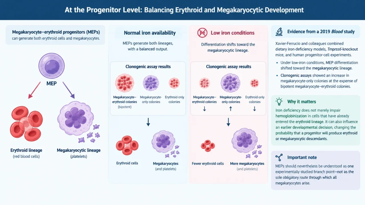
Megakaryocyte–erythroid progenitors, or MEPs, can generate both erythroid cells and megakaryocytes. These progenitors provide an important experimental framework for investigating how iron availability influences hematopoietic lineage decisions.
In a 2019 study published in Blood, Xavier-Ferrucio and colleagues combined findings from dietary iron-deficiency models, Tmprss6-knockout mice, and human progenitor-cell experiments. Under low-iron conditions, MEP differentiation shifted toward the megakaryocytic lineage. Clonogenic assays showed an increase in megakaryocyte-only colonies at the expense of bipotent megakaryocyte–erythroid colonies.
This distinction matters. Iron deficiency does not merely impair hemoglobinization in cells that have already entered the erythroid lineage.
It can also influence an earlier developmental decision, changing the probability that a progenitor will produce erythroid or megakaryocytic descendants. MEPs should nevertheless be understood as one experimentally studied branch point—not as the sole obligatory route through which all megakaryocytes arise.
Low Iron, ERK Signaling, and Progenitor Proliferation
The same study identified several related changes in iron-deficient MEPs:
- Reduced intracellular labile iron—the metabolically accessible iron pool.
- Decreased ERK phosphorylation.
- Slower cellular proliferation.
- Increased megakaryocytic commitment.
ERK signaling participates in the regulation of cell proliferation and differentiation.
These findings support a model in which restricted iron availability changes the metabolic and signaling state of progenitor cells, reduces ERK activity, and favors megakaryocytic development.
They do not, however, establish a single, fully validated causal pathway operating identically in every patient with iron deficiency.
The proposed sequence can be summarized as:
Reduced iron availability leads to altered progenitor cell metabolism and signaling, which results in slower proliferation, a bias toward megakaryocytic differentiation, and increased platelet production.
The apparent contradiction—slower progenitor proliferation but greater platelet output—becomes clearer when proliferation and differentiation are considered separately.
Final platelet production depends not only on the number of progenitors but also on their developmental trajectory and the maturation of their descendants.
Beyond Lineage Commitment: Effects on Megakaryocyte Maturation
Iron deficiency can also affect cells that have already entered the megakaryocytic lineage. Studies in rats and cell cultures have described increased megakaryocyte ploidy, larger cell size, and accelerated differentiation, accompanied by quantitative and functional changes in platelets.
Ploidy refers to a cell’s DNA content. During maturation, megakaryocytes can replicate their genome without undergoing conventional cell division. Changes in this process indicate that iron restriction influences not only how many progenitors enter the megakaryocytic lineage, but also how those cells subsequently mature.
Iron-deficiency–associated thrombocytosis may therefore develop through at least two interacting processes: a shift in progenitor lineage commitment and changes in downstream megakaryocyte maturation.
Does Thrombopoietin Explain the Entire Response?
Thrombopoietin, or TPO, is the principal regulator of platelet production. Yet iron-deficiency–associated thrombocytosis does not necessarily require an increase in TPO production. A 2014 experimental study found that iron deficiency promoted megakaryopoiesis without increased production of TPO, interleukin-6, or interleukin-11.
This finding requires careful interpretation. The absence of increased TPO production does not mean that megakaryocytes become independent of its biological effects. Rather, it indicates that the response to iron deficiency cannot be explained simply by a quantitative rise in TPO.
It also helps distinguish iron-deficiency–associated thrombocytosis from a purely inflammatory model. Restricted iron availability can alter megakaryopoiesis even without increased production of the inflammatory growth factors examined in these experiments.
The HIF-2α–VEGF Axis: An Additional Mechanism, Not a Complete Explanation
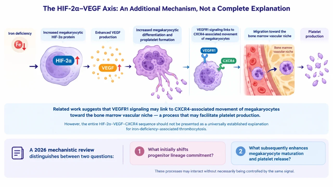
The HIF-2α–VEGF axis has also been investigated as a link between iron deficiency and megakaryocyte maturation. Experimental studies have reported increased megakaryocytic HIF-2α protein, enhanced VEGF production, and increased megakaryocytic differentiation and proplatelet formation under iron-deficient conditions.
Related work links VEGFR1 signaling to CXCR4-associated movement of megakaryocytes toward the bone marrow vascular niche—a process that may facilitate platelet production. However, the entire HIF-2α–VEGF–CXCR4 sequence should not be presented as a universally established explanation for iron-deficiency–associated thrombocytosis.
A 2026 mechanistic review likewise distinguishes between two questions: what initially shifts progenitor lineage commitment, and what subsequently enhances megakaryocyte maturation and platelet release.
These processes may interact without necessarily being controlled by the same signal.
An Adaptive Response—or a Thrombotic Liability?
One hypothesis is that increased platelet production during iron deficiency represents an adaptive response. When iron loss results from ongoing bleeding, a response that supports hemostasis could theoretically be beneficial. This remains a biological interpretation, however, rather than proof that iron-deficiency–associated thrombocytosis is consistently protective.
Clinical observations also suggest a potential downside. In the large retrospective study described above, thrombosis was reported in 7.8 percent of patients with iron-deficiency anemia without thrombocytosis and in 15.8 percent of those with thrombocytosis. These findings establish an association; they do not prove that the platelet count itself accounts for the entire difference. Nor should these percentages be used directly to estimate an individual patient’s risk.
Animal experiments provide biological support for the association. Iron-deficient rats developed larger thrombi in arterial and venous thrombosis models, while iron replacement reversed thrombocytosis and several prothrombotic changes. These findings do not establish that iron treatment reduces clinical thrombotic events to the same extent in humans.
What Does This Mean for Clinical Interpretation?
Iron Status Belongs in the Evaluation of Thrombocytosis
Persistent thrombocytosis should prompt assessment of iron status alongside the blood film, inflammatory markers, and clinical context.
The platelet count alone cannot reliably distinguish a reactive process from a clonal hematologic disorder.
Treatment Response Includes More Than Hemoglobin Recovery
Iron-deficiency–associated thrombocytosis generally decreases as iron deficiency is corrected.
Monitoring should therefore include the platelet count as well as hemoglobin and iron parameters.
The trajectory of the complete blood count can help reassess whether the thrombocytosis behaves as an iron-responsive reactive process.
If thrombocytosis persists despite adequate correction of iron deficiency, other or coexisting causes should be reconsidered rather than attributing the finding indefinitely to the previous iron deficit.
A Thrombotic Association Is Not an Automatic Indication for Antithrombotic Therapy
The reported association with thrombosis does not mean that every patient with iron-deficiency–associated reactive thrombocytosis should receive aspirin or anticoagulation.
Management centers on identifying and correcting the underlying cause; antithrombotic treatment depends on separate clinical indications and an assessment of bleeding risk.
Current Clinical Guidance: Translating Mechanisms Into Practice
Guidance reviewed through October 27, 2026.
Current guidelines inform the diagnosis of iron deficiency, the evaluation of thrombocytosis, and the selection of iron therapy. They should be distinguished from mechanistic studies: experimental findings on ERK signaling, progenitor commitment, and megakaryocyte maturation explain possible biological pathways, but do not themselves establish treatment recommendations.
ASH 2026: Updated Guidance on Diagnosing Iron Deficiency
The American Society of Hematology’s 2026 guidelines introduce population-specific ferritin thresholds and emphasize that iron deficiency can occur with or without anemia. For adults, including menstruating and pregnant individuals, the panel generally suggests a serum ferritin threshold of 30 ng/mL or lower. A threshold of 50 ng/mL or lower may be appropriate in selected symptomatic or higher-risk settings, including heavy menstrual bleeding and pregnancy complicated by anemia. These thresholds require clinical context rather than application as a single universal rule.
For adults with anemia of inflammation, ASH suggests assessing both ferritin and transferrin saturation rather than ferritin alone. Importantly, the panel did not make a formal recommendation for a specific diagnostic threshold in this setting because of limited evidence. Ferritin less than 100 ng/mL or transferrin saturation less than 20 percent may help identify concomitant iron deficiency, but these values should be interpreted as contextual guidance rather than definitive cutoffs for every inflammatory disorder.
BSH 2025/2026: Direct Recommendations for Reactive Thrombocytosis
The British Society for Haematology guideline, Investigation and management of thrombocytosis without JAK2, CALR or MPL mutations, was published online in December 2025 and appeared in the February 2026 journal issue. Although its principal focus is mutation-negative thrombocytosis, it includes explicit recommendations for reactive thrombocytosis.
The guideline recommends:
Treating the underlying cause of reactive thrombocytosis—Grade 1B.
Not routinely prescribing antiplatelet or anticoagulant medication unless other clinical factors provide an indication—Grade 1C.
Monitoring the complete blood count to confirm that thrombocytosis resolves as the reactive condition is treated.onlinelibrary.wiley
It also highlights a useful diagnostic safeguard: patients with iron deficiency, thrombocytosis, and an initially normal hematocrit should have repeat blood counts after iron replacement.
An abnormal increase in hematocrit warrants investigation for polycythemia vera or hereditary erythrocytosis.
AGA 2024: Practical Guidance on Iron Replacement
The American Gastroenterological Association’s 2024 Clinical Practice Update is an expert review providing best-practice advice, rather than a formally graded clinical practice guideline. It advises giving oral iron no more than once daily; every-other-day dosing may improve tolerability for some patients while providing similar or equal iron absorption.
Intravenous iron is advised when oral iron is not tolerated, ferritin fails to improve after a trial of oral treatment, or the patient has a condition in which oral iron is unlikely to be adequately absorbed. Thrombocytosis alone does not establish a separate indication for intravenous treatment under this advice.
The practical priority is therefore to confirm iron deficiency, identify its cause, replenish iron appropriately, and reassess the blood count—not to treat an isolated platelet number as if it automatically represented a myeloproliferative neoplasm.
Conclusion
Iron availability does more than supply the raw material for hemoglobin synthesis. It helps shape progenitor-cell metabolism, signaling, proliferation, and differentiation. When iron becomes scarce, the balance may shift toward megakaryocytic development, while changes in subsequent maturation can further support platelet production.
This framework helps explain how impaired erythropoiesis and thrombocytosis can coexist. It also highlights an important boundary in interpreting the evidence: experimental changes in progenitor-cell behavior, clinically observed thrombocytosis, and thrombotic outcomes are connected but distinct findings. Keeping those levels separate allows iron-deficiency–associated thrombocytosis to be understood as a clinically relevant example of how nutrient availability can influence hematopoietic cell fate—without overstating what current evidence proves.
Frequently Asked Questions
1. Why can iron deficiency cause thrombocytosis?
Low iron availability can favor megakaryocytic differentiation and alter megakaryocyte maturation, increasing platelet production.
2. Does everyone with iron-deficiency anemia develop thrombocytosis?
No. Platelet counts may remain normal and, rarely, decrease in severe cases.
3. What ferritin threshold does ASH recommend for adults?
ASH 2026 generally suggests ferritin ≤30 ng/mL, with higher thresholds appropriate in selected clinical settings.
4. Can normal ferritin exclude iron deficiency?
Not reliably during inflammation. Ferritin should be interpreted alongside transferrin saturation and the clinical context.
5. Is increased thrombopoietin the main mechanism?
Not necessarily. Experimental studies show enhanced megakaryopoiesis without increased thrombopoietin production.
6. Does this type of thrombocytosis increase thrombotic risk?
Observational studies suggest an association, but they do not prove that elevated platelets alone cause the increased risk.
7. Is aspirin routinely recommended?
No. BSH advises against routine antiplatelet or anticoagulant treatment for reactive thrombocytosis without another indication.
8. Does thrombocytosis require intravenous iron?
No. Treatment route depends on oral iron tolerance, absorption, response, and clinical circumstances—not thrombocytosis alone.
9. Should platelet counts be monitored after treatment?
Yes. Repeat blood counts help confirm that thrombocytosis resolves as iron deficiency improves.
10. What if thrombocytosis persists after iron correction?
Reassess for other reactive causes and consider evaluation for a clonal hematologic disorder.
Written by Anna Stepanyan, MD
Stay updated with Hemostasis Today.
-
Oct 27, 2026, 16:02Ahmad G. Alzahrani: AF and Vascular Risk Factors – Should We Add Antiplatelet Therapy?
-
Oct 27, 2026, 15:46Christiane Berger-Schaffitzel: New PhD Project Explores Snake Venom and Haemostasis
-
Oct 27, 2026, 13:51Muhammed Hajmusa: ESMO 2026 Highlights the Challenge of Anticoagulation in ROS1-Positive Lung Cancer
-
Oct 27, 2026, 13:39Luca Galassi: Retrieved Venous Thrombi as a Tool for Predicting Post-Thrombotic Syndrome
-
Oct 27, 2026, 11:43Tareq Abadl: Hidden Interferences Behind Unexpected Laboratory Results
-
Oct 27, 2026, 11:42Cece Calhoun: ASH Promotes Clinical Research Training and Mentorship for Emerging Hematologists
-
Oct 27, 2026, 11:41Ifeanyichukwu Ifechidere: The Laboratory Challenges of Measuring Direct Factor Xa Inhibitors
-
Oct 27, 2026, 11:39Gene Therapy Offers New Possibilities for Patients with Thalassaemia in the UAE – Thalassaemia International Federation
-
Oct 27, 2026, 11:35Alan Nurden: rFVIIa Prophylaxis May Reduce Bleeding-Related Healthcare Burden in Glanzmann Thrombasthenia