


Ifeanyichukwu Ifechidere: Anaemia is not a Diagnosis – It’s a Symptom
Ifeanyichukwu Ifechidere, Biomedical Scientist at Sheffield Teaching Hospitals NHS Foundation Trust, posted on LinkedIn:
”’Anaemia is not a diagnosis – it’s a symptom.’
As biomedical scientists, one of the most important mindset shifts we can make is this:
- Anaemia doesn’t tell you what the problem is.
- It tells you there is a problem.
Low haemoglobin is the starting point – not the conclusion.
The real work begins when we ask:
What type of anaemia is this? And why?
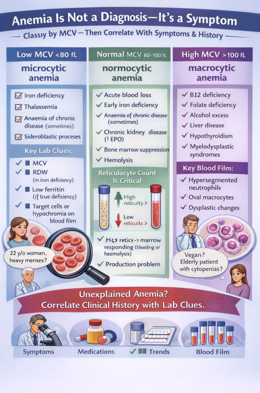
Step 1: Classify by MCV – Microcytic, Normocytic, or Macrocytic
The Mean Corpuscular Volume (MCV) is more than a number.
It’s a diagnostic compass.
Microcytic Anaemia (Low MCV)
Think: Impaired haemoglobin production
Most common causes:
- Iron deficiency
- Thalassaemia trait
- Anaemia of chronic disease (sometimes)
- Sideroblastic processes
Key lab clues:
- Increased MCV
- Deceased RDW (in iron deficiency)
- Low ferritin (if true deficiency)
- Target cells or hypochromia on blood film
But here’s where clinical-lab correlation matters:
A 22-year-old woman with heavy menses?
Very different from a 68-year-old man with new microcytosis.
Same indices. Very different implications.
Normocytic Anaemia (Normal MCV)
This is where many clinicians get stuck.
Normocytic does not mean normal physiology.
Think:
- Acute blood loss
- Early iron deficiency
- Anaemia of chronic disease
- Chronic kidney disease (decrease EPO)
- Bone marrow suppression
- Haemolysis
Now the reticulocyte count becomes critical:
- High retics to marrow responding (bleeding or haemolysis)
- Low retics to production problem
Without reticulocytes, you’re only seeing half the picture.
Macrocytic Anaemia (High MCV)
Think: DNA synthesis or marrow dysfunction
Common causes:
- Vitamin B12 deficiency
- Folate deficiency
- Alcohol excess
- Liver disease
- Hypothyroidism
- Myelodysplastic syndromes
Blood film can be transformative here:
- Hypersegmented neutrophils
- Oval macrocytes
- Dysplastic changes
And again — context matters.
Macrocytosis in a vegan patient?
Different from macrocytosis in a 72-year-old with cytopenias.
The Most Important Step: Clinical–Laboratory Correlation
A haemoglobin value in isolation is a number.
Add:
- Symptoms (fatigue, weight loss, bleeding)
- Medication history
- Renal function
- Inflammatory markers
- Serial trends
- Blood film morphology
Now it becomes a narrative.
As biomedical scientists, our role is not just to validate results – it’s to interpret patterns, question inconsistencies, and know when something doesn’t fit.
Because:
- Not all iron deficiency is dietary.
- Not all macrocytosis is B12.
- Not all normocytic anaemia is ‘chronic disease.’
Anaemia is the smoke.
Our job is to help locate the fire.
How do you approach unexplained anaemia?
Do you start with indices, reticulocytes, iron studies – or the blood film first?
Let’s discuss the frameworks that have shaped your practice.”
Stay updated with Hemostasis Today.
-
Sep 17, 2026, 09:56Marianne Yee: MySCDstory Expands Access to Multilingual Sickle Cell Education
-
Sep 17, 2026, 09:36Enewa Onoja: Platelet Satellitism Phenomenon, Three Misleading Counts
-
Sep 17, 2026, 09:22Emmanuel J Favaloro: Hemostasis and Thrombosis in Exercise and Athletes
-
Sep 17, 2026, 09:18Updated Iron Deficiency Diagnostic Guidelines – American Society of Hematology
-
Sep 17, 2026, 09:14Angela Weyand: ASH 2026 Iron Deficiency Guidelines
-
Sep 17, 2026, 08:18Anirban Sen Gupta: From Flow to Clot Insights Into Hemostasis and Thrombosis
-
Sep 17, 2026, 08:03Aya Sherif: HEMO COP – Building a Collaborative Future for Hemophilia Care
-
Sep 17, 2026, 07:50Applications Are Now Open for the 2026 WFH IHTC Fellowships
-
Sep 17, 2026, 07:37Jim Hoffman: Could NET Removal Help Restore Vascular Homeostasis?