


Dhananjaya Kotebagilu Narayana Vamyanmane: High-Resolution Ultrasound Evaluation of Bowel Wall Ischemia
Dhananjaya Kotebagilu Narayana Vamyanmane, Pediatric Radiologist and Al Imaging Innovator, Consultant at Manipal Hospitals, shared a post on LinkedIn:
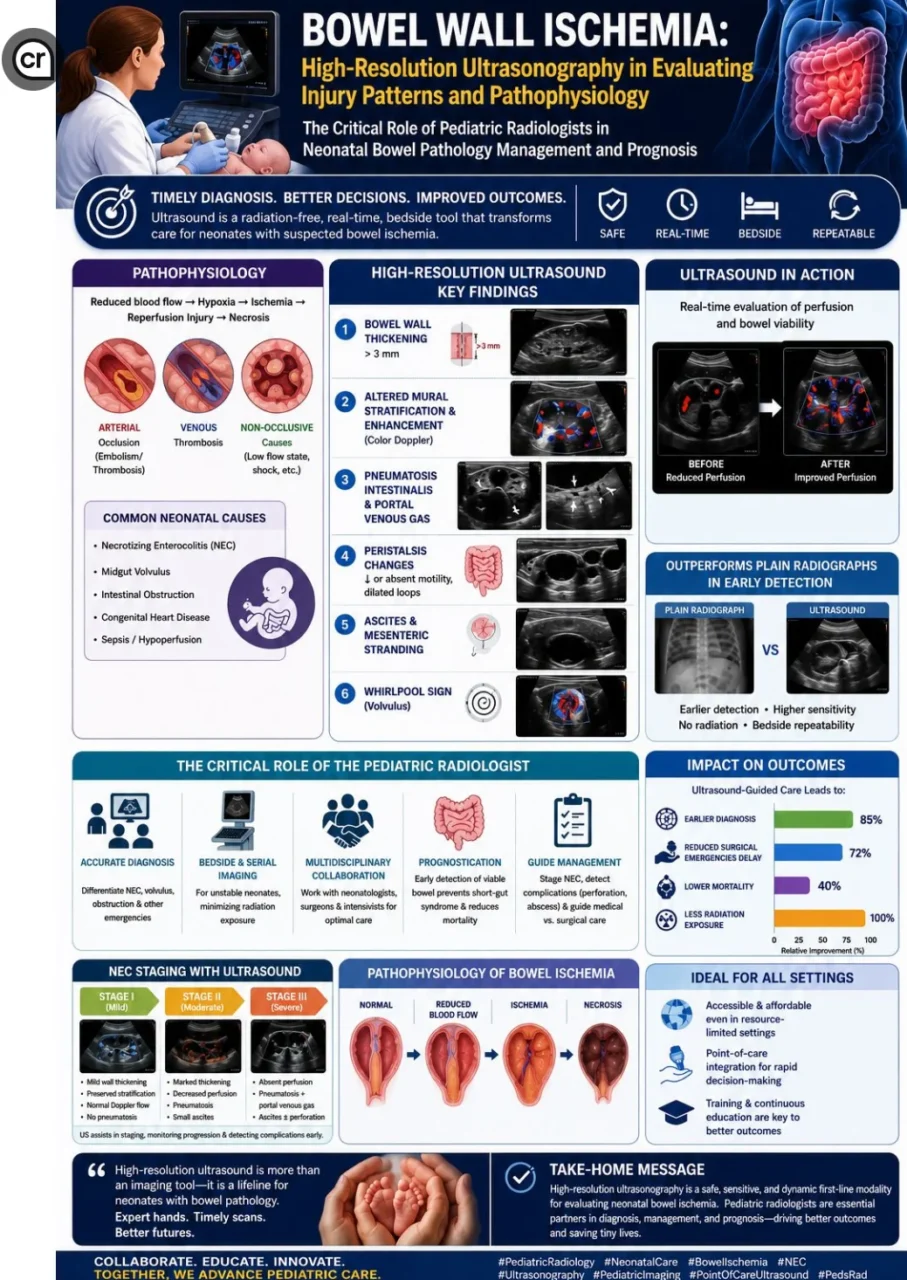
“Bowel Wall Ischemia: High-Resolution Ultrasonography in Evaluating Injury Patterns and Pathophysiology
The Critical Role of Pediatric Radiologists in Neonatal Bowel Pathology Management and Prognosis.
Bowel wall ischemia represents a serious condition, particularly in neonates, where timely diagnosis can significantly impact outcomes. High-resolution ultrasonography (HRUS) has emerged as a valuable, radiation-free tool for assessing injury patterns, understanding pathophysiology, and guiding management in pediatric patients.
Pathophysiology and Ultrasound Patterns:
Intestinal ischemia occurs due to reduced blood flow—often from arterial occlusion (embolism/thrombosis), venous thrombosis, or non-occlusive causes—leading to hypoxia and potential reperfusion injury. In neonates, this is frequently linked to conditions like necrotizing enterocolitis (NEC), midgut volvulus, or other obstructions.
Key HRUS findings include:
- Bowel wall thickening (more than 3 mm) or thinning (‘paper-thin’ appearance in advanced arterial ischemia).
- Altered mural stratification and enhancement (assessed with color Doppler for vascularity).
- Pneumatosis intestinalis (gas in the bowel wall) and portal venous gas — highly concerning for necrosis.
- Peristalsis changes: Reduced or absent motility, dilated loops with fluid stasis.
- Ascites, mesenteric stranding, or whirlpool sign (in volvulus).
HRUS excels in real-time evaluation of perfusion, wall viability, and dynamic changes, often outperforming plain radiographs in early detection and monitoring progression.
Role of the Pediatric Radiologist:
Pediatric radiologists play a pivotal role beyond diagnosis.
- Differentiate high versus low obstructions and identify surgical emergencies (e.g., volvulus vs. NEC).
- Provide serial bedside imaging for unstable neonates, minimizing radiation exposure.
- Collaborate in multidisciplinary teams to predict prognosis — early detection of viable bowel can prevent short-gut syndrome and reduce mortality.
In NEC, ultrasound helps stage disease, detect complications like perforation, and guide conservative versus surgical management.
Clinical Implications:
High-resolution US offers a safe, accessible first-line modality, especially in resource-variable settings. Its integration into point-of-care protocols by trained neonatologists and radiologists enhances rapid decision-making.
Advancing expertise in these techniques is essential for improving neonatal survival and quality of life in bowel pathologies.
This review underscores the transformative potential of ultrasound in pediatric gastrointestinal emergencies. Collaboration and continuous education in pediatric radiology remain key.”
Stay updated with Hemostasis Today.
-
Oct 16, 2026, 16:01Obi Light Ogbonnia: The Greatest Reward Is Changing Lives, Not Winning Awards
-
Oct 16, 2026, 15:18Carmine Liberatore: Limited Accuracy of VTE Risk Scores in Multiple Myeloma
-
Oct 16, 2026, 14:55Alex Borstein and the Bleeding Phenotype of Hemophilia A Carriers
-
Oct 16, 2026, 13:59Aya Akmal Amin: Antiphospholipid Syndrome Management and Risk Stratification
-
Oct 16, 2026, 13:10Emanuel Sueldo: CAHT 2026 Celebrates Scientific Progress and Team Achievements
-
Oct 16, 2026, 13:05Maha Othman: Queen’s Health Sciences Advances CAT Risk Models and Pediatric ISTH-BAT Cut-Offs
-
Oct 16, 2026, 13:03Thrombosis Awareness Must Lead to Timely Action – Roche
-
Oct 16, 2026, 12:49Wolfgang Miesbach: 5-Year CBDR Data Confirms Emicizumab Safety and Efficacy in Hemophilia A
-
Oct 16, 2026, 12:40Edward Lee Carter: How Factor XI Inhibition Could Transform Anticoagulation Therapy