RBC Inclusions Decoded: Diagnostic Pearls from William Aird
William Aird, Professor of Medicine at Harvard Medical School, posted on X:
”1/7
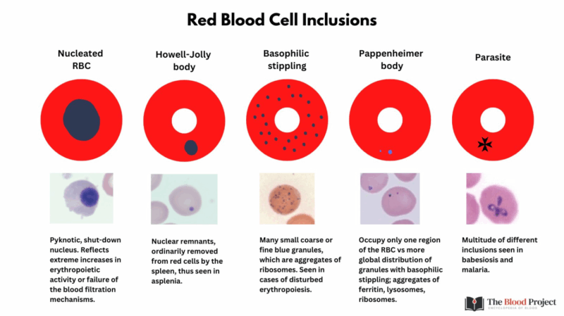
RBC INCLUSIONS
Red blood cell (RBC) inclusions: small structures with big diagnostic value. Each one points to a different process, from marrow stress to iron overload to infection. Here’s a tour, one inclusion at a time.
2/7
Nucleated RBCs: immature erythroid precursors that still carry their nucleus.
- Normally confined to the marrow (except neonates).
- In circulation, signal marrow under stress (hemolysis, hypoxia, marrow infiltration)
Think of them as a sign of the BM “pushing hard.”
3/7
Howell–Jolly bodies: small, round basophilic nuclear remnants (DNA) DNA found within circulating RBCs.
- Appear when the spleen is absent or not functioning (asplenia, hyposplenism).
- If you see them, ask about the spleen.
See here.
4/7
Basophilic stippling: numerous fine or coarse punctate, blue-purple granules (aggregates of ribosomal RNA).
- Causes include thalassemia, lead poisoning, sideroblastic anemia.
- Coarse stippling is especially associated with lead.
5/7
Pappenheimer bodies: iron-containing granules (siderotic granules) in RBCs.
- Stain positive with Prussian blue
- Seen in sideroblastic anemia, iron overload, post-splenectomy.
See here.
6/7
Intraerythrocytic parasites: malaria, babesiosis.
- Morphology varies (rings, trophozoites, schizonts, gametocytes).
- Always correlate with travel/exposure history; finding parasites in RBCs is a diagnostic emergency.
7/7
Red cell inclusions provide direct windows into pathophysiology: nuclear remnants, iron deposits, ribosomal aggregates, or pathogens. Recognizing them is a high- yield skill at the microscope.”

Stay updated with Hemostasis Today.
-
Jan 9, 2026, 12:17Louise St Germain Bannon Invites You to Self-Nominate for the ISTH Council
-
Jan 9, 2026, 09:25Emma Groarke Shares A Comprehensive Review on VEXAS Syndrome
-
Jan 9, 2026, 09:13Anirban Sen Gupta Presents PlateChek
-
Jan 9, 2026, 08:58Oscar Pena Shares KDIGO 2026 Update: 5 Critical Changes for Anemia in CKD Management
-
Jan 9, 2026, 07:38Bartosz Hudzik on Subacute Coronary Occlusions: Navigating the Gray Zone in CAD Care
-
Jan 9, 2026, 07:27Gregory Piazza on The Role of D-Dimer in VTE Evaluation
-
Jan 9, 2026, 07:19Emmanuel J Favaloro Shares the 1st Article in a VWD Series Marking the 100-Year Anniversary
-
Jan 9, 2026, 07:07Taylor Robichaux on Recombinant ADAMTS13 Therapy in cTTP
-
Jan 9, 2026, 06:25Sara Ng: VTE Continues to Plague Modern Oncology
