


Samwel Mikaye Breaks Down Peripheral Blood Smear
Samwel Mikaye, CEO of SaMik Medical Center, shared on LinkedIn:
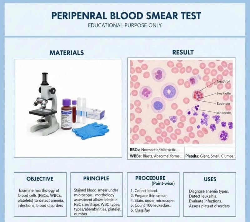
”Peripheral Blood Smear (PBS)
Definition:
A laboratory test where a thin layer of blood is spread on a slide, stained, and examined under a microscope to evaluate red blood cells (RBCs), white blood cells (WBCs), and platelets.
Purpose:
• Assess morphology of blood cells
• Detect anemia, infections, leukemia, thrombocytopenia, and other hematologic disorders
1. Indications
• Unexplained anemia, leukocytosis, leukopenia, thrombocytopenia
• Suspected malaria, hemoglobinopathies, parasites
• Monitoring chemotherapy or bone marrow disorders
• Abnormal complete blood count (CBC) findings
2. Sample Collection
• Peripheral venous blood in EDTA anticoagulant (lavender-top tube)
• Mix gently; do not shake
• Process within 2–4 hours for best morphology
3. Preparation of Smear
• Place small drop of blood near one end of a clean slide
• Use a second slide at 30–45° angle to spread the blood
• Make a thin, even smear with a feathered edge
• Air dry
4. Staining
• Romanowsky stains (most common):
• Wright stain
• Giemsa stain
• Leishman stain
• Staining highlights:
• RBC morphology
• WBC nuclei and cytoplasm
• Platelets
5. Microscopic Examination
• Low power (10×): assess smear quality, overall cellularity
• High power (40×–100× oil immersion): detailed cell morphology
• Examine:
1. Red blood cells (RBCs): size, shape, color, inclusions
2. White blood cells (WBCs): type, count, abnormalities
3. Platelets: number, size, clumping
4. Parasites: malaria, microfilaria
6. Key Findings
RBC Abnormalities
• Microcytic, macrocytic, normocytic
• Hypochromic, polychromasia
• Poikilocytosis (target cells, sickle cells, spherocytes, elliptocytes)
• Inclusions: Howell–Jolly bodies, basophilic stippling
WBC Abnormalities
• Leukocytosis, leukopenia
• Immature forms (blasts) → leukemia
• Toxic granulation → infection
Platelet Abnormalities
• Thrombocytopenia / thrombocytosis
• Giant platelets
Parasites
• Malaria: trophozoites, schizonts, gametocytes
• Microfilaria: sheathed worm in RBCs
7. Advantages
• Rapid, inexpensive
• Detects morphologic changes not visible on automated CBC
• Guides further tests
8. Limitations
• Subjective interpretation
• Requires skill and experience
• Cannot quantify accurately (use CBC for counts)
• Artifacts possible due to poor smear or staining
• Peripheral blood smear evaluates morphology, not just counts
• Thin feathered edge is examined for cell morphology
• RBC morphology → anemia type
• WBC abnormalities → leukemia or infection
• Platelets → count and morphology
• Malaria diagnosis → confirmed on smear”
Stay updated with Hemostasis Today.
-
Oct 16, 2026, 13:10Emanuel Sueldo: CAHT 2026 Celebrates Scientific Progress and Team Achievements
-
Oct 16, 2026, 13:05Maha Othman: Queen’s Health Sciences Advances CAT Risk Models and Pediatric ISTH-BAT Cut-Offs
-
Oct 16, 2026, 13:03Thrombosis Awareness Must Lead to Timely Action – Roche
-
Oct 16, 2026, 12:49Wolfgang Miesbach: 5-Year CBDR Data Confirms Emicizumab Safety and Efficacy in Hemophilia A
-
Oct 16, 2026, 12:40Edward Lee Carter: How Factor XI Inhibition Could Transform Anticoagulation Therapy
-
Oct 16, 2026, 12:40Marc Carrier: Looking Ahead to ISTH 2027 in Vancouver
-
Oct 16, 2026, 12:30Bob Lempke: How One Blood Donation Can Save Up to Three Lives
-
Oct 16, 2026, 12:30Why VWF Therapies Belong on the EU Critical Medicines List – EHC
-
Oct 16, 2026, 12:17Exploring Endothelial Cells, Genetic Fate Mapping, and Revascularization – ISTH