


Amy Rivera: Understanding Lymphedema Imaging
Amy Rivera, Founder and Executive Director of the Ninjas Fighting Lymphedema Foundation, shared a post on LinkedIn:
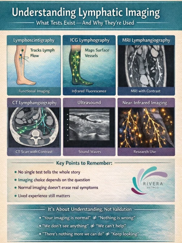
“When lymphedema is suspected, progressing, or not responding as expected, imaging can help answer an important question:
What is actually happening inside the lymphatic system? There isn’t just one imaging option and not every test looks at the same thing.
Lymphoscintigraphy:
What it shows:
- How lymph fluid moves through the body over time.
How it works:
- A small radioactive tracer is injected (usually into the hands or feet), and images track how it travels through lymphatic vessels.
Why it’s used:
- Confirms impaired lymph flow
- Helps diagnose lymphedema
- Often required for insurance or documentation
Limitations:
- It shows function, but not fine structural detail.
Indocyanine Green (ICG) Lymphography:
What it shows:
- Superficial lymphatic vessels and flow patterns just under the skin.
How it works:
- A fluorescent dye is injected and viewed with a near-infrared camera.
Why it’s used:
- Maps lymphatic pathways
- Identifies dermal backflow
- Guides surgical planning
Limitations:
- Only shows superficial lymphatics not deeper structures.
MRI / MR Lymphangiography:
What it shows:
- Soft tissue changes, fluid accumulation, fat deposition, and sometimes lymphatic vessels.
How it works:
- MRI imaging, sometimes with contrast, creates high-resolution images.
Why it’s used:
- Evaluates tissue composition
- Helps differentiate fluid vs fat-dominant swelling
- Useful for surgical planning
Limitations:
- Availability and expertise vary by center.
CT Lymphangiography:
What it shows:
- Anatomical detail of lymphatic structures and surrounding tissues.
How it works:
- CT imaging with contrast.
Why it’s used:
- Specific surgical or anatomical questions
- Less common for routine lymphedema diagnosis
Limitations:
- Radiation exposure; limited functional insight.
Ultrasound:
What it shows:
- Tissue thickness, fibrosis, and fluid characteristics.
How it works:
Sound waves visualize soft tissue.
Why it’s used:
- Evaluates tissue quality
- Monitors changes over time
- Rules out other causes of swelling
Limitations:
- Does not directly visualize lymphatic flow.
Near-Infrared Fluorescence Imaging (Research / Advanced Centers):
What it shows:
- Dynamic lymphatic flow and vessel function.
Why it’s used:
- Research settings
- Specialized surgical centers
- Advanced diagnostic insight”
Stay updated with Hemostasis Today.
-
Oct 21, 2026, 03:36Steve Watson: Ligand Epitope Ratios Influence Antagonist Inhibition
-
Oct 21, 2026, 03:18Stephen D. Coombs: Superficial Venous Thrombosis in Athletes and Importance of Early Recognition and Referral
-
Oct 21, 2026, 02:52Aymar Akilimali: Bleeding During Anticoagulation as a Potential Signal of Occult Cancer
-
Oct 21, 2026, 00:58Anees Ur Rahman: Completing a Two-Week Clinical Rotation at Haemophilia Treatment Centre Rawalpindi
-
Oct 21, 2026, 00:56Caitlyn Mooney: Pulmonary Embolism in Athletes, Recognition and Return to Sport
-
Oct 21, 2026, 00:54Hani Karrar: Pleased to Share Our Review on Emicizumab in Hemophilia A Management
-
Oct 21, 2026, 00:53Akshay Sharma: HSCT Leads to Sustained Improvements in Sickle Cell Cardiomyopathy for Up to 3 Years
-
Oct 21, 2026, 00:51Prateek Meena: The F. A. T. R. N. Mnemonic for Recognizing TTP
-
Oct 20, 2026, 23:51Registration Is Open for WFH CCS 2027 – World Federation of Hemophilia