

Tareq Abadl: Differentiating Cold Agglutination from Hemolysis
Tareq Abadl, Medical Laboratory Specialist and Director of the Blood Bank at Dr. Abdelkader Al-Mutawakkil Hospital, shared a post on LinkedIn:
“Differentiating Cold Agglutination from Hemolysis.
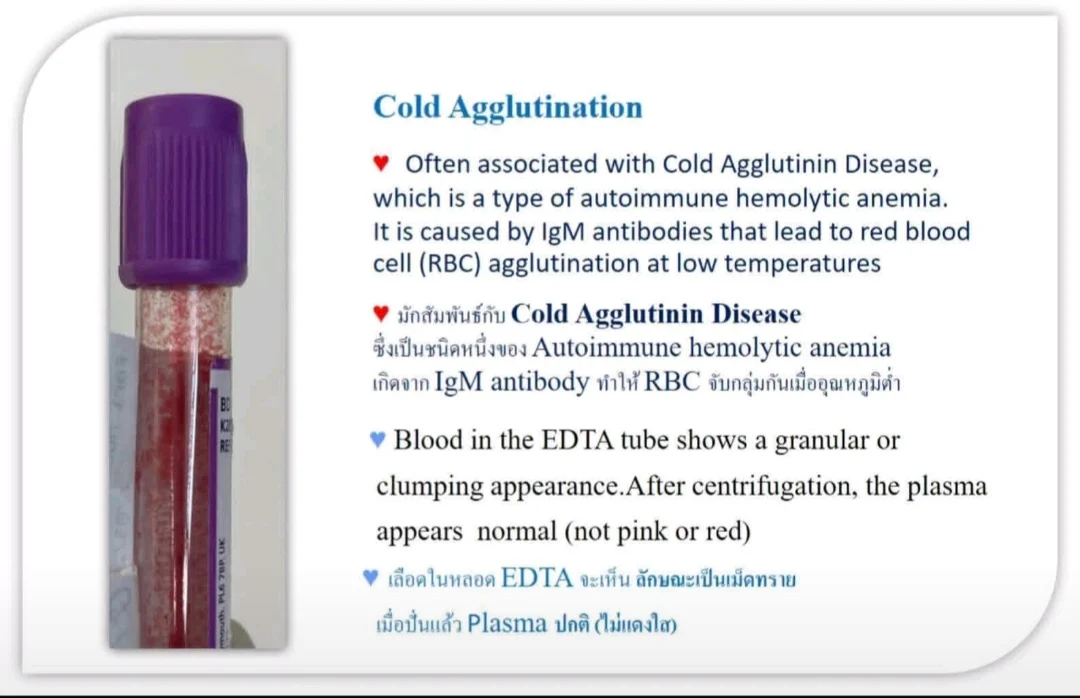
1.Cold Agglutination
Cold agglutination is commonly associated with Cold Agglutinin Disease, a type of autoimmune hemolytic anemia caused by IgM antibodies that bind to red blood cells (RBCs) at low temperatures, leading to RBC clumping.
Specimen Appearance
- EDTA blood shows a granular or clumping appearance
- After centrifugation, the plasma remains clear and normal (not red)
CBC Findings
- Decreased RBC count (falsely low)
- Decreased hematocrit (Hct)
- Hemoglobin (Hb) remains normal or near normal
- Hb and Hct are inconsistent
- MCV falsely increased
- MCHC falsely increased
Cause: The analyzer counts clumped RBCs as a single cell, producing erroneous results.
Blood Smear Findings
- RBCs appear in irregular clumps (agglutination)
Confirmation Test
- Warm the specimen at 37°C for 15–30 minutes
Re-analyze CBC:
- CBC parameters return closer to normal
- RBC clumping disappears on smear
2.Hemolysis
Hemolysis refers to the destruction of RBCs and can occur in vitro (in the tube) or in vivo (in the patient).
A. In Vitro Hemolysis (Specimen Hemolysis)
Specimen Appearance
- Plasma appears clear red after centrifugation (hemolyzed plasma)
- No granular or clumping appearance
CBC Findings
- Hb may decrease slightly
- Hct decreases proportionally
- Hb and Hct remain consistent
Blood Smear
- May see ghost cells
- No agglutination
B. In Vivo Hemolysis
Commonly seen in conditions such as:
- Autoimmune hemolytic anemia
- G6PD deficiency
- Thalassemia
Plasma Findings
- Plasma may appear pink
- Increased bilirubin
- Increased LDH
- Decreased haptoglobin
Blood Smear Findings
- Spherocytes – autoimmune hemolytic anemia
- Bite cells – G6PD deficiency
- Schistocytes – fragmentation hemolysis
Key Laboratory Points (Very Important)
Suspect Cold Agglutination if:
- Granular/clumping blood appearance
- Hb and Hct are inconsistent
- Markedly high MCHC (more than 36–37 g/dL)
Action:
- Warm the sample at 37 degrees Celsius and repeat CBC
Suspect Hemolysis if:
- Plasma is red after centrifugation
- Hb and Hct remain consistent
- No RBC clumping on smear”

More posts featuring Tareq Abadl on Hemostasis Today.
-
Jun 24, 2026, 16:40William Wallace: Vitamin C and Collagen Work as One Biological System
-
Jun 24, 2026, 16:32Arlindo Nascimento de Lemos Junior: Thrombus Echogenicity as a Predictor of Post-Thrombotic Syndrome Severity After DVT
-
Jun 24, 2026, 16:09Jimi Olaghere: The Future of Sickle Cell Disease Research Starts With Collaboration
-
Jun 24, 2026, 16:03Von Willebrand Disease as a Common Yet Under-Recognised Bleeding Disorder – EHC
-
Jun 24, 2026, 15:57Maia Meier: Women with Bleeding Disorders Reach the Summit of Mont Blanc
-
Jun 24, 2026, 15:40Annette Bowyer: The Emerging Role of Extravascular Factor VII in Haemostasis
-
Jun 24, 2026, 13:50Tijjani Balas: Why Early Detection of DVT Matters
-
Jun 24, 2026, 13:43Brandon Michael Henry: Interpreting Complement Activation After AAV Gene Therapy
-
Jun 24, 2026, 13:37Heba Youssef: Heparin-Induced Thrombocytopenia – The Prothrombotic Emergency You Cannot Miss