
Abhijit D: Reticulocyte Count Methods in Hematology Laboratories
Abhijit D., Laboratory Supervisor at NSK Hospital, shared a post on LinkedIn:
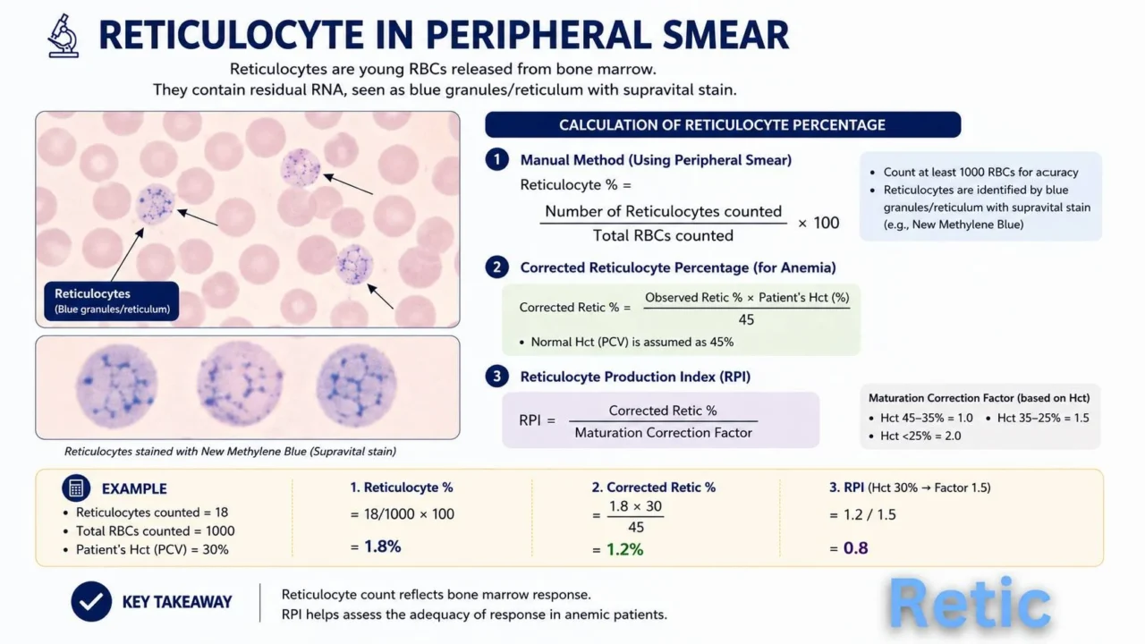
”Reticulocyte Count Methods in Hematology Laboratories
Manual Reticulocyte Count Method
Manual reticulocyte counting is performed using a supravital stain such as New Methylene Blue (NMB) or Brilliant Cresyl Blue. The stain precipitates residual ribosomal RNA inside immature RBCs, appearing as blue granules or reticular networks under the microscope.
Procedure Summary:
- Mix equal volumes of EDTA blood and supravital stain.
- Incubate for 10–15 minutes.
- Prepare peripheral smear and examine under oil immersion.
- Count reticulocytes among 1000 RBCs.
Calculation:
Reticulocyte percentage is determined by dividing the number of reticulocytes counted by the total number of red blood cells counted, then multiplying the result by 100.
Advantages: Low cost, useful in low-resource settings.
Limitations: Subjective, labor-intensive, inter-observer variation.
Fully Automated Reticulocyte Analysis
Modern hematology analyzers use fluorescent flow cytometry for rapid and accurate reticulocyte enumeration. Immature RBCs are stained with fluorescent dyes targeting RNA, and cells are analyzed by laser optics and scatter signals.
Sysmex Asia Pacific Automated Reticulocyte Analysis
Sysmex Global
Sysmex analyzers (XN/XE series) use:
- Fluorescent flow cytometry
- RNA-specific fluorescent dye
- Forward scatter plus side fluorescence detection
Parameters Reported:
- Reticulocyte percent
- Absolute reticulocyte count
- RF (Immature Reticulocyte Fraction)
- RET-He (Reticulocyte Hemoglobin Equivalent)
Widely used for anemia evaluation, marrow response, and iron therapy monitoring.
Mindray Automated Reticulocyte Analysis
Mindray India
Mindray BC series analyzers use:
- Semiconductor laser technology
- Fluorescent staining methods
- Multi-angle light scatter analysis
Features:
- Fast turnaround time
- Automated retic scattergrams
- Reduced manual errors
- Useful for routine hematology workflow
Siemens Automated Reticulocyte Analysis
Siemens Healthineers
Siemens ADVIA analyzers perform reticulocyte analysis using:
- Oxazine fluorescent dyes
- Flow cytometric cell classification
- Dual-angle laser light scatter
Clinical Utility:
- Bone marrow activity assessment
- Monitoring chemotherapy recovery
- Evaluation of hemolytic anemia
Beckman Coulter Automated Reticulocyte Analysis
Beckman Coulter Diagnostics
analyzers use:
- VCS technology (Volume, Conductivity, Scatter)
- Fluorescent nucleic acid staining
- Automated maturity fraction analysis
Advantages:
- High precision and reproducibility
- Integrated CBC plus reticulocyte workflow
- Advanced reticulocyte maturity parameters
Key Clinical Importance of Reticulocyte Count
- Evaluates bone marrow response
- Helps differentiate anemia types
- Monitors treatment response
- Assesses erythropoietic activity”
Stay updated with Hemostasis Today.
-
Jun 28, 2026, 19:05Jack Shuang Hou: Novo Nordisk to Present New Haemophilia Data on Investigational Denecimig at ISTH 2026
-
Jun 28, 2026, 19:05Pinar Demirtepe: Not All “Blood Thinners” Are the Same
-
Jun 28, 2026, 19:05Andrea Cesari: Comparing Catheter-Based Reperfusion Strategies for Intermediate-High-Risk PE
-
Jun 28, 2026, 19:02Nassim Emteir: Interpreting TSAT with Ferritin to Guide Iron Deficiency Diagnosis and Treatment
-
Jun 28, 2026, 19:00Andrew Jackson: Predicting PICC-Related Venous Thrombosis in Patients Undergoing Radiotherapy
-
Jun 28, 2026, 10:34Heghine Khachatryan: Menarche as the First ‘Bleeding Challenge’
-
Jun 28, 2026, 10:13Mohammed Alharbi: Assessing Warfarin Management and Anticoagulation Quality in Older Populations
-
Jun 28, 2026, 10:03Rick Kapur: Current Understanding of FNAIT Regarding Diagnostics, Pathophysiology and Management
-
Jun 28, 2026, 09:55Utkarsh Pandey: Japan Approves Wayrilz for ITP Patients