


Dip Ghosh: Understanding Blood – From Sample to Diagnosis
Dip Ghosh, Student at North South University, shared a post on LinkedIn:
“Understanding Blood – From Sample to Diagnosis
A single drop of blood can reveal anemia, hemolysis, infections, nutritional deficiencies, liver disease, and even life-threatening systemic disorders.
In hematology, every smear tells a diagnostic story.
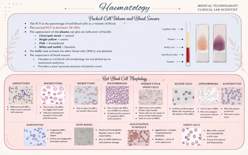
Two essential laboratory› tools remain fundamental in patient diagnosis: Packed Cell Volume (PCV) and Peripheral Blood Smear Examination.
PCV measures the percentage of red blood cells in blood, with normal values generally ranging between 30–50% depending on age and sex.
Even plasma appearance itself provides valuable clinical clues:
- Bright yellow plasma: cterus/jaundice
- Pink plasma: hemolysis
- Milky plasma: lipemia
According to the World Health Organization, anemia affects nearly 1.62 billion people globally, making it one of the world’s most common health disorders (WHO, 2023).
Peripheral smear examination remains essential for identifying its underlying cause.
Key Blood Smear Findings and Clinical Significance:
- Anisocytosis – variation in RBC size, commonly seen in anemia
- Macrocytosis – linked with vitamin B12/folate deficiency and marrow disorders
- Microcytosis and Hypochromia – associated with iron deficiency anemia and thalassemia
- Polychromasia – indicates increased reticulocyte response from bone marrow
- Spherocytes and Ghost Cells – suggest hemolytic conditions
- Schistocytes and Blister Cells – indicators of microangiopathic hemolysis or shear injury
- Target Cells and Acanthocytes – associated with liver disease and membrane abnormalities
Despite advances in automation, studies show that automated hematology analyzers may miss clinically significant morphological abnormalities without manual smear correlation (Barnes et al., 2017).
The International Council for Standardization in Hematology (ICSH) also emphasizes that peripheral smear review remains critical for diagnostic accuracy in hematology laboratories (ICSH, 2021).
Research further demonstrates that schistocyte detection is vital in diagnosing thrombotic microangiopathies, where delayed recognition can significantly increase mortality (Zini et al., 2012). Iron deficiency alone accounts for nearly 50% of global anemia cases .
Even in the era of AI and automation, the microscope remains one of the most powerful diagnostic tools in medicine.
Because behind every stained slide lies:
- A hidden pathology
- A physiological response
- Or the first clue that changes a patient’s life
For medical technologists and clinical laboratory scientists, hematology is more than identifying cells — it is transforming microscopic evidence into clinical decisions that directly impact patient care.
What is the most interesting blood smear finding you’ve encountered during laboratory practice or training?”
Find more posts featuring Dip Ghosh on Hemostasis Today.
-
Oct 20, 2026, 16:51Suzan Nakasendwa: A Growing Stroke Challenge in Uganda
-
Oct 20, 2026, 16:38Steve Moon: How Does Smoking Cessation Duration Affect Ischemic Stroke Risk
-
Oct 20, 2026, 16:25Cedrice Mentor: Reducing Platelet Wastage Through Cold Storage
-
Oct 20, 2026, 16:09Thirunavukkarasu Angappan: Can a Purified Fibrinogen Concentrate Replace Cryoprecipitate
-
Oct 20, 2026, 16:01Kalyan Roy: Platelet Conservation Through Evidence-Based Transfusion Practice
-
Oct 20, 2026, 15:28Stacy E. Croteau: 33 Countries, One Mission – Better Bleeding Disorder Care
-
Oct 20, 2026, 15:19Teresa Barnes: What Does a Pulmonary Embolism Mean for Jalen Hurts?
-
Oct 20, 2026, 15:04Laura S.: Could Speaking Up Prevent Maternal Deaths from Thrombosis?
-
Oct 20, 2026, 14:44Pete Stibbs: VTE in Professional Athletes – When Fitness Is Not Enough