
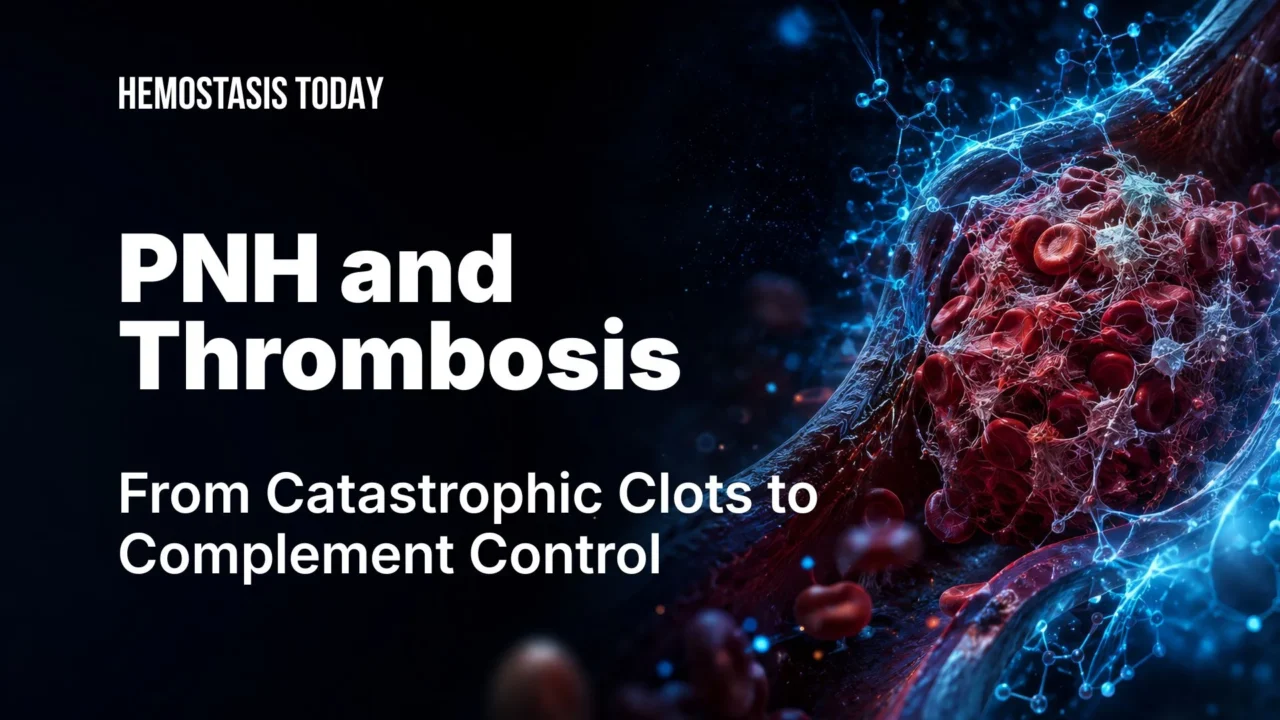
PNH and Thrombosis: From Pathogenesis to Complement Inhibition
When clinicians think of paroxysmal nocturnal hemoglobinuria (PNH), they often picture dark urine, fatigue, and hemolysis. Yet for many patients, the first and most feared manifestation is not hemolysis at all, but a catastrophic thrombosis in an unusual site, such as the hepatic, mesenteric, or cerebral veins.
Before complement inhibitors, thrombotic events accounted for the majority of PNH‑related deaths, and up to one‑third to one‑half of patients experienced a clot during their lifetime.
Today, the paradigm has shifted: complement inhibition is not only about controlling hemolysis, it is the cornerstone of thrombosis prevention in PNH.
What Makes PNH So Thrombogenic?
Paroxysmal nocturnal hemoglobinuria is a rare but potentially life‑threatening hematologic disorder caused by an acquired mutation in hematopoietic stem cells affecting the PIG‑A gene.
The disease is characterized by the loss of glycosylphosphatidylinositol (GPI)‑anchored proteins on the surface of blood cells, particularly the complement regulators CD55 and CD59.Without these protective proteins, red blood cells become vulnerable to uncontrolled complement‑mediated destruction, leading to chronic intravascular hemolysis.
However, hemolysis represents only one component of the disease pathophysiology. Although hemolysis is the hallmark of PNH, thrombosis remains its most serious and feared complication and has historically been responsible for the majority of PNH‑related deaths.
One of the most distinctive features of PNH‑associated thrombosis is its tendency to occur in unusual vascular territories, often in young or otherwise low‑risk individuals.
Which Mechanisms Drive Thrombosis in PNH?
Thrombosis in PNH is not driven by a single abnormality but by multiple interacting pathways, most of them directly linked to complement‑mediated hemolysis.
- Nitric oxide depletion: Free hemoglobin released during intravascular hemolysis scavenges nitric oxide, leading to vasoconstriction, endothelial dysfunction, smooth muscle dystonia, and platelet activation. This contributes to pain crises, esophageal spasm, pulmonary hypertension, and a prothrombotic milieu.
- Platelet activation and microparticles: Complement activation damages platelets, promotes their activation, and drives the release of procoagulant microparticles rich in phosphatidylserine, accelerating thrombin generation.
- Loss of GPI‑anchored regulators: Loss of GPI‑anchored proteins such as tissue factor pathway inhibitor (TFPI) and urokinase‑type plasminogen activator receptor (u‑PAR) impairs endogenous anticoagulant and fibrinolytic mechanisms, shifting the hemostatic balance toward clot formation.
- Inflammation and C5a: C5a‑driven release of inflammatory cytokines (IL‑6, IL‑8, TNF‑α) activates endothelial cells and leukocytes, sustaining a chronic prothrombotic state.
- Neutrophil extracellular traps (NETs): More recently, NET formation has been recognized as an additional contributor, providing a scaffold for platelets and coagulation factors and further enhancing thrombin generation and clot stability.
Together, these mechanisms create a powerful prothrombotic network that explains why PNH sits among the most thrombogenic conditions in hematology.

Thrombosis in PNH: Unusual Sites, Complex Presentations
Thrombosis in PNH most often affects unusual vascular sites. Mesenteric, hepatic, portal, cerebral, and dermal veins are commonly involved, while classic lower‑limb deep vein thrombosis and pulmonary embolism are less frequent than in other thrombophilic states.
Intra‑abdominal thrombosis is the predominant pattern, accounting for roughly two‑thirds of events in historical cohorts, including hepatic, portal, mesenteric, and splenic veins as well as the inferior vena cava.
Hepatic vein thrombosis (Budd–Chiari syndrome) may present acutely with abdominal pain, ascites, and rapidly progressive liver dysfunction, or insidiously with chronic portal hypertension.
Acute presentations often coincide with episodes of brisk hemolysis, while recurrent hepatic vein thrombosis can lead to cirrhosis, portal hypertension, and progressive rerouting of portal circulation; extension into the portal or splenic veins may result in splenic congestion and hypersplenism, and microvascular splanchnic thrombosis may manifest as abdominal pain and mucosal injury.
Cerebral venous thrombosis represents about 10–20% of thrombotic events in PNH, with remaining events occurring in dermal veins and the lower extremities. Cerebral venous thrombosis in PNH may present suddenly with focal deficits or seizures, or more subtly with headache and visual symptoms, making early diagnosis challenging.

Can We Predict Thrombosis Risk in PNH?
Thrombosis risk in PNH is driven primarily by PNH clone size and the degree of intravascular hemolysis. In untreated patients, a granulocyte clone size ≥50–60% and an LDH level ≥1.5 times the upper limit of normal are associated with a substantially increased risk of thromboembolic events.
At a biological level, risk reflects the combined effects of nitric oxide depletion, platelet activation, loss of GPI-anchored anticoagulant proteins, and complement-mediated endothelial inflammation.
Although these factors help identify high-risk patients, thrombosis in PNH remains only partially predictable at the individual level.
Modern Treatment of PNH and Its Impact on Thrombosis
The management of PNH has shifted dramatically with the advent of complement inhibition.
Today, treatment is no longer focused solely on controlling hemolysis but also on preventing one of its most serious complications – thrombosis.
Terminal pathway (C5) inhibitors: The backbone of thrombosis prevention
Eculizumab and ravulizumab, monoclonal antibodies that block complement component C5, have transformed PNH from a highly fatal disease into a largely controllable chronic condition.
In pivotal trials such as TRIUMPH and SHEPHERD, and in large observational cohorts, C5 inhibition significantly reduced intravascular hemolysis, transfusion requirements, and the incidence of major thrombotic events, establishing complement blockade as the cornerstone of thrombosis prevention in PNH.
Ravulizumab, with its extended half‑life and dosing every 8 weeks, provides more sustained complement suppression and stable disease control compared with eculizumab, reducing the risk of breakthrough hemolysis between infusions. Real‑world data suggest that both agents are associated with a consistently low rate of thrombotic complications when adequate complement inhibition is maintained.
Proximal complement inhibitors: Addressing residual hemolysis and prothrombotic drivers
Newer agents that act upstream in the complement cascade have further refined PNH management:
Pegcetacoplan, a C3 inhibitor, controls both intravascular and extravascular hemolysis and is particularly useful in patients with residual hemolysis despite C5 inhibition. Clinical studies have reported low rates of thrombotic events with sustained treatment.
Iptacopan, an oral factor B inhibitor and the first approved oral monotherapy for PNH, provides effective hemolysis control, improves hemoglobin levels, and has been associated with very low thrombotic event rates in clinical trials.
Danicopan, an oral factor D inhibitor used as add-on therapy to C5 inhibitors, helps control persistent extravascular hemolysis and may reduce residual prothrombotic drivers in patients who remain symptomatic despite treatment.
Additional agents, such as the subcutaneous anti‑C5 monoclonal antibody crovalimab, are expanding the therapeutic landscape and may further improve convenience and access.
With effective complement inhibition, thrombosis in PNH has become far less common. In well‑treated patients on modern regimens, thrombotic risk now approaches that of the general population, and PNH‑related mortality has declined dramatically.
The key is early diagnosis, timely initiation of complement blockade in patients with hemolysis or thrombosis, and careful monitoring of LDH, clone size, and breakthrough hemolysis.
Anticoagulation Strategy in PNH‑Associated Thrombosis
Acute thrombosis in PNH requires immediate therapeutic anticoagulation, with thrombolysis or interventional procedures reserved for selected life‑threatening cases depending on the site and severity (for example, massive pulmonary embolism or extensive cerebral venous thrombosis with neurological deterioration).
Management must always combine anticoagulation with effective complement inhibition, which targets the underlying driver of thrombosis.
Duration of anticoagulation is individualized:
In patients with well‑controlled disease on complement inhibitors, no additional major risk factors, and a first provoked thrombosis , anticoagulation may be considered for 3–6 months, followed by careful reassessment of hemolysis, clone size, and ongoing risk.
In recurrent or unprovoked thrombosis, ongoing hemolysis, large PNH clone (≈50–60% or more), or additional thrombophilic risks (e.g., inherited thrombophilia, immobilization, malignancy), long-term or indefinite anticoagulation is generally preferred.
Primary anticoagulation is not routinely recommended in PNH with access to complement inhibitors. It may be considered in patients without complement therapy or in those with large clones and ongoing hemolysis, especially when the granulocyte clone exceeds ~30–50% or additional risk factors are present.
Short‑term prophylaxis should also be strongly considered in high‑risk settings where thrombotic risk increases sharply:
- Major surgery (especially abdominal, hepatic, or neurosurgical procedures)
- Hospitalization with significant immobility
- Pregnancy and the postpartum period
Procedures should ideally be timed close to complement inhibitor dosing to minimize breakthrough hemolysis and peri‑procedural thrombosis. In pregnancy, eculizumab and ravulizumab have an extensive safety record and are generally continued throughout gestation, with anticoagulation tailored to individual thrombotic risk and obstetric considerations.
FAQ
1. Why is thrombosis the most feared complication of PNH?
Thrombosis has historically been the leading cause of death in PNH. Before complement inhibitors became available, up to 40–50% of patients experienced a thrombotic event during their lifetime.
2. What makes PNH so thrombogenic?
Complement-mediated hemolysis drives nitric oxide depletion, platelet activation, endothelial dysfunction, release of procoagulant microparticles, inflammation, and neutrophil extracellular trap (NET) formation. Together these processes create a highly prothrombotic environment.
3. Why do clots in PNH occur in unusual sites?
Complement activation, endothelial injury, and low-flow venous circulation predispose patients to thrombosis in the hepatic, portal, mesenteric, splenic, and cerebral veins rather than typical lower-limb veins.
4. Which patients with PNH are at highest thrombotic risk?
Risk is highest in patients with large PNH granulocyte clones greater than 50–60%, elevated LDH levels at least 1.5 times the upper limit of normal, active intravascular hemolysis, high disease activity, and a history of prior thrombosis.
5. Can thrombosis be the first manifestation of PNH?
Yes. Budd–Chiari syndrome, splanchnic vein thrombosis, and cerebral venous thrombosis can precede diagnosis and occasionally represent the first clinical presentation of PNH.
6. How do C5 inhibitors prevent thrombosis?
Eculizumab and ravulizumab block terminal complement activation, suppress intravascular hemolysis, reduce platelet activation, and lower thrombotic risk by approximately 80% or more.
7. What role do proximal complement inhibitors play in thrombosis prevention?
Agents targeting C3, factor B, or factor D provide broader complement control, reduce residual hemolysis, and maintain very low thrombotic event rates during treatment.
8. How is acute thrombosis managed in PNH?
Management combines immediate therapeutic anticoagulation with prompt initiation or optimization of complement inhibition. Selected severe cases may require thrombolysis or endovascular intervention depending on the site and severity.
9. When is long-term anticoagulation indicated?
Extended or indefinite anticoagulation is generally recommended for patients with recurrent thrombosis, persistent hemolysis, large PNH clones, or additional thrombophilic risk factors.
10. How has the prognosis of PNH changed in the complement inhibitor era?
Effective complement inhibition has dramatically reduced thrombotic complications, improved survival, and transformed PNH from a frequently fatal disease into a manageable chronic condition.
Written by Elen Avetisyan, MD
Stay updated with Hemostasis Today.
-
Jul 17, 2026, 23:05Leonardo Roever: New Perspectives on Thromboinflammation in Acute Ischemic Stroke
-
Jul 17, 2026, 22:36Maeva May: Start Your Stroke Research Career with a Stroke Association Fellowship
-
Jul 17, 2026, 20:19Thomas Muenzel: Proud to Deliver the Sylviane Lévy-Toledano Memorial Plenary Keynote Lecture at ISTH 2026
-
Jul 17, 2026, 20:16Karin Leiderman: Proud of Jamie Madrigal for Receiving the JTH Editors’ Award
-
Jul 17, 2026, 20:14Lily Bull: Presenting New Research and Building Connections at ISTH 2026
-
Jul 17, 2026, 20:12Modern Concepts in Traumatic Disseminated Intravascular Coagulation – JTH
-
Jul 17, 2026, 20:10Nahid A. Qushmaq: Science, Collaboration, and Inspiration at ISTH 2026
-
Jul 17, 2026, 18:20Sarah Jones: Bringing Vessel-on-a-Chip Research to ISTH 2026
-
Jul 17, 2026, 18:06Virginia Barras Sánchez: United by a Commitment to Hemophilia at ISTH 2026