


Ifeanyichukwu Ifechidere: Why Fibrinolysis Matters as Much as Clot Formation
Ifeanyichukwu Ifechidere, Specialist Biomedical Scientist at Sheffield Teaching Hospitals NHS Foundation Trust, shared a post on LinkedIn:
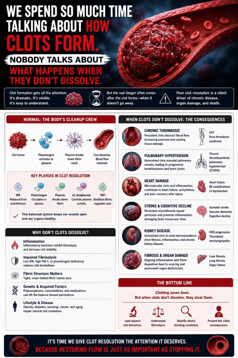
“We spend so much time talking about how clots form. Nobody talks about what happens when they don’t dissolve.
Haemostasis has two sides.
Clot formation gets all the attention — the factors, the cascade, the platelets, the thrombin burst.
But fibrinolysis — the system that breaks clots down is equally critical. And when it fails, patients bleed catastrophically or clot relentlessly.
This is haemostasis’s forgotten half. Let’s fix that.
The Fibrinolytic System — how it works:
At the centre of fibrinolysis is a single, elegant mechanism:
Plasminogen is converted to plasmin, which then degrades fibrin.
But the elegance is in the regulation.
- Tissue Plasminogen Activator (tPA) is released from endothelial cells in response to thrombus formation. It binds fibrin within the clot — converting plasminogen to plasmin with extraordinary specificity. Precise. Targeted. Intentional.
- Plasmin is the engine. Once activated, it degrades fibrin into fibrin degradation products — D-dimers among them. It also degrades fibrinogen, Factor V, and Factor VIII if insufficiently regulated.
- PAI-1 (Plasminogen Activator Inhibitor-1) is the primary brake on tPA. Elevated PAI-1 suppresses fibrinolysis — seen in obesity, metabolic syndrome, sepsis, and acute thrombotic states. High PAI-1 means clots persist longer than they should.
- Alpha-2 Antiplasmin neutralises circulating plasmin instantly — confining fibrinolytic activity to the clot surface. When alpha-2 antiplasmin is deficient, systemic plasmin runs unchecked.
When fibrinolysis fails — in both directions:
Hyperfibrinolysis — the system accelerates beyond control:
- Seen in trauma, liver disease, DIC, post-thrombolysis, major obstetric haemorrhage
- Presents as uncontrolled bleeding, rapidly dissolving clots, plummeting fibrinogen
- TEG/ROTEM signatures show characteristic lysis patterns — LY30, ML, FIBTEM amplitude loss
Hypofibrinolysis — the system is suppressed:
- Seen in thrombotic APS, elevated PAI-1 states, COVID-19 coagulopathy, obesity
- Clots form and persist — driving DVT, PE, microvascular thrombosis
- Increasingly recognised as a distinct thrombotic phenotype missed by standard coagulation screens
What your standard laboratory panel misses:
PT. APTT. Fibrinogen. D-dimer.
None of these directly assess fibrinolytic capacity. D-dimer tells you fibrinolysis has occurred — not whether it is functioning optimally or pathologically.
A coagulation screen without fibrinolytic assessment is half a haemostatic picture.
Quick Poll — I want to know where you stand:
In your clinical or laboratory practice, how often is fibrinolytic function formally assessed?
- Regularly — we use TEG/ROTEM routinely
- Occasionally — in specific bleeding scenarios
- Rarely — standard coagulation panel only
- Never — we don’t have access to viscoelastic testing
Drop your answer in the comments. Let’s map the real-world gap together.
Has hyperfibrinolysis or hypofibrinolysis ever changed your clinical management?
Share below in the comment section.”
Find more posts featuring Ifeanyichukwu Ifechidere on Hemostasis Today.
-
Sep 18, 2026, 16:51Adarsh Krishnan Sundara Rajan: Is VTE the Most Underdiagnosed Critical Threat in Modern Healthcare?
-
Sep 18, 2026, 16:19CRISPR and Hemophilia: The Next Frontier of In Vivo Gene Editing
-
Sep 18, 2026, 16:13Christian Irsara: PF4 Immunothrombosis Following Natural Adenovirus Infection
-
Sep 18, 2026, 16:10Leo Nicolai։ The Spleen Emerges as a Key Regulator of Thromboinflammation
-
Sep 18, 2026, 16:05Targeting Factor XI to Reduce Thrombosis Without Compromising Hemostasis – ISTH
-
Sep 18, 2026, 15:54Swapnil Pawar: Why Aren’t We Using Cryopreserved Platelets Everywhere Yet?
-
Sep 18, 2026, 15:37Abdul Mannan: Evaluating Longitudinal Platelet Trends Beyond Standard Reference Intervals
-
Sep 18, 2026, 15:30Peter A. Everts։ Platelet Phenotype May Hold the Key to More Consistent PRP Therapy
-
Sep 18, 2026, 15:25Brandon Temple: Turning Personal Experience Into Advocacy for wAIHA Research