

Chokri Ben Lamine: Septic Embolism in Hematology – Postgraduate Thread
Chokri Ben Lamine, Assistant Consultant at King Faisal Specialist Hospital and Research Center, shared a post on X:
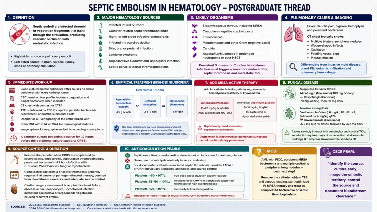
“Septic embolism in hematology – postgraduate thread
1. Septic emboli are infected thrombi or vegetation fragments that travel through the circulation, producing vascular occlusion with metastatic infection.
- Right-sided source – pulmonary emboli
- Left-sided source – brain, spleen, kidney, limbs or coronary circulation.
2.Major hematology sources:
- Infected PICC/CVC/port
- Catheter-related septic thrombophlebitis
- Right- or left-sided infective endocarditis
- Infected intracardiac device
- Skin, oral or perianal infection
- Lemierre syndrome
- Angioinvasive Candida and Aspergillus infection
- Septic pelvic or portal thrombophlebitis.
3.Likely organisms:
- Staphylococcus aureus, including MRSA
- Coagulase-negative staphylococci
- Enterococcus
- Pseudomonas and other Gram-negative bacilli
- Candida
- Aspergillus/Mucorales in prolonged neutropenia or post-HSCT.
Persistent S. aureus or Candida bloodstream infection must trigger a search for endocarditis, septic thrombosis and metastatic foci.
4.Pulmonary clues: fever, pleuritic pain, hypoxia, hemoptysis and persistent bacteremia.
CT chest typically shows:
- Multiple bilateral peripheral nodules
- Wedge-shaped infarcts
- Cavitation
- Feeding-vessel sign
- Pleural effusion.
Differentiate from invasive mold disease, bland PE, leukemic infiltration and pulmonary hemorrhage.
5.Immediate work-up:
- Blood cultures before antibiotics if this causes no delay: peripheral with every catheter lumen
- CBC, renal or liver profile, lactate, coagulation and fungal biomarkers when indicated
- CT chest with contrast or CTPA
- TTE should be followed by TEE if suspicion persists, bacteremia is persistent, or prosthetic material exists
- Doppler or CT venography of the catheterized vein
- Brain MRI with CTA or MRA for neurological features
Image spleen, kidney, spine and joints according to symptoms. A catheter culture becoming positive for at least 2 hours before the peripheral culture supports CRBSI.
6.Empirical treatment in high-risk neutropenia – give within approximately 1 hour:
- Piperacillin–tazobactam (Tazocin) 4.5 g IV q6h, or
- Cefepime (Maxipime) 2 g IV q8h, or
- Meropenem (Meronem) 1 g IV q8h
Use local antibiogram, previous colonization and renal adjustment. Meropenem is favored when ESBL infection, septic shock or a resistant Gram-negative pathogen is likely.
7.Add MRSA-active therapy for catheter infection, skin focus, pneumonia, hemodynamic instability or known MRSA:
- Vancomycin (Vancocin) 15–20 mg/kg IV q8–12h, preferably AUC-guided target 400–600
- Alternative: daptomycin (Cubicin) 8–10 mg/kg IV q24h for bacteremia or right-sided endocarditis
Daptomycin is inactivated by pulmonary surfactant – do not use for primary pneumonia.
Vancomycin: nephrotoxicity; avoid unnecessary nephrotoxic combinations.
8.Fungal disease:
- Suspected Candida CRBSI: micafungin (Mycamine) 100 mg IV daily or caspofungin (Cancidas) 70 mg loading, then 50 mg daily
- Invasive aspergillosis: voriconazole (Vfend) 6 mg/kg IV q12h ×2, followed by 4 mg/kg q12h, or isavuconazole (Cresemba) 372 mg q8h ×6 dosesdoses, followed by 372 mg daily
Azoles strongly interact with venetoclax and several TKIs; venetoclax requires major dose reduction. Voriconazole prolongs QT, whereas isavuconazole shortens QT.
9. Source control is essential: remove the catheter when CRBSI is complicated by severe sepsis, endocarditis, suppurative thrombophlebitis, persistent bacteremia beyond 72 hours, or infection with S. aureus, Pseudomonas, fungi or mycobacteria.
Complicated bacteremia or septic thrombosis generally requires 4–6 weeks of pathogen-directed therapy, counted from bloodstream clearance and adequate source control.
Cardiac surgery assessment is required for heart failure, abscess or pseudoaneurysm, uncontrolled infection, persistent bacteremia or large or mobile vegetations causing recurrent emboli.
10.Anticoagulation pearls:
- Septic embolism or endocarditis alone is not an indication for anticoagulation
- Never use thrombolysis routinely in septic embolism
- For documented catheter-associated septic thrombosis, consider LMWH or UFH individually alongside antibiotics and source control
- Platelets count greater than 50×10⁹/L: full-dose anticoagulation usually feasible
- Platelets 25–50: reduced-dose LMWH or transfusion-supported treatment for high-risk thrombosis
- Platelets count less than 25: generally hold anticoagulation
Intracranial hemorrhage or mycotic aneurysm overrides these thresholds.
MCQ: AML with PICC, persistent MRSA bacteremia and multiple cavitating peripheral lung nodules – best next step?
Remove the catheter, obtain TEE and venous imaging, start optimized IV MRSA therapy and treat as complicated bacteremia or septic thrombophlebitis.
OSCE pearl: ‘Identify the source, culture early, image the embolic territory, control the source and document bloodstream clearance.’
Sources: 2023 ESC endocarditis guideline, ESC guideline summary, IDSA catheter-related infection guideline, 2024 AGIHO febrile-neutropenia update, cancer-associated thrombosis with thrombocytopenia.”
Other posts featuring Chokri Ben Lamine on Hemostasis Today.
-
Oct 2, 2026, 00:33Marwa Mostafa: Hemoglobinopathies – When Globin Genes Go Wrong
-
Oct 1, 2026, 23:46Behnood Bikdeli: Global Burden of Venous Thromboembolism
-
Oct 1, 2026, 23:46Sthanu Subramanian: Clinical Presentation, Risk Factors, Diagnosis, and Neuroimaging Findings in CVT
-
Oct 1, 2026, 23:45Martha Gulati: Oral MHT Is Associated With Increased Thrombotic Risk
-
Oct 1, 2026, 23:44Priya Sriskandarajah: HEMA-MED Trial Exploring Roginolisib Plus JAK Inhibitors in Myelofibrosis
-
Oct 1, 2026, 23:44Inge Vanhomwegen: Join Our Upcoming Webinar on D-dimer Testing
-
Oct 1, 2026, 23:43Bianca Rocca: Beyond API-CAT – Who Should Continue Anticoagulation, And for How Long?
-
Oct 1, 2026, 23:26Sara Gaafar Ibnauf Suliman: The Changing Role of Bariatric Surgery in the GLP-1 Era
-
Oct 1, 2026, 23:16Ahmed Reda: New Ferritin Cutoffs For Iron Deficiency – Ash 2026