

Maryam Alaa Alsaid: Diagnostic Puzzle of Sickle Beta Thalassemia
Maryam Alaa Alsaid, Surgical Pathology Trainee at Children’s Cancer Hospital, shared a post on LinkedIn:
“Diagnostic Puzzle: Sickle-Beta Thalassemia (S/β-Thalassemia)
When Sickle Cell Disease and Beta Thalassemia Trait overlap, the clinical and laboratory picture becomes a fascinating diagnostic challenge.
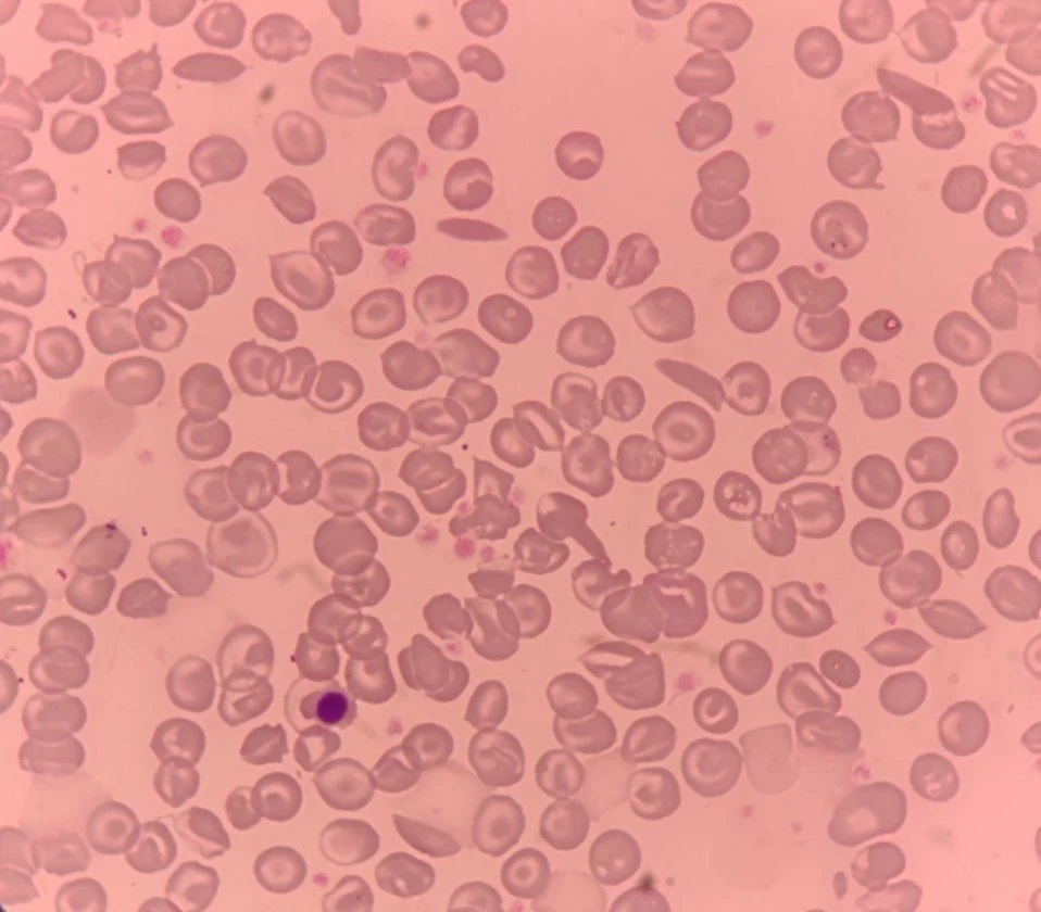
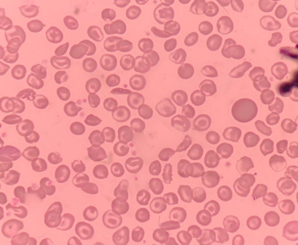
- Peripheral Blood Smear and Lab Highlights:
- RBC Morphology: Marked Microcytosis and Hypochromia (driven by the Thalassemia component), along with prominent Sickle cells (Drepanocytes), Target cells (Codocytes), Anisopoikilocytosis, and Polychromasia.
- CBC Indices: Markedly reduced MCV and MCH, setting it apart from classic Homozygous Sickle Cell Anemia (HbSS) where MCV is typically normocytic unless iron deficiency co-exists.
Diagnostic Subtypes and Differentiation:
1.Hb S / β⁰-Thalassemia:
No HbA synthesized (HbA: 0 percent).
Clinically severe, closely mimicking Homozygous HbSS.
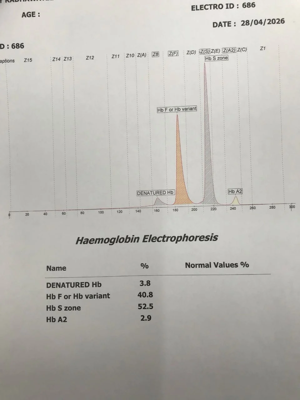
Electrophoresis shows high HbS (~80–90 percent), elevated HbA₂ (elevated above 3.5 percent), and variable HbF.
2.Hb S / β⁺-Thalassemia:
Reduced synthesis of normal beta chains (HbA: 5–30 percent).
Generally milder clinical course compared to S/β⁰.
Electrophoresis reveals the presence of a small HbA fraction.
Key Takeaway for Hematology Labs:
Always evaluate MCV and Hb Electrophoresis/HPLC concurrently!
A patient with sickle cell features and disproportionately low MCV/MCH should strongly raise suspicion for co-inherited Beta-Thalassemia or Alpha-Thalassemia trait.
To my Hematology and Pathology colleagues:
What is your primary screening protocol when differentiating Hb S/β⁰ from classic HbSS in routine clinical practice?”
Stay updated with Hemostasis Today.
-
Oct 20, 2026, 16:51Suzan Nakasendwa: A Growing Stroke Challenge in Uganda
-
Oct 20, 2026, 16:38Steve Moon: How Does Smoking Cessation Duration Affect Ischemic Stroke Risk
-
Oct 20, 2026, 16:25Cedrice Mentor: Reducing Platelet Wastage Through Cold Storage
-
Oct 20, 2026, 16:09Thirunavukkarasu Angappan: Can a Purified Fibrinogen Concentrate Replace Cryoprecipitate
-
Oct 20, 2026, 16:01Kalyan Roy: Platelet Conservation Through Evidence-Based Transfusion Practice
-
Oct 20, 2026, 15:28Stacy E. Croteau: 33 Countries, One Mission – Better Bleeding Disorder Care
-
Oct 20, 2026, 15:19Teresa Barnes: What Does a Pulmonary Embolism Mean for Jalen Hurts?
-
Oct 20, 2026, 15:04Laura S.: Could Speaking Up Prevent Maternal Deaths from Thrombosis?
-
Oct 20, 2026, 14:44Pete Stibbs: VTE in Professional Athletes – When Fitness Is Not Enough