

Omar Adwan: Summary of Blood Smear Examination Process
Omar Adwan, Medical Laboratory Technologist at Modawah lab center, shared a post on LinkedIn:
“Summary of Blood Smear Examination Process
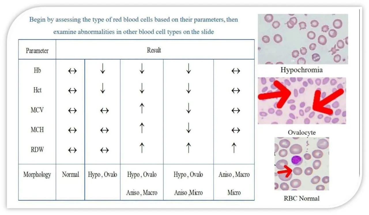
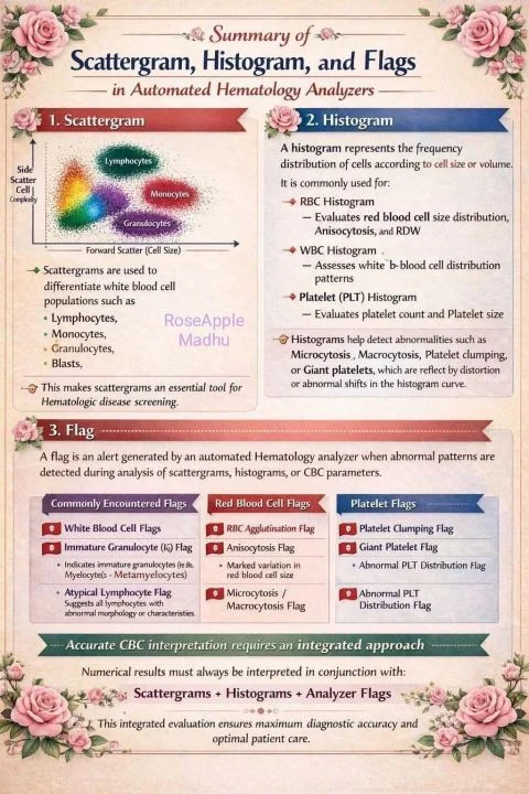
1.Start with Basic Cell Parameters
Review WBC, Platelet, and RBC values from the automated analyzer (e.g., Count, MCV, MCH, MCHC, RDW, Hb, Hct).
Use these parameters to anticipate possible abnormalities:
- High WBC – Possible infection or leukemia
- Low Platelets – Risk of abnormal bleeding
- Abnormal RBC parameters – Anemia or red cell disorders
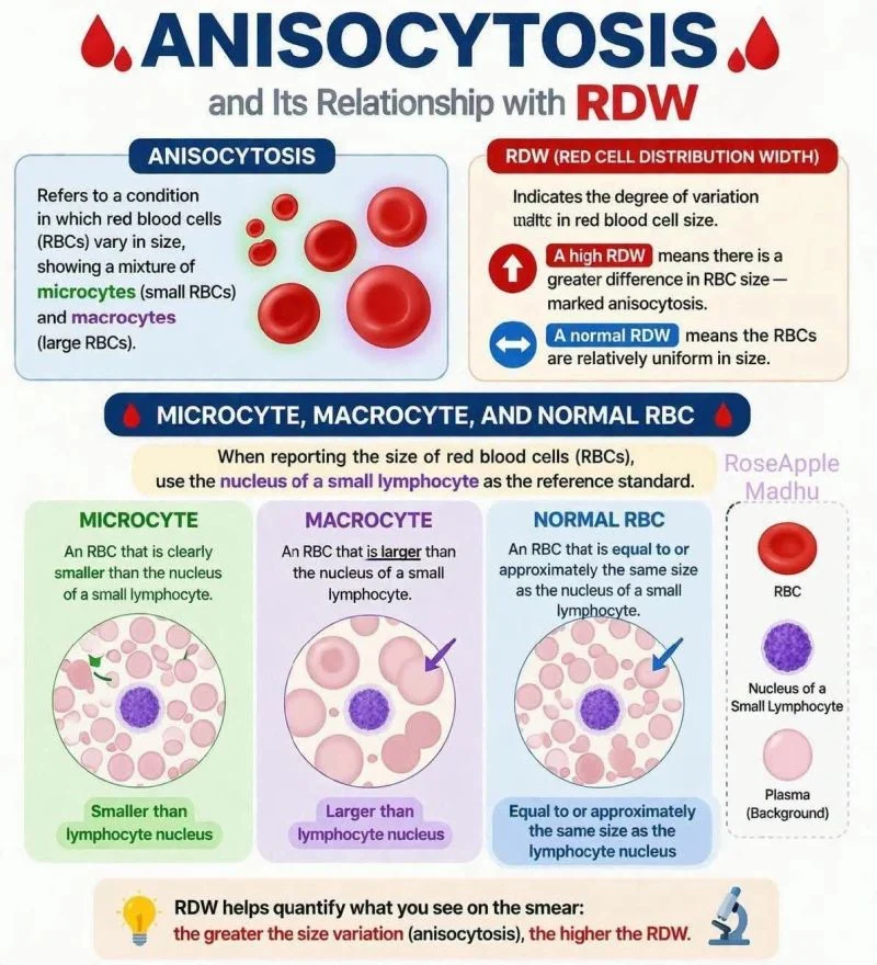
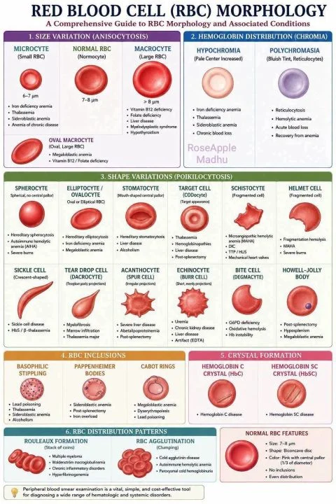
2.Evaluate Red Blood Cell (RBC) Morphology
Assess size, color, shape, and uniformity of red blood cells.
Key classification criteria:
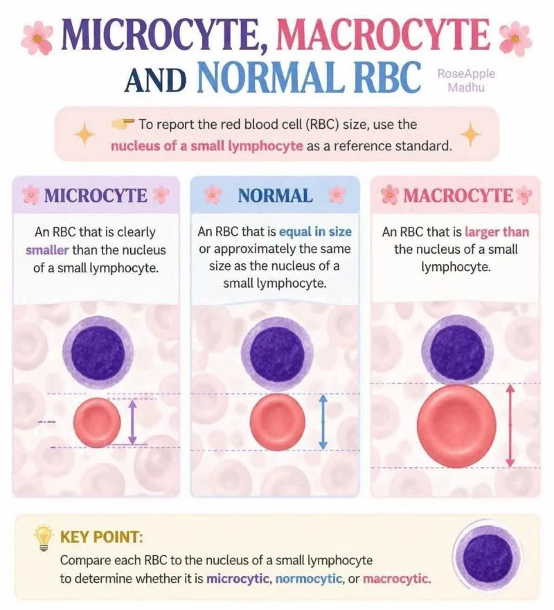
By size:
- Normocytic (normal)
- Microcytic (small)
- Macrocytic (large)
By color:
- Normochromic (normal)
- Hypochromic (pale)
Important:
Begin by determining the RBC type based on analyzer parameters to identify the predominant pattern. Then, examine the smear under the microscope for additional abnormalities.
Following this sequence provides a structured approach and improves diagnostic accuracy.
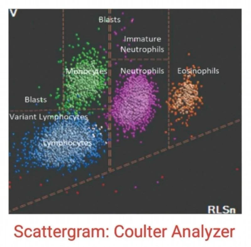
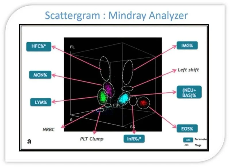
3.Examine All Cell Lines for Abnormalities on the Slide
WBC (White Blood Cells):
- Immature forms
- Blast cells
- Hypersegmented neutrophils
Platelets:
- Platelet clumping
- Giant platelets
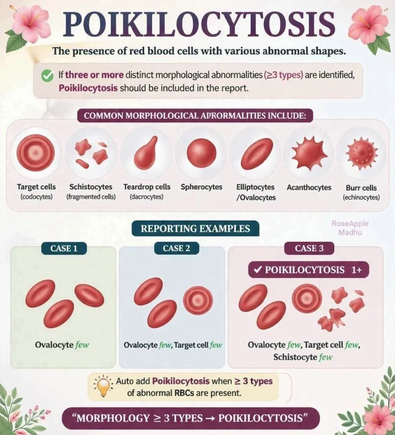
RBC (Red Blood Cells):
- Target cells
- Spherocytes
- Schistocytes
- Anisocytosis
Key Takeaway:
Review analyzer results, than classify RBC type, after that identify all morphological abnormalities.
This systematic approach ensures comprehensive, accurate reporting and good correlation with automated analyzer findings.”

Other posts featuring Omar Adwan on Hemostasis Today.
-
Jun 30, 2026, 16:35Shabeeha Ranna: Improving Cancer Care Through Venous Thromboembolism Risk Assessment and Prevention
-
Jun 30, 2026, 16:20Fiona Robinson: ”Living with Glanzmann Thrombasthenia” Live on Next Week’s NBDF Wednesday Webinar
-
Jun 30, 2026, 16:18Martha Kilner: What a Deep Vein Thrombosis Taught Me About Life Structure
-
Jun 30, 2026, 15:59Alejandro González Veliz: Understanding the Mechanisms Driving Stent Thrombosis
-
Jun 30, 2026, 15:46Omid Seidizadeh: Honored to Join NordCoag 2026 to Celebrate 100 Years of von Willebrand Disease
-
Jun 30, 2026, 15:42Obstetric Disseminated Intravascular Coagulation: Early Recognition and Life-Saving Management Strategies
-
Jun 30, 2026, 15:38Ammir Abuzahra: A Case Report of Upper Extremity Deep Vein Thrombosis Presenting as Cellulitis
-
Jun 30, 2026, 15:33Francois Delvoye: Thrombo-Inflammation and NETs May Represent the Next Therapeutic Frontier in SAH
-
Jun 30, 2026, 15:23The DETECT-VTE Study Reminds Us That VTE Doesn’t Always Look Typical – RPTH