


Seema Dawood: Morphologic Differences Between Platelet Types
Seema Dawood, Medical Laboratory Technologist at The Aga Khan University Hospital, shared a post on LinkedIn:
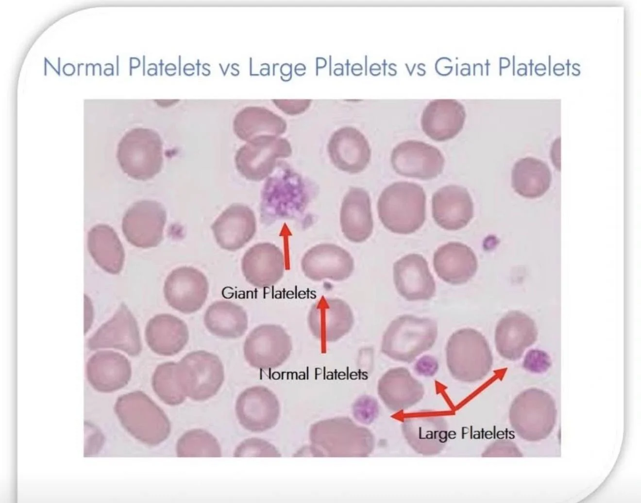
”Differences Between Normal Platelets, Large Platelets, and Giant Platelets
Platelets are essential components of blood that play a major role in blood clotting and hemostasis.
They can appear in various sizes and morphologic forms, as described below:
Normal Platelets
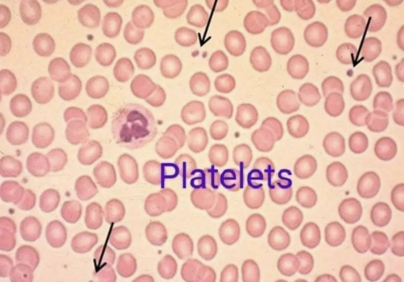
- Normal platelets are small, measuring approximately 2–3 micrometers in diameter.
- They are clearly smaller than red blood cells (RBCs).
- Morphologically, they are usually round to oval with evenly distributed purple granules within the cytoplasm.
- The presence of platelets with normal size and adequate numbers generally reflects balanced platelet production and normal bone marrow function.
Large Platelets
- Large platelets are bigger than normal platelets but are generally still smaller than red blood cells.
- They are commonly seen in conditions associated with increased platelet production, such as excessive platelet destruction in Immune Thrombocytopenia (ITP) or during recovery after blood loss.
- These platelets are often referred to as ‘young platelets’ because newly released platelets from the bone marrow tend to be larger and contain more prominent granules.
- Therefore, the presence of large platelets usually indicates an active and appropriate bone marrow response to platelet loss.
Giant Platelets
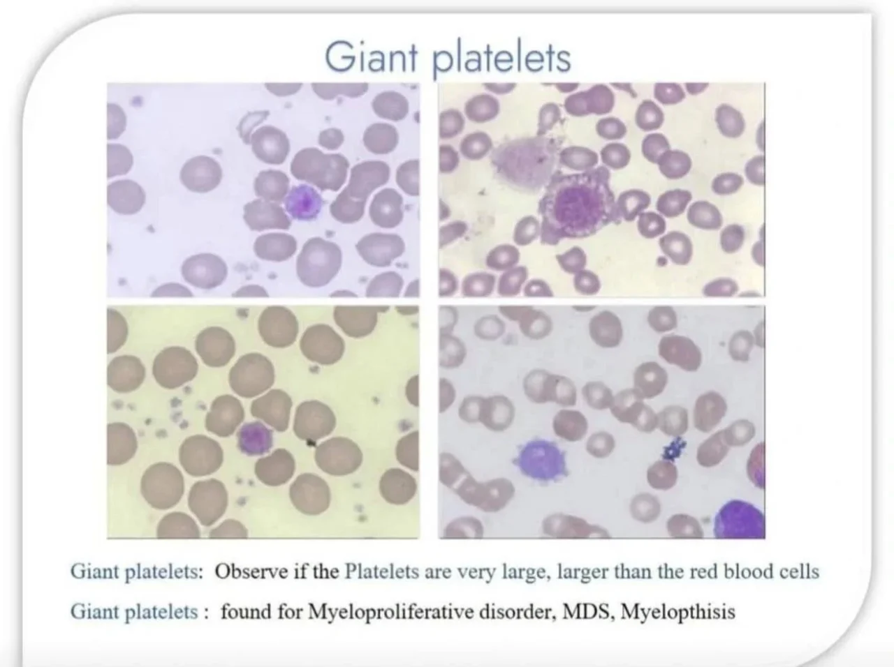
- Giant platelets are extremely large platelets that may approach or even exceed the size of red blood cells.
- They are considered clinically significant abnormalities and are often associated with inherited platelet disorders or abnormalities in megakaryocyte maturation.
Conditions associated with giant platelets include:
- Bernard–Soulier Syndrome
- May–Hegglin Anomaly
- Gray Platelet Syndrome
- Myeloproliferative disorders
- Severe platelet destruction or abnormal platelet production states
Laboratory Significance
The presence of large or giant platelets is highly important in hematology because automated hematology analyzers may mistakenly classify these oversized platelets as red blood cells. This can result in falsely low platelet counts or pseudothrombocytopenia.
Therefore, peripheral blood smear examination under the microscope remains essential for accurate platelet morphology assessment.
Summary
- Normal Platelets – Small, uniform in size, and clearly smaller than RBCs
- Large Platelets – Larger than normal, usually associated with increased platelet production
- Giant Platelets – Extremely large platelets, commonly associated with hematologic and inherited platelet disorders.”
Stay updated with Hemostasis Today.
-
Sep 29, 2026, 22:45Victor Agunyego: Paracetamol – The Most Common Drug We Underestimate
-
Sep 29, 2026, 22:42Nova Sinclair: Advancing the Future of Hematology – Meet Our 2027 Organizing Committee
-
Sep 29, 2026, 22:41Lorenz Räber: Intracoronary Imaging in PCI – Why, When, How, and Which Modality?
-
Sep 29, 2026, 22:31Selma Turkovic: Pleased to Deliver the Opening Presentation at Kingston Hospital
-
Sep 29, 2026, 22:31Miguel Teran: Ultrasound and Simulation in Arterial Blood Sampling
-
Sep 29, 2026, 21:47Javed Anees: DADA2 – When a Young Stroke Is More Than a Stroke
-
Sep 29, 2026, 22:28Mohammad Refaei: Navigating Your Career in Hematology From Training to Practice
-
Sep 29, 2026, 22:27Matteo Foschi: 10 Clinical Pearls for Antithrombotic Therapy in Secondary Stroke Prevention
-
Sep 29, 2026, 22:26A Global Call for Action on Cardiovascular Health on World Heart Day – ISTH