


Gurpreet Kaur Sagoo: The Power of AI Based Morphological Fingerprinting in Predicting Myelofibrosis Progression
Gurpreet Kaur Sagoo, Professor at Armed Forces Medical College, shared a post on LinkedIn about a recent article she and her colleagues co-authored, published in Indian Journal of Hematology and Blood Transfusion, adding:
“Morphometric analysis of megakaryocytes plays a central role in diagnosing and subclassifying Philadelphia-chromosome negative Myeloproliferative Neoplasms (MPNs), as specific cellular and spatial abnormalities distinguish Essential Thrombocythemia (ET), Polycythemia Vera (PV), and Primary Myelofibrosis (PMF).
While manual histopathological evaluation is a standard World Health Organization (WHO) requirement, digital morphometry and machine learning workflows on bone marrow trephine biopsies provide precise, reproducible, and automated quantification of these parameters to avoid subjective variation.
Primary Morphometric Parameters Analyzed Digital image analysis systems typically measure several core quantitative indicators from H&E or immunohistochemically stained sections:
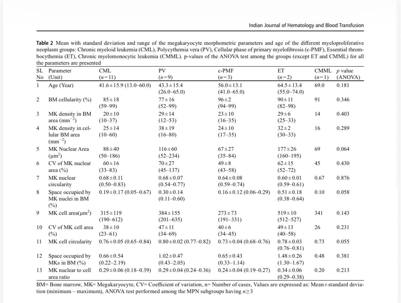
- Cellular Size and Area: Calculated via the cytoplasmic major diameter and total cytoplasmic area.
- Nuclear Area and Perimeter: Measures the total footprint of the nucleus and its boundary length.
- Nuclear-to-Cytoplasmic (N:C) Ratio: Derived by dividing the nuclear area by the overall cytoplasmic area.
- Nuclear Shape Indices: Quantified via the Nuclear Roundness Factor and Nuclear Contour Ratio to capture the irregularity or lobation profile.
- Topographical Clustering and Density: Quantitative spatial alignment mapping whether cells form loose or tight clusters (3 or more juxtaposed cells) and their spatial distance from bone trabeculae or blood sinuses.
- Advanced and Predictive Morphometric markers:
Megakaryocytic Activation (M-ACT Index):
A specialized morphometric index scored on the coexistence of megakaryocytic emperipolesis (intact neutrophils passing through megakaryocyte cytoplasm), tight cluster count, and dense collagen fiber perimeters.
High M-ACT indices mathematically correlate with higher JAK2 V617F allele burden and predict rapid progression to secondary myelofibrosis.
AI-Based Morphological Fingerprinting: Advanced neural networks extract up to 9 distinct megakaryocytic phenotypes from bone marrow images based on shape variations, generating cohort-wide radar plots.
This high-throughput tool isolates true MPNs from reactive mimics (like immune thrombocytopenia or reactive thrombocytosis) with an area under the curve (AUC) accuracy of up to 0.95.”
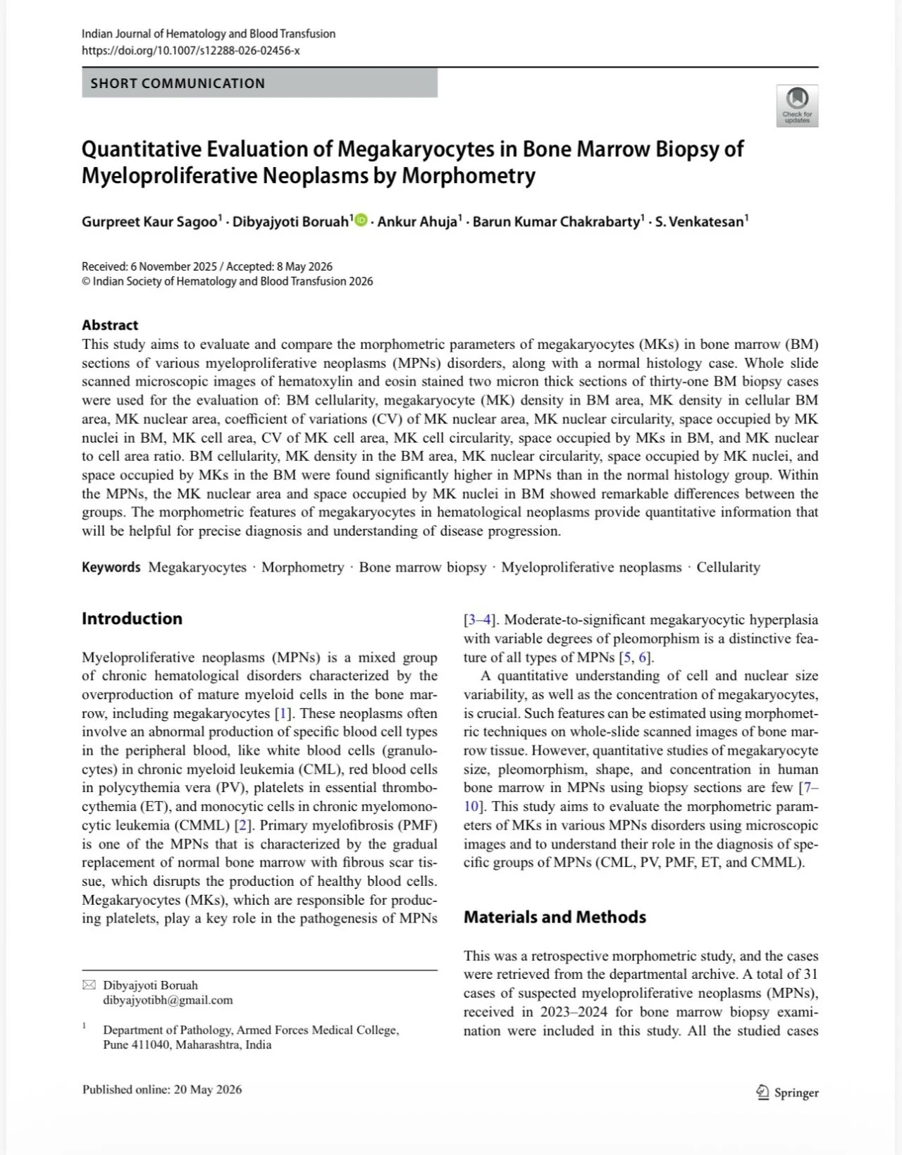
Title: Quantitative Evaluation of Megakaryocytes in Bone Marrow Biopsy of Myeloproliferative Neoplasms by Morphometry
Authors: Gurpreet Kaur Sagoo, Dibyajyoti Boruah, Ankur Ahuja, Barun Kumar Chakrabarty, S. Venkatesan
Stay updated on all scientific advances with Hemostasis Today.
-
Oct 20, 2026, 16:51Suzan Nakasendwa: A Growing Stroke Challenge in Uganda
-
Oct 20, 2026, 16:38Steve Moon: How Does Smoking Cessation Duration Affect Ischemic Stroke Risk
-
Oct 20, 2026, 16:25Cedrice Mentor: Reducing Platelet Wastage Through Cold Storage
-
Oct 20, 2026, 16:09Thirunavukkarasu Angappan: Can a Purified Fibrinogen Concentrate Replace Cryoprecipitate
-
Oct 20, 2026, 16:01Kalyan Roy: Platelet Conservation Through Evidence-Based Transfusion Practice
-
Oct 20, 2026, 15:28Stacy E. Croteau: 33 Countries, One Mission – Better Bleeding Disorder Care
-
Oct 20, 2026, 15:19Teresa Barnes: What Does a Pulmonary Embolism Mean for Jalen Hurts?
-
Oct 20, 2026, 15:04Laura S.: Could Speaking Up Prevent Maternal Deaths from Thrombosis?
-
Oct 20, 2026, 14:44Pete Stibbs: VTE in Professional Athletes – When Fitness Is Not Enough